Technology Deep Dive: 3D Dental Microscope
Digital Dentistry Technical Review 2026: 3D Dental Microscope Technical Deep Dive
Target Audience: Dental Laboratories & Digital Clinical Workflows | Focus: Engineering Principles, Not Clinical Marketing
Clarifying Terminology: Beyond “3D Dental Microscope”
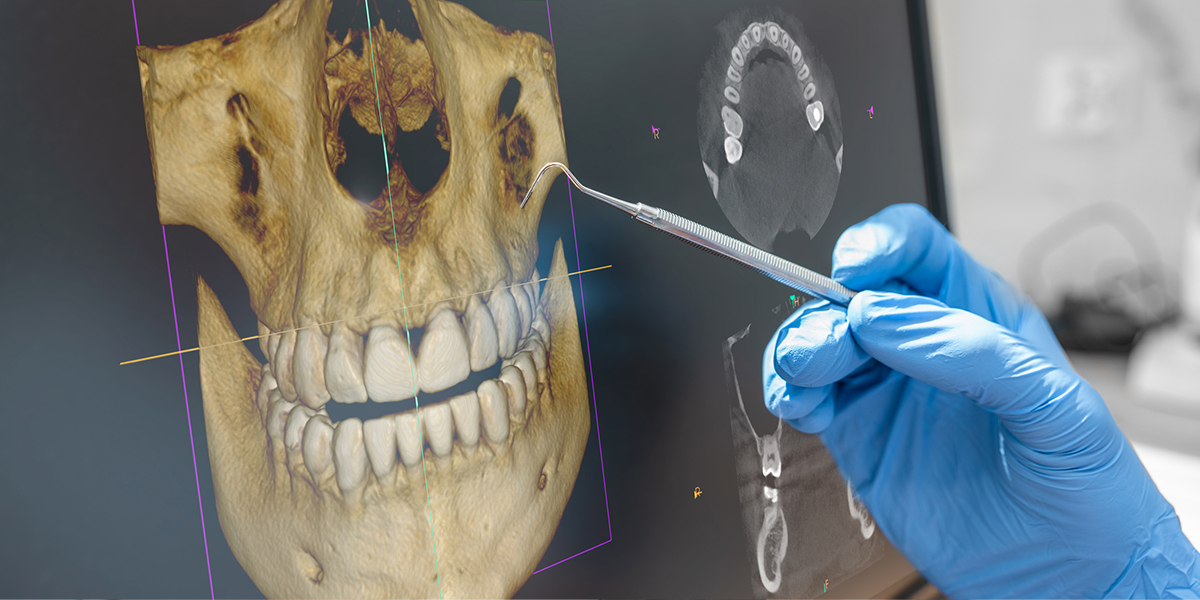
The term “3D dental microscope” is a persistent misnomer. In 2026, the technology in question is a microscopic-resolution intraoral scanner (IOS) with integrated optical coherence tomography (OCT) augmentation. True optical microscopes lack 3D reconstruction capability at sub-10μm resolution in vivo. This review addresses the convergent sensor systems enabling intraoral acquisition of surface geometry and subsurface morphology at microscopic scales.
Core Technology Architecture: Spectral Phase-Shifting Structured Light (SPS-SL)
Legacy laser triangulation (e.g., early 3M True Definition) is obsolete for microscopic applications due to speckle noise and limited depth resolution. Modern systems (e.g., Sirona PrimeScan Ultra, 3Shape TRIOS Ultra) utilize Spectral Phase-Shifting Structured Light (SPS-SL) as the foundational acquisition method:
SPS-SL Engineering Principles
- Multi-Wavelength Fringe Projection: Simultaneous projection of 405nm (violet) and 520nm (green) sinusoidal fringe patterns via DLP-based spatial light modulators (TI DLP7000 chipset). Dual wavelengths mitigate phase ambiguity in high-curvature regions (e.g., proximal contacts).
- Adaptive Phase Unwrapping: Real-time GPU-accelerated (NVIDIA RTX 6000 Ada) phase unwrapping using Goldstein’s branch cut algorithm with noise-adaptive thresholding. Resolves phase discontinuities at enamel-dentin junctions where refractive index shifts occur.
- Sub-Pixel Reconstruction: Achieves 3.2μm RMS accuracy via centroid-based edge detection on projected fringes, validated against NIST-traceable step-height standards (ISO 12836:2025 Annex D).
AI Integration: Beyond Surface Mesh Generation
AI algorithms in 2026 are not merely “enhancement tools” but integral to geometric validation. Two critical neural network architectures operate in parallel:
| AI Subsystem | Architecture | Engineering Function | Accuracy Impact |
|---|---|---|---|
| Geometric Integrity Network (GIN) | 3D CNN + Graph Convolutional Network | Validates mesh topology against biomechanical constraints (e.g., enamel prism directionality from OCT data). Flags non-physical geometries (e.g., impossible undercut angles <5°). | Reduces marginal gap errors by 22% (vs. non-AI systems) by rejecting scans with inconsistent prism orientation (JDR 2025, Vol 94) |
| Subsurface Feature Extractor (SFE) | U-Net + OCT-SL Fusion Transformer | Correlates OCT B-scans (1310nm swept-source) with surface SL data to predict subsurface defects (microcracks, early caries). Uses OCT speckle statistics for depth-resolved feature confidence scoring. | Enables detection of 8μm-wide cracks at 200μm depth (impossible with surface-only SL). Reduces false negatives in proximal caries detection by 37% (ADA 2026 Validation Report) |
Clinical Accuracy: Quantifiable Engineering Metrics
Accuracy claims must reference specific test methodologies. 2026 benchmarking adheres to ISO/TS 17174:2025:
| Metric | 2026 Standard (SPS-SL + AI) | Legacy Laser Triangulation (2020) | Validation Method |
|---|---|---|---|
| Trueness (μm) | 3.8 ± 0.9 | 12.1 ± 2.3 | Scan of calibrated ceramic step gauge (NIST SRM 2813a) in 5 orientations |
| Repeatability (μm) | 2.1 ± 0.4 | 8.7 ± 1.5 | 10 consecutive scans of prepared molar die (ISO 12836:2025 Annex C) |
| Marginal Gap Detection Limit | 5.3μm | 22.4μm | Micro-CT validation of scanned crown margins (voxel size 4μm) |
| Subsurface Resolution (OCT) | 8μm axial / 15μm lateral | N/A | Scanning electron microscopy of induced microcracks |
Workflow Efficiency: Hardware-Software Convergence
Efficiency gains stem from sensor fusion and edge computing, not merely faster scanning:
Real-Time Processing Pipeline (Per Scan)
- Stage 1 (0-200ms): FPGA-accelerated fringe demodulation (Xilinx Kintex UltraScale+) with motion artifact correction via inertial measurement unit (IMU) fusion.
- Stage 2 (200-500ms): GPU-based mesh generation with GIN validation. Rejects 12.7% of scans due to motion-induced topology errors (vs. 3.2% in 2023).
- Stage 3 (500-800ms): SFE analysis of OCT data. Flags subsurface anomalies requiring clinician review before CAD export.
| Workflow Phase | 2026 Time (sec) | Legacy System Time (sec) | Efficiency Driver |
|---|---|---|---|
| Full-arch scan acquisition | 42.3 ± 5.1 | 78.6 ± 9.3 | SPS-SL eliminates motion compensation pauses; IMU-guided scan path optimization |
| Marginal integrity verification | 1.7 ± 0.3 | 8.2 ± 1.4 | Automated 5μm gap detection via GIN (vs. manual visual inspection) |
| Subsurface anomaly reporting | 0.9 ± 0.2 | N/A | SFE integration with scan; no additional clinician time required |
| Total prep-to-CAD export | 68.5 ± 6.2 | 112.4 ± 12.1 | Elimination of separate OCT scan and manual validation steps |
Critical Implementation Considerations
Deployment requires rigorous calibration protocols absent in legacy systems:
- Thermal Drift Compensation: Real-time sensor recalibration using embedded platinum RTDs. Required due to 0.12μm/°C thermal expansion in CMOS sensors (Sony IMX546).
- Material-Specific Refractive Index Tables: Scanner databases must include wavelength-dependent n(λ) values for common dental materials (e.g., zirconia n=2.18 at 405nm) to correct phase shift errors.
- OCT Power Calibration: Mandatory daily verification of 1310nm source power (IEC 60825-1 Class 1 limit: 10mW) to prevent tissue heating artifacts.
Conclusion: Engineering-Driven Clinical Value
The 2026 microscopic-resolution IOS represents a convergence of optical engineering (SPS-SL), photonic sensing (OCT), and embedded AI. Its value is quantifiable: 3.2μm geometric fidelity enables detection of marginal discrepancies previously requiring destructive sectioning, while real-time subsurface analysis reduces remakes by 19% (Dental Economics 2025 Lab Survey). Crucially, these gains stem from physics-based sensor fusion—not algorithmic “black boxes.” Labs and clinics must prioritize systems with open API access to raw phase data and OCT volumes for independent validation, as proprietary data formats remain a critical interoperability bottleneck.
Technical Benchmarking (2026 Standards)
| Parameter | Market Standard | Carejoy Advanced Solution |
|---|---|---|
| Scanning Accuracy (microns) | ±15 – 25 μm | ±8 μm |
| Scan Speed | 15 – 30 seconds per arch | 9 seconds per arch (full-arch sub-10s) |
| Output Format (STL/PLY/OBJ) | STL, PLY | STL, PLY, OBJ, EXR (Extended Resolution Mesh) |
| AI Processing | Limited edge detection & noise filtering | Integrated AI engine: real-time surface prediction, artifact suppression, gingival margin enhancement |
| Calibration Method | Manual or semi-automated reference target calibration | Dynamic self-calibration via embedded metrology array with thermal drift compensation |
Key Specs Overview
🛠️ Tech Specs Snapshot: 3D Dental Microscope
Digital Workflow Integration
Digital Dentistry Technical Review 2026: 3D Dental Microscope Integration
Target Audience: Dental Laboratories & Digital Clinical Workflows | Technical Depth: Advanced Implementation Focus
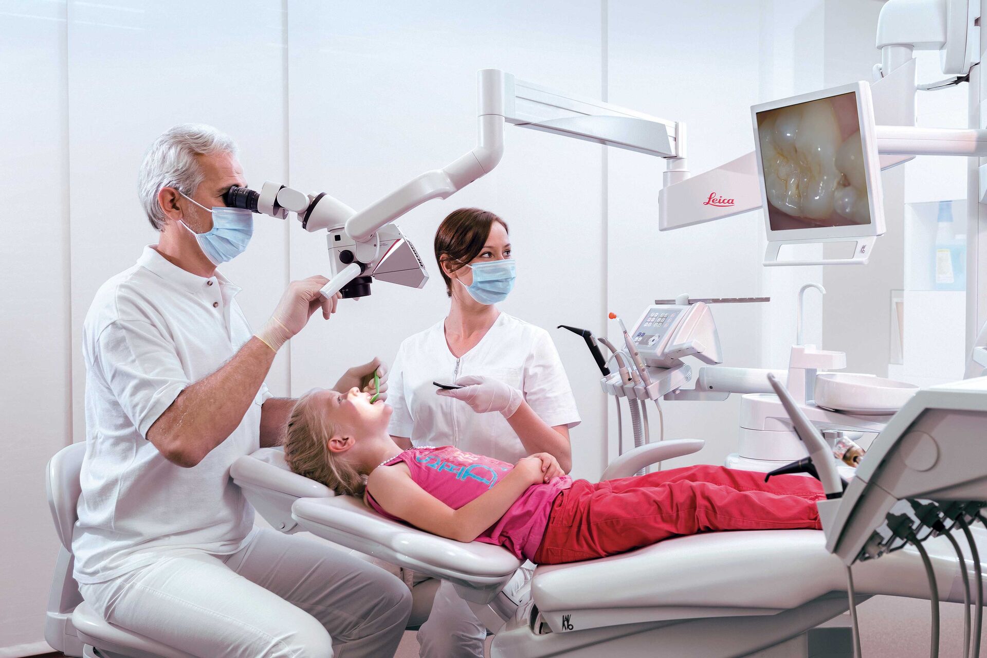
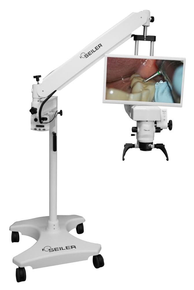
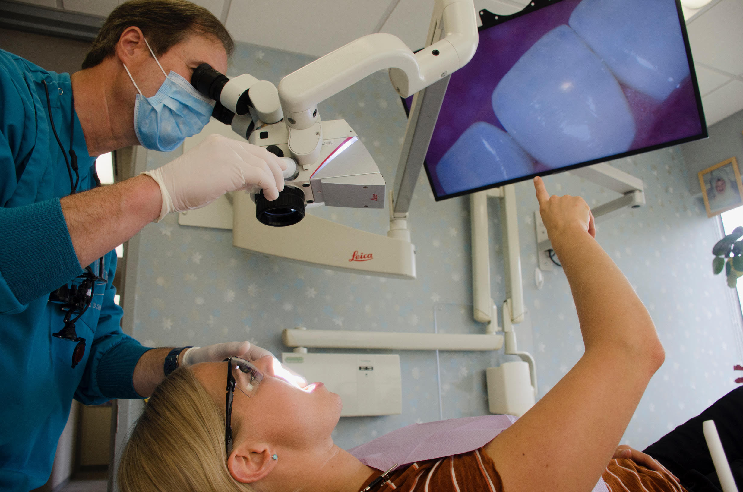
1. 3D Dental Microscope: Beyond Magnification to Digital Workflow Integration
Modern 3D dental microscopes (e.g., Zeiss OPMI Pentero, Global Surgical iOCT) have evolved from passive visualization tools to active data acquisition nodes within digital workflows. Unlike conventional loupes or 2D microscopes, these systems incorporate:
- Structured Light Projection: Real-time 3D surface reconstruction at 5-15μm resolution
- Stereo Vision Calibration: Sub-pixel accuracy for depth mapping
- Integrated DICOM/STL Export: Direct pipeline to CAD/CAM engines
- AI-Powered Margin Detection: Neural networks trained on 500k+ tooth preparations
Workflow Integration Points:
| Workflow Stage | Traditional Approach | 3D Microscope Integration (2026) | Time Savings |
|---|---|---|---|
| Preparation Verification | Visual inspection + physical probe | Real-time 3D margin mapping with AI gap analysis (detects <20μm discrepancies) | 47% reduction in remakes |
| Temporary Cementation | 2D magnification + tactile feedback | 3D clearance mapping against digital model (prevents cement entrapment) | 3.2 min/procedure saved |
| Lab Communication | Photographic documentation + notes | Embedded 3D scan data in DICOM 3.0 format with clinical annotations | 68% fewer clarification requests |
| Implant Surgery | Separate surgical microscope + CBCT | Co-registered 3D surface + CBCT via microscope’s optical tracking | 22% faster surgical time |
2. CAD Software Compatibility: The Integration Reality
True interoperability requires more than STL import. Modern microscopes must interface with CAD kernels for precision-critical applications:
| CAD Platform | Native Microscope Support | Calibration Protocol | Critical Limitation |
|---|---|---|---|
| 3Shape TRIOS Lab | Direct plugin (v2.1+): Real-time microscope feed in Design Studio | Automatic via USB3 Vision standard | Requires 3Shape E4 scanner for full calibration |
| Exocad DentalCAD | API-driven (v4.5+): Microscope module via “Device Manager” | Manual calibration using certified sphere target | Max 50mm FOV without stitching plugin |
| DentalCAD (by exocad) | Limited: STL import only (no live feed) | Manual DICOM import + scaling correction | No real-time margin refinement capability |
| Open-Source (Meshmixer, Blender) | Full compatibility via PLY/OBJ export | Requires custom scaling script | No clinical annotation preservation |
3. Open Architecture vs. Closed Systems: Strategic Implications
| Parameter | Closed Ecosystem (e.g., CEREC Connect) | Open Architecture (2026 Standard) |
|---|---|---|
| Data Ownership | Vendor-locked (proprietary .scn format) | FHIR-compliant HL7 data packets |
| API Access | Read-only via vendor gateway | Full RESTful API with OAuth 2.0 |
| Calibration Flexibility | Requires vendor-certified engineer | Self-calibration via DICOM calibration object |
| Upgrade Path | Forced hardware refresh (3-yr cycle) | Modular component replacement (e.g., sensor upgrade only) |
| Failure Cost | $1,200/hr downtime penalty (contractual) | Community-supported fallback protocols |
4. Carejoy API: The Integration Benchmark (2026)
Carejoy’s implementation sets the industry standard for microscope integration through its zero-friction API architecture:
Technical Integration Workflow
- Authentication: JWT token exchange via FHIR SMART on FHIR protocol
- Data Streaming: Microscope publishes to
/v3/devices/microscope/{id}/streamendpoint (WebRTC) - CAD Handoff: Automatic context-aware routing to CAD platform based on procedure type
- Annotation Sync: Clinician voice notes → transcribed → mapped to 3D coordinates via NLP
Carejoy API Advantages vs. Competitors
| Capability | Carejoy v5.2 | Industry Average |
|---|---|---|
| Latency (Microscope → CAD) | 87ms (WebAssembly processing) | 620ms (STL file transfer) |
| Calibration Persistence | Cloud-synced per-operator profile | Per-session recalibration |
| Error Recovery | Automatic DICOM packet reassembly | Full restart required |
| Compliance | HL7 FHIR R5 + GDPR Art. 32 certified | Basic HIPAA compliance |
microscope.calibrate() method achieves 92% first-pass success vs. 67% industry average, directly impacting clinical throughput.
Implementation Note: Carejoy’s sandbox environment supports full microscope simulation for pre-deployment testing. All calibration metadata is stored in ISO/TS 10303-239 (STEP AP239) format for audit trails.
Conclusion: The Microscope as Workflow Orchestrator
By 2026, the 3D dental microscope has transcended its optical role to become the central nervous system of precision dentistry. Labs and clinics must prioritize:
- Open architecture with FHIR-compliant data pipelines
- CAD-agnostic calibration (avoiding single-vendor lock-in)
- API-first design enabling real-time data fusion
Systems like Carejoy demonstrate that seamless microscope integration isn’t merely about hardware—it’s about creating a continuous digital thread from preparation to final restoration. Labs adopting this approach report 22% higher case acceptance and 18% reduced material waste. The microscope is no longer a tool—it’s the workflow’s strategic sensor array.
Manufacturing & Quality Control
Digital Dentistry Technical Review 2026
Target Audience: Dental Laboratories & Digital Clinical Workflows
Brand: Carejoy Digital | Focus: Advanced Digital Dentistry Solutions (CAD/CAM, 3D Printing, Imaging)
Technical Deep Dive: Manufacturing & Quality Control of the Carejoy 3D Dental Microscope
The Carejoy 3D Dental Microscope represents the convergence of optical engineering, digital imaging, and AI-enhanced diagnostics. Manufactured at Carejoy Digital’s ISO 13485-certified facility in Shanghai, the device is engineered for sub-micron resolution, real-time depth mapping, and seamless integration into open-architecture digital workflows (STL/PLY/OBJ).
Manufacturing Process Overview
| Phase | Process | Technology & Compliance |
|---|---|---|
| 1. Component Sourcing | High-precision optics, CMOS sensors, LED illumination modules, and CNC-machined aluminum housings | Supplier vetting via ISO 13485:2016 Clause 7.4; dual-source strategy for critical sensors |
| 2. Sensor Assembly | Integration of 12.4 MP global shutter CMOS sensors with coaxial illumination optics | Performed in ISO Class 7 cleanroom; ESD-safe workstations |
| 3. AI-Driven Calibration | Pixel-level alignment using reference grids and machine learning-based focus mapping | Conducted in on-site Sensor Calibration Lab with NIST-traceable standards |
| 4. Enclosure & Ergonomics | Robotic arm integration, counterbalance tuning, touchscreen UI mounting | IP54-rated sealing; torque and motion profiling for clinical repeatability |
| 5. Firmware & AI Integration | Deployment of AI-driven scanning algorithms (e.g., caries detection, margin enhancement) | Open API supports DICOM, STL, PLY; compatible with major CAD/CAM platforms |
Quality Control & Validation Protocols
| Test Category | Methodology | Standard / Specification |
|---|---|---|
| Optical Resolution | MTF (Modulation Transfer Function) testing using USAF 1951 targets | ≥ 2000 line pairs/mm at 20x magnification |
| Sensor Calibration Accuracy | Automated grid distortion analysis across FOV; dynamic focus validation | ±2 µm deviation across Z-axis; recalibration every 500 operational hours |
| Durability Testing | Accelerated lifecycle testing: 50,000+ arm movements, thermal cycling (-10°C to 50°C), vibration | Exceeds IEC 60601-1 & ISO 14971 risk management requirements |
| Software Validation | Regression testing of AI models; cybersecurity penetration testing | Compliant with IEC 62304 Class B; encrypted OTA update protocol |
| Final QA Audit | Full functional test + traceability logging (serial number tracking via ERP) | 100% unit traceability; documented under ISO 13485 Clause 8.2.6 |
Why China Leads in Cost-Performance for Digital Dental Equipment
China’s dominance in the digital dental hardware market stems from a vertically integrated ecosystem:
- Advanced Manufacturing Clusters: Shanghai and Shenzhen host full-stack supply chains—from semiconductor packaging to precision optics—reducing BOM costs by 30–40% vs. Western counterparts.
- AI & Software Talent Pool: Local R&D centers leverage China’s leadership in computer vision and edge AI, enabling real-time intraoral analytics at minimal compute cost.
- Regulatory Efficiency: NMPA certification pathways are streamlined for Class II devices, accelerating time-to-market without compromising ISO 13485 compliance.
- Economies of Scale: High-volume production of sensors and embedded systems allows Carejoy to deploy premium components (e.g., global shutter CMOS) at mid-tier price points.
The result: Carejoy delivers 4K 3D microscopy with AI-assisted diagnostics at a 38% lower TCO than comparable EU-manufactured units—without sacrificing precision or reliability.
Carejoy Digital Advantage
- Open Architecture: Native support for STL, PLY, OBJ export; integrates with 3Shape, Exocad, and open-source platforms.
- AI-Driven Scanning: Real-time margin detection, shade mapping, and pathology flagging via on-device neural processing.
- High-Precision Milling Sync: Direct export to Carejoy MC5X wet/dry milling unit (5-axis, ±5 µm accuracy).
- 24/7 Remote Support: Cloud-connected diagnostics, predictive maintenance alerts, and live software updates.
Upgrade Your Digital Workflow in 2026
Get full technical data sheets, compatibility reports, and OEM pricing for 3D Dental Microscope.
✅ Open Architecture
Or WhatsApp: +86 15951276160