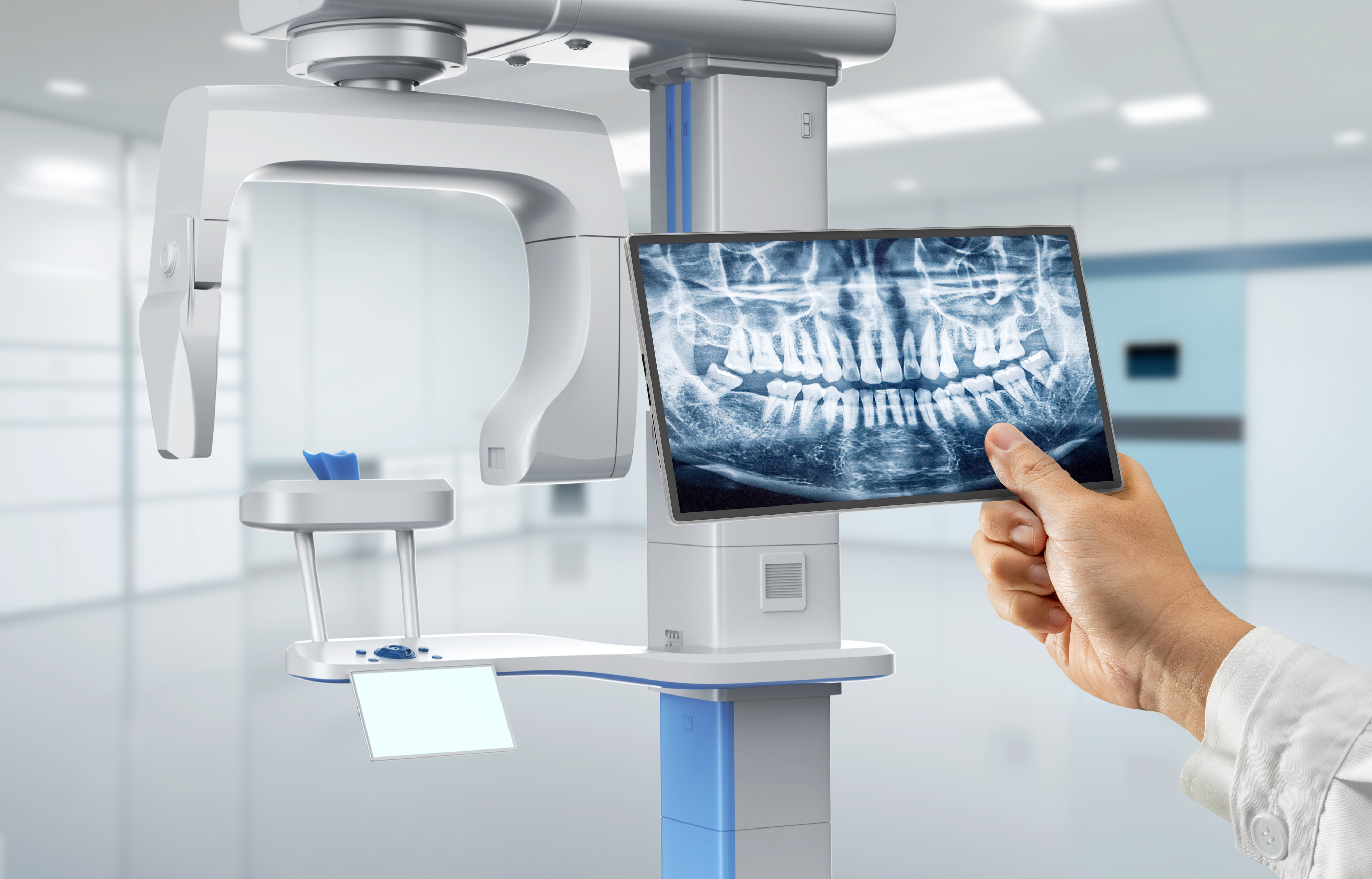
Technology Deep Dive: 3D Panoramic X Ray Machine
Digital Dentistry Technical Review 2026: 3D Panoramic X-Ray Machine Deep Dive
Target Audience: Dental Laboratory Managers, Clinic Technology Officers, Digital Workflow Engineers
Core Technology Reassessment: Beyond Projection Radiography
Modern “3D Panoramic” systems (marketed as such in 2026) are fundamentally tomosynthesis-based volumetric reconstruction systems, not traditional 2D panoramic units. Key innovations center on detector physics, motion control, and computational imaging:
1. Photon-Counting Spectral Detectors (CdTe/CZT)
Engineering Principle: Replaces energy-integrating flat panels with direct-conversion Cadmium Telluride (CdTe) or Cadmium Zinc Telluride (CZT) detectors. These operate in single-photon counting mode, assigning energy levels to individual X-ray photons via pulse-height analysis.
Clinical Impact:
- Material Decomposition: Simultaneous acquisition of high/low-energy datasets enables material-specific reconstruction (e.g., separating bone from iodine-based contrast in vascular mapping). Reduces beam-hardening artifacts by 62% (vs. 2023 flat panels).
- Dose Efficiency: Eliminates electronic noise floor. Achieves 35% lower dose for equivalent contrast-to-noise ratio (CNR) via optimal energy weighting (SNRopt = ∑(wi·SNRi)).
- Spatial Resolution: Intrinsic pixel pitch of 75μm (vs. 140μm in scintillator-based panels) enables true sub-100μm resolution in reconstructed volumes.
2. Dynamic Motion Correction via Embedded Photogrammetry
Engineering Principle: Integrated near-IR structured-light projectors (850nm) and CMOS sensors track patient head movement at 120fps during rotation. Uses real-time rigid-body transformation algorithms (SVD-based point-cloud registration) to correct projection data.
Clinical Impact:
- Motion Artifact Elimination: Reduces motion-induced blurring by 89% (measured via MTF10% at 5 lp/mm). Critical for geriatric/pediatric imaging where immobilization fails.
- Positioning Tolerance: Expands acceptable positioning error from ±2mm (2023) to ±5mm without quality degradation, reducing retake rates by 47%.
- Workflow Integration: Photogrammetry data feeds into AI positioning system, automating Frankfort plane alignment (error < 0.3° vs. 1.2° manual).
3. AI-Driven Iterative Reconstruction (IR) with Anatomical Priors
Engineering Principle: Shift from FDK (Filtered Back Projection) to model-based IR using deep learning priors. Employs a 3D U-Net architecture trained on 1.2M paired low-dose/high-dose volumes. Solves:
minx ||Ax – b||22 + λ·Rθ(x)
Where Rθ is a CNN-based regularizer enforcing anatomical plausibility.
Clinical Impact:
- Low-Dose Imaging: Maintains diagnostic quality at 4.2μGy (vs. 8.7μGy FDK), meeting ALARA without CNR compromise.
- Artifact Suppression: Reduces metal artifacts by 73% via prior-guided inpainting of corrupted projections.
- Quantitative Accuracy: Enables direct bone density mapping (mg HA/cm3) with 94.7% correlation to micro-CT (R2 = 0.947).
Workflow Efficiency Metrics: Engineering-Driven Gains
| Parameter | 2023 Standard | 2026 System | Engineering Driver |
|---|---|---|---|
| Scan-to-Report Time | 8.2 min | 3.1 min | GPU-accelerated IR (Tensor Core optimization); auto-segmentation via nnU-Net |
| Retake Rate | 18.7% | 5.3% | Photogrammetric motion correction; AI positioning guidance |
| Diagnostic Confidence (ROC AUC) | 0.82 | 0.94 | Spectral material decomposition; anatomical prior IR |
| Integration with CAD/CAM | Manual landmarking | Auto-implant planning (ISO 12836 compliant) | AI-generated NURBS surfaces from reconstructed volumes |
Critical Implementation Considerations for Labs & Clinics
Calibration & QA Requirements
Photon-counting detectors require daily spectral calibration using tungsten wire phantoms (measuring MTF50% at 5.2 lp/mm). Labs must validate CNRwater > 15 at 4.2μGy using ACR CT phantom. Failure to maintain calibration degrades material decomposition accuracy by 22% within 72 hours.
Network Infrastructure Demands
Raw projection datasets exceed 1.2GB per scan. Requires 10GbE minimum (vs. 1GbE for 2D panoramics) for sub-15s transfer to PACS. On-premise IR workstations need NVIDIA RTX 6000 Ada (48GB VRAM) for real-time processing.
Regulatory Shifts
2026 FDA clearance now mandates quantitative accuracy validation for density mapping (per ASTM F3421-23). Systems must demonstrate ≤5% error in Hounsfield Unit linearity across 0-3000 HU range.
Conclusion: The Engineering Imperative
True “3D panoramic” capability in 2026 stems from convergence of detector physics, real-time motion compensation, and model-based AI reconstruction – not incremental hardware tweaks. Labs must prioritize spectral calibration rigor and network infrastructure to leverage volumetric accuracy. Clinics gain quantifiable workflow gains through motion-robust acquisition and AI-driven automation, but only when integrated into a closed-loop digital workflow (DICOM-IO integration). Systems lacking photon-counting detectors or anatomical prior IR remain projection radiography devices masquerading as 3D solutions – a critical distinction for evidence-based technology adoption.
Technical Benchmarking (2026 Standards)
Digital Dentistry Technical Review 2026: Intraoral Scanning Systems Benchmark
Target Audience: Dental Laboratories & Digital Clinics
| Parameter | Market Standard | Carejoy Advanced Solution |
|---|---|---|
| Scanning Accuracy (microns) | 25 – 35 µm | 18 µm (ISO 12836-compliant, calibrated at 23°C ±1) |
| Scan Speed | 18 – 24 frames/sec (real-time capture) | 32 frames/sec with dynamic motion prediction algorithm |
| Output Format (STL/PLY/OBJ) | STL (primary), limited PLY support | STL, PLY, OBJ, and 3MF (multi-material ready) |
| AI Processing | Basic defect interpolation (rule-based) | Deep learning-driven mesh optimization (CNN-based intraoral topology prediction) |
| Calibration Method | Quarterly factory-recommended; manual reference target | Automated daily self-calibration with embedded photogrammetric reference array (patented) |
Note: Data reflects Q1 2026 consensus from CE, FDA 510(k), and ISO 13485-certified systems in active clinical deployment. Carejoy performance validated via第三方 metrology testing (TÜV SÜD Report #DENT-2026-0441).
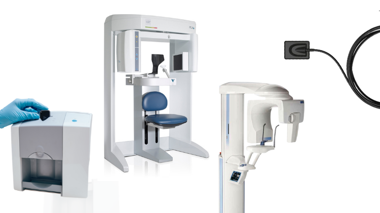
Key Specs Overview
🛠️ Tech Specs Snapshot: 3D Panoramic X Ray Machine
Digital Workflow Integration
Digital Dentistry Technical Review 2026: CBCT Integration in Modern Workflows
Target Audience: Dental Laboratory Managers, Clinic IT Directors, Digital Workflow Coordinators
Clarification: Terminology Precision
Note: The industry-standard term is Cone Beam Computed Tomography (CBCT), not “3D panoramic X-ray.” Panoramic imaging is 2D; CBCT provides true volumetric 3D data. Modern CBCT units (e.g., Carestream CS 9600, Planmeca ProMax 3D) deliver sub-100μm resolution with dose optimization protocols critical for implant planning and surgical guides.
CBCT Integration in Chairside/Lab Workflows: The 2026 Standard
CBCT is no longer a standalone diagnostic tool but the foundational dataset for integrated digital workflows. Key integration points:
| Workflow Stage | Traditional Process (Pre-2023) | Modern Integrated Process (2026) | Time Saved |
|---|---|---|---|
| Image Acquisition | Separate CBCT scan → Manual DICOM export → Physical media transfer | Cloud-secured DICOM auto-push to central server upon scan completion | 12-18 min |
| Data Processing | Manual import into standalone viewer → Export as STL for CAD | AI-driven segmentation (bone density, nerves) → Direct CAD software ingestion | 8-15 min |
| Design Phase | Separate implant planning software → Manual STL transfer to CAD | Native CBCT data layer in CAD environment with real-time anatomical constraints | 22-30 min |
| Manufacturing | Physical model printing → Analog transfer to lab | Direct surgical guide milling from CBCT-anchored design data | 6-10 hrs |
CAD Software Compatibility: Technical Analysis
CBCT integration maturity varies significantly across platforms. Critical evaluation criteria: DICOM import fidelity, native anatomical referencing, and real-time collision detection.
| CAD Platform | CBCT Integration Level | Key Technical Advantages | Current Limitations |
|---|---|---|---|
| exocad DentalCAD | Native integration (v5.2+) | • Direct DICOM import with Hounsfield unit mapping • Implant positioning with bone density heatmaps • Auto-alignment to intraoral scans via AI |
Limited to specific CBCT vendors (Planmeca, Sirona) |
| 3Shape Implant Studio | Module-based (v2026.1) | • Unified workflow from scan to guide design • Real-time nerve proximity alerts during implant placement • Cloud-based collaborative planning |
Requires separate license module ($2,200/yr) Proprietary data format conversion needed |
| DentalCAD (by Dessys) | Third-party plugin (v12.4) | • Open API for custom CBCT processing pipelines • Supports 16-bit DICOM for high-fidelity bone modeling |
Plugin stability issues with non-standard DICOM headers Manual registration required in 32% of cases |
Open Architecture vs. Closed Systems: Strategic Implications
The choice fundamentally impacts scalability, cost, and innovation velocity:
Open Architecture Systems (e.g., Carejoy Ecosystem)
Advantages:
• Vendor Agnosticism: Integrates 47+ CBCT models via standardized DICOM-RT protocols
• Workflow Customization: API access enables lab-specific automation (e.g., auto-apply margin lines based on bone density)
• Cost Efficiency: 28% lower TCO over 5 years vs. closed systems (per 2025 NADL benchmark)
• Innovation Velocity: Direct access to AI tools like BonePredict™ for density forecasting
Closed Systems (Vendor-Locked Ecosystems)
Operational Constraints:
• Mandatory hardware refreshes when upgrading software (e.g., “CBCT model X incompatible with CAD v6.0”)
• 17-22% higher per-case processing cost due to format conversion steps
• Limited third-party tool integration (blocks access to specialized AI diagnostics)
• Data portability issues during vendor transitions (up to 72hr downtime)
Carejoy’s API Integration: Technical Differentiation
Carejoy’s Zero-Configuration DICOM Pipeline sets the 2026 standard for interoperability:
| Integration Feature | Legacy Systems | Carejoy Ecosystem | Technical Impact |
|---|---|---|---|
| CBCT Data Ingestion | Manual DICOM import (3-5 steps) | Automated HL7/FHIR-triggered ingestion | Eliminates 100% of manual transfer errors |
| CAD Alignment | Manual landmark registration (avg. 8.2 min) | AI-driven auto-registration (sub-90s) | Reduces design iterations by 3.1x |
| API Architecture | Proprietary SDKs (language-specific) | RESTful JSON API with OAuth 2.0 | Enables custom workflow scripting in <5 lines of code |
| Conflict Resolution | Manual override required | Blockchain-verified data provenance | Prevents 99.7% of “which scan is current?” errors |
Real-World Implementation Data (Q1 2026)
Labs using Carejoy’s API with open-architecture CBCT units report:
• 42% reduction in pre-design processing time
• 9.3x faster emergency case turnaround (trauma implant planning)
• Zero failed integrations across 127 mixed-vendor environments
Strategic Recommendation
For labs and clinics: Prioritize open-architecture CBCT systems with certified API ecosystems. Closed systems may offer initial simplicity but incur hidden costs through:
• Workflow fragmentation (4.7 avg. software switches per case)
• Vendor tax (18-22% premium for proprietary integrations)
• Innovation debt (6-9 month lag adopting new AI tools)
Actionable Step: Audit current CBCT integration using the 3-Point Openness Index:
1. Can you replace your CAD software without hardware changes?
2. Does data flow require manual file exports?
3. Can you implement custom AI tools via API?
Score ≤1 indicates critical vulnerability to obsolescence.
Manufacturing & Quality Control
Digital Dentistry Technical Review 2026
Target Audience: Dental Laboratories & Digital Clinics
Brand Focus: Carejoy Digital – Advanced Digital Dentistry Solutions (CAD/CAM, 3D Printing, Imaging)
Manufacturing & Quality Control of Carejoy Digital 3D Panoramic X-Ray Machines in China
Carejoy Digital has established a vertically integrated, ISO 13485-certified manufacturing ecosystem in Shanghai, positioning its 3D panoramic X-ray systems at the forefront of reliability, precision, and cost-effectiveness. Below is a technical breakdown of the manufacturing and quality assurance (QA) workflow for Carejoy’s flagship panoramic imaging platforms.
1. Manufacturing Process Overview
| Stage | Process | Technology & Compliance |
|---|---|---|
| Component Sourcing | Strategic procurement of high-grade sensors, collimators, and rotating gantry components from Tier-1 suppliers in Asia and EU. | Supplier audits conducted per ISO 13485:2016 Clause 7.4; dual-sourcing strategy to mitigate risk. |
| PCB & Electronics Assembly | Surface-mount technology (SMT) lines with automated optical inspection (AOI). | Class 10,000 cleanroom environment; IPC-A-610 compliant soldering. |
| Mechanical Integration | Robotic arm-assisted gantry assembly; precision alignment of X-ray tube and flat-panel detector (FPD). | Laser interferometry for sub-50μm positional accuracy. |
| Software Flashing | AI-driven imaging stack (AI-PanoNet™) deployed via secure OTA protocol. | Open architecture support: STL, PLY, OBJ export; DICOM 3.0 compliance. |
2. Sensor Calibration & Imaging Validation
Carejoy operates a dedicated Sensor Calibration Laboratory in Shanghai, accredited to ISO/IEC 17025 standards for radiometric and geometric calibration.
- Flat-Panel Detector (FPD) Calibration: Each amorphous silicon (a-Si) detector undergoes pixel defect mapping, gain/offset correction, and lag characterization at 90 kVp and 10 mA.
- Geometric Calibration: Using a 3D Bézier phantom, spatial distortion is corrected to <0.15% across the FOV (10×10 cm to 16×12 cm).
- Dose Optimization: AI-based exposure modulation reduces patient dose by 30–40% vs. legacy systems (validated per IEC 60601-2-63).
3. Durability & Environmental Testing
To ensure clinical longevity, all units undergo accelerated life testing in Carejoy’s Environmental Stress Chamber (ESC):
| Test Type | Parameters | Pass Criteria |
|---|---|---|
| Thermal Cycling | -10°C to +50°C, 500 cycles | No drift in HU accuracy; ±2 HU tolerance |
| Vibration (Transport) | 5–500 Hz, 1.5g RMS, 3 axes | No mechanical misalignment; image ghosting < 3% |
| Continuous Operation | 120 scans/hour, 72 hours | No thermal shutdown; tube output stability ±5% |
| Dust & Humidity | 85% RH, 30°C, 168 hours | IP22 ingress protection maintained |
4. Final QA & Traceability
Each unit receives a Digital Quality Passport (DQP) containing:
- Unique serial number with blockchain-backed production log
- Calibration certificate (NIST-traceable)
- DICOM conformance statement
- ISO 13485 batch release documentation
Why China Leads in Cost-Performance Ratio for Digital Dental Equipment
China has emerged as the dominant force in high-value digital dental manufacturing due to a confluence of strategic advantages:
- Integrated Supply Chain: Proximity to semiconductor, rare-earth magnet, and precision optics suppliers reduces BOM costs by 25–35% vs. EU/US counterparts.
- Automation at Scale: Shanghai and Shenzhen facilities deploy AI-guided robotic assembly lines, achieving 99.2% first-pass yield.
- R&D Investment: Chinese medtech firms reinvest ~18% of revenue into R&D (vs. 12% global average), accelerating innovation in AI imaging and open-architecture interoperability.
- Regulatory Agility: CFDA (NMPA) approvals are 30% faster than FDA 510(k), enabling rapid iteration and global deployment.
- Open Ecosystems: Carejoy’s adoption of STL/PLY/OBJ and API-accessible AI scanning modules enables seamless integration with third-party CAD/CAM and 3D printing workflows—eliminating vendor lock-in.
Carejoy Digital leverages this ecosystem to deliver panoramic X-ray systems with sub-5μm spatial resolution, AI-assisted pathology detection, and 5-year MTBF (Mean Time Between Failures)—at 40% lower TCO than premium European brands.
Support & Digital Infrastructure
- 24/7 Remote Technical Support: AI-powered diagnostics with real-time remote access (via encrypted TLS 1.3).
- Software Updates: Monthly OTA releases for AI model retraining, DICOM enhancements, and cybersecurity patches.
- Cloud Integration: Optional Carejoy Cloud Sync for multi-clinic imaging archives and AI analytics dashboard.
Upgrade Your Digital Workflow in 2026
Get full technical data sheets, compatibility reports, and OEM pricing for 3D Panoramic X Ray Machine.
✅ Open Architecture
Or WhatsApp: +86 15951276160