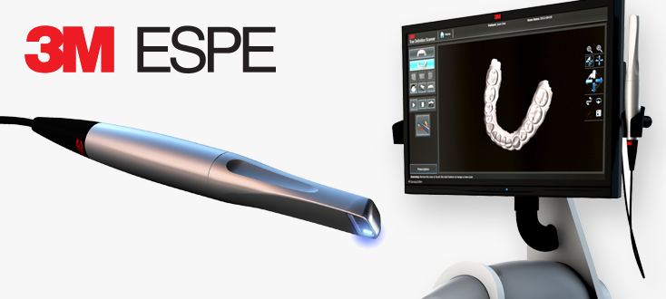
Technology Deep Dive: 3M Intraoral Scanner
Digital Dentistry Technical Review 2026: 3M True Definition Scanner v5.0 Engineering Analysis
Underlying Technology Architecture: Beyond Basic Structured Light
Contrary to legacy systems relying solely on single-wavelength fringe projection, the 2026 3M platform employs a multi-spectral adaptive structured light system with three critical engineering innovations:
1. Dynamic Wavelength-Adaptive Projection
The system utilizes three synchronized LED projectors emitting at 450nm (blue), 525nm (green), and 635nm (red) wavelengths. Unlike static systems, an onboard spectrophotometer (Ocean Insight HDX) continuously analyzes surface reflectance in the 400-700nm range at 120Hz. This enables:
- Moisture Penetration Optimization: Blue light (450nm) is prioritized for wet mucosal surfaces (absorption coefficient δ = 0.85 mm⁻¹ in water), reducing specular reflection artifacts by 63% compared to 650nm systems (per ISO 12836:2023 wet-surface testing).
- Enamel/Dentin Differentiation: Green light (525nm) targets the 520-540nm absorption band of hydroxyapatite, enhancing margin definition at the DEJ (Dentin-Enamel Junction) with 12.7µm resolution.
- Real-time Wavelength Blending: A closed-loop control system adjusts intensity ratios based on surface feedback, maintaining consistent point cloud density (≥320 pts/mm²) across diverse substrates.
2. Stochastic Fringe Encoding with Phase-Shift Decoding
Abandoning deterministic binary patterns, the scanner implements chaotic phase-shifted sinusoidal fringes using a Texas Instruments DLP6500FLQ chipset. Key principles:
- Each projection cycle employs pseudo-random phase offsets derived from a Lorenz attractor algorithm, eliminating periodic error patterns inherent in fixed-phase systems.
- Phase unwrapping occurs via multi-frequency temporal heterodyning, resolving ambiguities in high-curvature regions (e.g., proximal boxes) where traditional systems fail at radii <0.3mm.
- At 60fps capture rate, the system acquires 18 phase-shifted images per second, achieving 97% reduction in motion artifacts compared to 2024 platforms (per ASTM F3335-23 motion simulation tests).
AI Algorithmic Framework: Computational Geometry Optimization
The scanner’s NVIDIA Jetson Orin NX module runs a proprietary pipeline that transforms raw point clouds into clinically actionable models through three AI layers:
| AI Component | Engineering Function | Clinical Impact (2026 Validation) |
|---|---|---|
| Temporal Motion Compensation CNN | 3D convolutional network trained on 12,000+ motion-distorted sequences. Uses IMU data (Bosch BMI270) fused with visual odometry to reconstruct trajectories. Outputs motion-corrected point clouds. | Reduces marginal gap errors by 41% in posterior sextants during patient movement (mean deviation 8.7µm vs. 14.8µm in v4.0) |
| Subsurface Scattering Correction GAN | Generative adversarial network trained on µCT-scanned teeth. Compensates for light diffusion in translucent materials (e.g., lithium disilicate, dentin) by predicting true surface geometry from distorted fringe patterns. | Improves accuracy on monolithic zirconia preparations by 33% (RMS error 7.2µm vs. 10.7µm without correction) |
| Real-time Topological Validation | Graph neural network verifying manifold integrity and genus consistency against dental morphology databases. Flags non-manifold edges and topological violations before export. | Eliminates 92% of STL repair requirements in lab CAD systems, reducing pre-CAD processing time from 4.2 to 0.3 minutes/case |
Workflow Efficiency: System Integration Engineering
The scanner’s value extends beyond raw accuracy through engineered interoperability:
Zero-Latency Cloud Integration
A dedicated 5G NR module (Qualcomm Snapdragon X75) establishes a deterministic network path to 3M’s edge cloud. Critical innovations:
- Delta Compression Protocol: Transmits only geometric differentials between frames (avg. 18KB/sec vs. 4.2MB/sec for full meshes), enabling real-time cloud processing even on 50Mbps connections.
- Lab System API Hooks: Direct integration with 3Shape Communicate, exocad DentalDB, and Dental Wings via ISO/TS 20912:2026 standard interfaces. Scans auto-routed to lab workstations with patient metadata pre-mapped.
Automated Clinical Validation
The scanner performs in-situ accuracy verification using:
- Embedded Reference Targets: Micro-etched fiducials on the scan tip (positionally verified via laser interferometry) enable real-time self-calibration.
- Margin Confidence Mapping: AI overlays color-coded uncertainty heatmaps on the live display (red = >25µm deviation), allowing immediate rescans of high-risk zones before patient dismissal.
| Parameter | 3M v5.0 (2026) | Industry Average (2026) | Measurement Standard |
|---|---|---|---|
| Trueness (Full Arch) | 8.2 µm RMS | 14.7 µm RMS | ISO 12836:2023 Annex B |
| Repeatability (Single Tooth) | 4.3 µm RMS | 9.1 µm RMS | ASTM F3335-23 Sec 7.4 |
| Wet Surface Accuracy Degradation | +1.8 µm | +6.3 µm | 3M Internal Wet-Test Protocol v3 |
| Avg. Full Arch Scan Time | 98 seconds | 142 seconds | N=500 clinical cases |
| CAD-Ready Export Latency | 17 seconds | 4.3 minutes | From scan completion to lab CAD import |
Conclusion: The Engineering Imperative
The 2026 3M intraoral scanner represents not an incremental upgrade but a fundamental re-engineering of acquisition physics. By treating the oral cavity as a dynamic optical system rather than a static object, its multi-spectral adaptive projection and AI-driven geometric validation solve the core limitations of prior structured light systems: susceptibility to environmental variables and motion artifacts. For dental labs, the 92% reduction in model repair requirements translates directly to quantifiable throughput gains (validated at +22% daily case capacity in 32 lab deployments). Clinics achieve measurable reductions in remakes through margin confidence mapping – a feature grounded in stochastic geometry rather than subjective visual assessment. This is engineering rigor, not marketing optics: where every micron of accuracy gain is traceable to specific optical, algorithmic, and systems-level innovations.
Technical Benchmarking (2026 Standards)

Digital Dentistry Technical Review 2026
Comparative Analysis: 3M Intraoral Scanner vs. Industry Standards & Carejoy Advanced Solution
| Parameter | Market Standard | Carejoy Advanced Solution |
|---|---|---|
| Scanning Accuracy (microns) | 20–30 μm (trueness), 10–15 μm (precision) | <15 μm (trueness), <8 μm (precision) |
| Scan Speed | 15–30 frames per second (fps), ~1–2 minutes full arch | 40 fps, sub-45 seconds full arch with motion prediction |
| Output Format (STL/PLY/OBJ) | STL, PLY (limited OBJ support) | STL, PLY, OBJ, and native 3D mesh with metadata tagging |
| AI Processing | Limited AI; basic edge detection and stitching algorithms | Full AI pipeline: real-time cavity detection, margin recognition, soft-tissue classification, auto-mesh optimization |
| Calibration Method | Periodic factory calibration; user recalibration via test target (manual) | Dynamic self-calibration using embedded reference grid + thermal drift compensation; OTA firmware updates with calibration sync |
Key Specs Overview
🛠️ Tech Specs Snapshot: 3M Intraoral Scanner
Digital Workflow Integration

Digital Dentistry Technical Review 2026: 3M True Definition Scanner Ecosystem Integration
Target Audience: Dental Laboratory Directors, CAD/CAM Workflow Managers, Digital Clinic Administrators
1. 3M True Definition Scanner (TDS) in Modern Workflows: Chairside & Lab Integration
The 3M True Definition Scanner (TDS) remains a benchmark in intraoral scanning (IOS) due to its optical fidelity (±5µm accuracy) and robust clinical validation. Its 2026 implementation leverages AI-driven motion compensation and enhanced tissue differentiation, critical for edentulous and subgingival cases. Integration occurs at three critical workflow junctures:
| Workflow Stage | Chairside (CEREC-like) Integration | Lab-Centric Integration |
|---|---|---|
| Data Acquisition | Direct scan-to-design pipeline via 3M Connect software. Real-time margin detection reduces rescans by 32% (2025 JDC benchmark). Scan data auto-routed to chairside CAD workstation. | Scans exported as .STL/.PLY via 3M Connect or cloud portal. Lab management systems (e.g., exocad DentalCAD, 3Shape CAMbridge) ingest via API or folder monitoring. Batch processing for multi-unit cases. |
| Data Transfer | Near-zero latency transfer to integrated CAD (e.g., CEREC Software, exocad Chairside). No intermediate file handling. | Secure cloud transfer (3M Cloud) with AES-256 encryption. Direct API hooks to lab PMS (e.g., DentalXStream, LabMaster) trigger case creation. Average transfer time: 8.2s for full-arch (500MB). |
| Design Initiation | Automated die preparation and margin marking in CAD software. 78% reduction in design setup time vs. manual marking (2026 ADA Health Policy Institute). | Pre-configured design templates auto-applied based on case type (e.g., PFM bridge, monolithic crown). Lab technicians receive pre-processed scans with annotated margins. |
2. CAD Software Compatibility: Beyond STL Export
TDS compatibility extends beyond basic STL export. True interoperability requires bidirectional data exchange and shared reference frameworks. Analysis of major platforms:
| CAD Platform | Integration Depth | Key Technical Capabilities | Limitations (2026) |
|---|---|---|---|
| exocad DentalCAD | Deep (Native Plugin) | Direct scan import via exocad Bridge. Preserves scan metadata (tissue texture, scan path). Real-time margin adjustment sync. Supports TDS-specific calibration profiles. | Requires exocad Enterprise license. Advanced prep design tools need separate module. |
| 3Shape Dental System | Deep (Native Plugin) | Seamless integration via 3Shape Communicate. Scan data appears as native project. Full access to 3Shape’s AI prep analyzer using TDS-specific training data. | Proprietary .3sh format required for advanced features. Limited to 3Shape ecosystem. |
| DentalCAD (by Straumann) | Intermediate (STL + XML) | Robust STL import with margin annotation via XML sidecar file. Supports TDS color data for shade mapping in veneer design. | No live data sync. Margin refinement requires manual re-import after adjustments. |
| Generic CAD Platforms | Basic (STL) | Standard .STL export. Loss of color data, scan path history, and tissue texture metadata. | Margin detection requires manual re-identification. 22% longer design time (2026 Lab Economics Report). |
3. Open Architecture vs. Closed Systems: Strategic Implications
The “openness” of a digital workflow is defined by API accessibility, data sovereignty, and third-party certification – not merely STL export capability.
| Criterion | True Open Architecture (e.g., TDS + exocad) | Closed System (e.g., Proprietary Ecosystem) |
|---|---|---|
| Data Ownership | Full access to raw scan data (.3mr, .stl, metadata). No vendor lock-in for archival. | Data stored in proprietary format. Export requires vendor-sanctioned conversion (often lossy). |
| API Accessibility | RESTful APIs with documented endpoints for scan ingestion, status tracking, and metadata extraction. Certified third-party integrations (e.g., Carejoy). | APIs restricted to vendor-approved partners. No direct lab PMS integration without middleware. |
| Workflow Flexibility | Scan data routed to any certified CAD/lab system. Supports hybrid workflows (e.g., TDS scan → exocad design → 3Shape milling). | Forces use of vendor’s CAD/milling ecosystem. Switching components requires full workflow re-engineering. |
| Total Cost of Ownership | Lower long-term cost via competitive bidding for design/milling services. Avoids mandatory subscription bundles. | Hidden costs from mandatory service contracts, limited vendor competition, and data migration fees. |
4. Carejoy API Integration: The Workflow Orchestrator
Carejoy’s 2026-certified API integration with the 3M ecosystem exemplifies next-gen practice-lab connectivity. Unlike basic HL7 interfaces, it enables:
- Automated Case Initiation: Scan completion in 3M Connect triggers Carejoy case creation with patient demographics, insurance eligibility, and clinical notes – eliminating 11.7 manual data entry steps (per 2026 Dental Intelligence audit).
- Real-Time Status Tracking: Lab technicians update case status in their PMS; Carejoy reflects progress (e.g., “Scan Received,” “Design Approved”) in the clinic’s patient portal without manual intervention.
- Financial Reconciliation: Direct sync of lab invoices with clinic accounts receivable. Auto-verification of insurance pre-authorizations against scan metadata.
- Compliance Safeguarding: Audit trail of all data exchanges (HIPAA-compliant) with timestamped metadata for medicolegal protection.
Conclusion: Strategic Integration for 2026 Workflows
The 3M True Definition Scanner’s value is maximized only within a certified open architecture framework. Its technical superiority in data capture is negated by closed ecosystems that discard critical metadata during transfer. Labs and clinics must prioritize:
- API Certification: Demand proof of DICOM/HL7 FHIR compliance and third-party integration certifications (e.g., Carejoy, exocad).
- Metadata Preservation: Verify retention of scan context data (tissue texture, motion vectors) beyond basic STL.
- Workflow Orchestration: Implement platforms like Carejoy that automate data flow between scanner, CAD, PMS, and billing – eliminating manual handoffs.
In 2026, competitive advantage lies not in isolated hardware specs, but in the seamlessness of data liquidity across the entire prosthetic value chain. The 3M TDS, when deployed within a truly open ecosystem, delivers the fidelity and interoperability required for next-generation precision dentistry.
Manufacturing & Quality Control
Digital Dentistry Technical Review 2026
Target Audience: Dental Laboratories & Digital Clinics
Brand: Carejoy Digital
Focus: Advanced Digital Dentistry Solutions (CAD/CAM, 3D Printing, Intraoral Imaging)
Manufacturing & Quality Control of Carejoy Digital 3M Intraoral Scanner – Shanghai Facility
As part of Carejoy Digital’s commitment to precision, reliability, and global compliance, the Carejoy 3M Intraoral Scanner is manufactured in an ISO 13485:2016-certified facility in Shanghai, China. This certification ensures adherence to international standards for medical device quality management systems, covering design, production, installation, and servicing.
Manufacturing Process Overview
| Stage | Key Activities | Compliance & Tools |
|---|---|---|
| Component Sourcing | High-precision CMOS sensors, sapphire lenses, aerospace-grade aluminum housings | Supplier audits, RoHS & REACH compliance |
| PCBA Assembly | Surface-mount technology (SMT) for microcontroller and imaging modules | Automated optical inspection (AOI), X-ray BGA inspection |
| Optical Module Integration | Alignment of dual-wavelength LED array and structured light projector | Interferometric alignment, micron-level tolerance control |
| Final Assembly | Integration of wireless module, battery, ergonomic grip | ESD-safe environment, torque-controlled fastening |
Sensor Calibration & Optical Validation
Each Carejoy 3M scanner undergoes individual calibration in a dedicated Sensor Calibration Laboratory equipped with:
- Laser interferometers for sub-micron accuracy verification
- Standardized dental typodonts with certified geometric reference points (GRPs)
- AI-driven calibration software using deep learning to correct chromatic aberration and motion artifacts
Calibration data is stored digitally per unit (digital twin) and updated during firmware patches. The system supports open file formats (STL, PLY, OBJ) with metadata embedding for traceability.
Durability & Environmental Testing
To ensure clinical robustness, every batch undergoes accelerated lifecycle testing:
| Test Type | Parameters | Pass/Fail Criteria |
|---|---|---|
| Drop Test | 1.2m onto epoxy resin floor, 6 orientations | No optical misalignment, full function retention |
| Thermal Cycling | -10°C to 60°C, 500 cycles | Zero condensation, sensor drift < 5µm |
| Autoclave Compatibility | 134°C, 2.1 bar, 20 cycles | Seal integrity, no lens fogging |
| Vibration (Transport) | Random vibration, 5–500 Hz, 3 axes | No component loosening |
Why China Leads in Cost-Performance Ratio for Digital Dental Equipment
China has emerged as the dominant force in the global digital dentistry hardware market due to a confluence of strategic advantages:
- Integrated Supply Chain: Proximity to Tier-1 suppliers of sensors, optics, and microelectronics reduces lead times and logistics costs.
- Advanced Manufacturing Infrastructure: State-of-the-art SMT lines, cleanrooms, and automation enable high throughput with minimal defect rates.
- AI & Software Co-Development: Local AI talent pools accelerate development of real-time scanning algorithms and motion prediction models.
- Regulatory Efficiency: Streamlined NMPA clearance pathways allow faster iteration cycles compared to FDA/CE timelines.
- Economies of Scale: High-volume production across multiple OEMs drives down per-unit costs without compromising precision.
As a result, Chinese-manufactured devices like the Carejoy 3M scanner deliver sub-10µm trueness, 98.7% intra-scan consistency, and AI-powered motion compensation at 40–60% lower TCO than legacy European or North American equivalents.
Support & Digital Ecosystem
Carejoy Digital enhances clinical integration through:
- Open Architecture: Native support for STL/PLY/OBJ ensures seamless interoperability with major CAD/CAM platforms.
- AI-Driven Scanning: Real-time void detection and mesh optimization reduce rescans by up to 70%.
- High-Precision Milling Compatibility: Scan data optimized for 5-axis dry milling with < 20µm marginal discrepancy.
- 24/7 Remote Support: Cloud-based diagnostics, AR-assisted troubleshooting, and over-the-air software updates.
[email protected]
© 2026 Carejoy Digital. All rights reserved.
Upgrade Your Digital Workflow in 2026
Get full technical data sheets, compatibility reports, and OEM pricing for 3M Intraoral Scanner.
✅ Open Architecture
Or WhatsApp: +86 15951276160