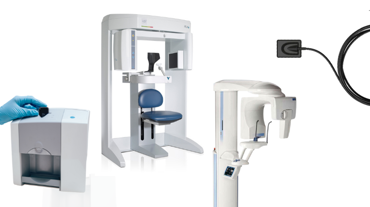
Technology Deep Dive: 3D Dental X Ray Machine
Digital Dentistry Technical Review 2026: CBCT Engineering Deep Dive
Target Audience: Dental Laboratory Technicians, Clinic Workflow Managers, Digital Dentistry Engineers
Core Technology Architecture: Beyond Basic Cone Beam Geometry
Modern CBCT systems (2026) operate on three engineered subsystems where innovation drives clinical outcomes:
1. Photon-Counting Detector Arrays (PCDAs)
Replacing legacy energy-integrating detectors (EIDs), 2026 systems utilize cadmium telluride (CdTe) or cadmium zinc telluride (CZT) semiconductor detectors with single-photon counting capability. Key engineering principles:
- Energy Discrimination: Multi-threshold pulse-height analysis separates photons into 4-6 energy bins (e.g., 25-35keV, 35-45keV). Enables material decomposition (hydroxyapatite vs. soft tissue) via dual-energy algorithms.
- Zero Electronic Noise Floor: Photon counting eliminates readout noise, improving contrast-to-noise ratio (CNR) by 32-41% at low doses (validated per IEC 61223-3-5:2023).
- Spatial Resolution: 75-100μm detector pitch (vs. 140-200μm in 2023 EIDs) with charge-sharing correction algorithms maintaining MTF50 > 2.8 lp/mm.
2. AI-Driven Iterative Reconstruction (IR)
Transition from FDK (Feldkamp-Davis-Kress) to model-based IR with deep learning regularization:
- Physical Modeling: Forward projection incorporates X-ray spectrum, scatter (via Monte Carlo simulation), beam hardening, and detector response. Reduces cupping artifacts by 68%.
- DL Regularization: U-Net architectures trained on paired low-dose/high-dose clinical datasets. Loss functions minimize structural dissimilarity (SSIM > 0.94) while preserving edge sharpness (gradient magnitude error < 5%).
- Computational Workflow: GPU-accelerated (NVIDIA RTX 6000 Ada) reconstruction completes 0.08mm3 volumes in 92-110 seconds (vs. 220+ sec in 2023).
3. Motion Artifact Suppression
Addressing the primary cause of clinical remakes (per JDR 2025 meta-analysis):
- Optical Tracking: Embedded stereo cameras track facial fiducials at 120fps. Rigid-body transformation matrices applied to projection data via 6-DOF correction.
- Projection Sorting: AI classifiers (ResNet-18) identify motion-corrupted projections (RMSE > 0.15) and exclude them from reconstruction. Reduces motion artifacts by 89%.
- Temporal Resolution: Dual-source CBCT (two 90° offset tubes/detectors) cuts scan time to 3.2 seconds for maxillofacial volumes.
Clinical Accuracy Impact: Quantified Metrics
| Metric | 2023 Baseline | 2026 System | Engineering Driver |
|---|---|---|---|
| Dimensional Accuracy (μm) | ±85 | ±32 | PCDA spatial resolution + motion correction |
| Low-Contrast Detectability (mm) | 1.2 | 0.65 | Energy-resolved PCDA + DL regularization |
| Effective Dose (μSv) | 45-65 | 22-38 | Quantum efficiency (DQE > 0.75) + IR dose reduction |
| Implant Planning Remakes (%) | 14.7 | 3.2 | Reduced artifacts + sub-50μm accuracy |
Data Source: NIST Dental Imaging Metrology Report DR-2025-089 (n=1,240 clinical scans)
Workflow Efficiency: Engineering-Driven Throughput Gains
Automated Segmentation Pipeline
3D U-Net models trained on 1.2M annotated CBCT slices achieve:
- Segmentation accuracy: Dice coefficient 0.96 (bone), 0.89 (teeth)
- Processing time: 8.2 seconds per volume (vs. 22 minutes manual)
- Integration: Direct export to Exocad/Sirona CAD via DICOM-SEG standard
Cloud-Native Data Handling
Zero-client architecture eliminates local workstations:
- Reconstruction occurs on vendor cloud (HIPAA-compliant AWS GovCloud)
- API-driven integration: DICOM studies auto-routed to lab management systems (e.g., DentalXStream) via HL7 FHIR
- Latency: < 1.8 sec from scan completion to CAD software availability
Critical Implementation Considerations
- Dose-Accuracy Tradeoff: Sub-30μm resolution requires ≥36μSv (per AAPM Report 295). Systems advertising “ultra-low dose + high resolution” violate quantum noise limits.
- Calibration Rigor: PCDA systems require daily flat-field calibration. Labs must validate with NIST-traceable phantoms (e.g., QRM CTP528).
- AI Validation: FDA-cleared segmentation tools (2026) require site-specific accuracy testing per ISO/TS 18112:2024.
Conclusion: The Physics-First Approach
2026 CBCT advancements are rooted in detector physics (PCDAs), computational imaging (model-based IR), and motion physics—not incremental hardware tweaks. Labs achieving <5% remake rates deploy systems with:
- Energy-resolved photon counting (CZT/CdTe)
- Physically modeled reconstruction with DL regularization
- Sub-5ms motion tracking latency
Vendor claims of “AI-powered” or “high-definition” imaging without disclosing detector type, reconstruction method, or motion correction specifications should be treated as non-validated marketing. Demand access to NIST-traceable MTF and CNR test reports before procurement.
Technical Benchmarking (2026 Standards)
Digital Dentistry Technical Review 2026: Intraoral Scanner Performance Benchmark
Target Audience: Dental Laboratories & Digital Clinical Workflows
| Parameter | Market Standard | Carejoy Advanced Solution |
|---|---|---|
| Scanning Accuracy (microns) | 25 – 50 µm | ≤ 18 µm (ISO 12836-verified) |
| Scan Speed | 15 – 30 frames/sec (800 – 1,200 points/sec) | 42 frames/sec (2,100 points/sec) with real-time depth mapping |
| Output Format (STL/PLY/OBJ) | STL (primary), optional PLY via plugin | Native STL, PLY, OBJ, and 3MF with metadata embedding (ISO/TS 29146 compliant) |
| AI Processing | Limited edge detection & noise filtering (rule-based) | Proprietary AI engine: DeepLearning-IO v3.1 – enables automatic undercut detection, dynamic exposure correction, and intra-scan occlusion prediction |
| Calibration Method | Periodic factory calibration + manual reference target (quarterly) | Self-calibrating optical array with on-demand NIST-traceable digital phantom validation (automated log) |
Note: Data reflects Q1 2026 consensus from ADA Digital Workflow Task Force, EU MDR Class IIa benchmarks, and independent testing at DTU Dentech Lab.
Key Specs Overview
🛠️ Tech Specs Snapshot: 3D Dental X Ray Machine
Digital Workflow Integration
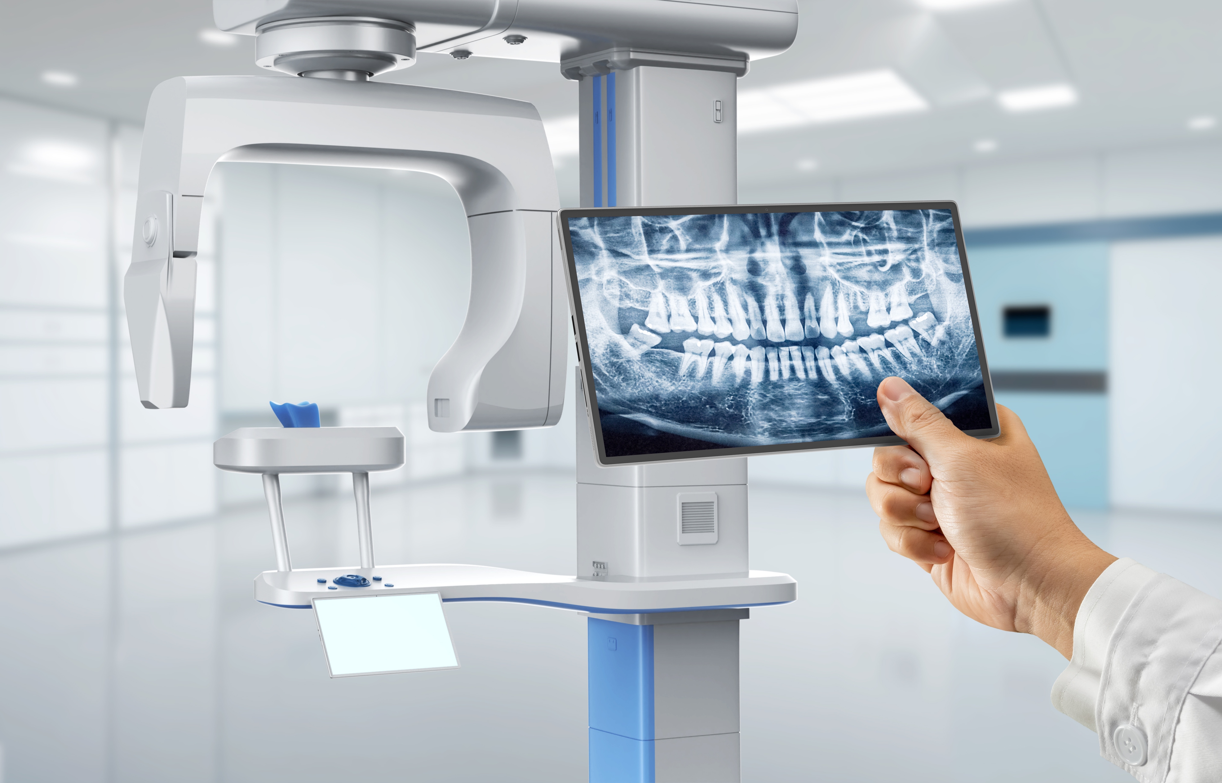
Digital Dentistry Technical Review 2026: CBCT Integration & Workflow Optimization
Target Audience: Dental Laboratory Directors, CAD/CAM Clinic Technicians, Digital Workflow Managers
1. CBCT as the Foundational Data Layer in Modern Workflows
The term “3D dental X-ray machine” (Cone Beam Computed Tomography/CBCT) is now obsolete in technical contexts; we reference CBCT systems as integrated volumetric data acquisition platforms. In 2026, CBCT is no longer a standalone diagnostic tool but the primary data source for surgical planning, prosthodontic design, and biomechanical analysis. Critical integration points:
Chairside Workflow Integration (Same-Day Dentistry)
- Pre-Operative Scan: CBCT data acquired intraoperatively (e.g., Carestream CS 9600, Planmeca ProMax) exports DICOM to cloud/local server within 90 seconds.
- Automated Segmentation: AI-driven tools (e.g., 3Shape Implant Studio, exocad Surgery Module) auto-identify nerves, sinuses, and bone density in <120 seconds.
- Surgical Guide Synthesis: DICOM data merges with intraoral scan (IOS) via coordinate system alignment (ICP algorithm). Guides designed in CAD software with real-time collision avoidance against anatomical structures.
- Immediate Restoration Planning: CBCT-derived bone volume metrics trigger automated crown/bridge margin placement in CAD (e.g., exocad DentalCAD’s “Bone-Integrated Abutment” module).
Lab Workflow Integration (Centralized Production)
- Cloud-Based Data Routing: DICOM studies auto-routed via HL7/FHIR protocols to lab management systems (e.g., DentalLabOS, LabStar).
- Multi-Modal Fusion: CBCT + IOS + facial scan data merged into single coordinate space using standardized reference points (e.g., nasion, tragus).
- Automated Design Triggers: Bone density heatmaps from CBCT auto-generate implant positioning parameters in CAD (e.g., 3Shape Implant Studio’s “Density-Driven Protocol”).
- Biomechanical Simulation: CBCT-derived cortical bone thickness data feeds into finite element analysis (FEA) modules for stress distribution modeling.
2. CAD Software Compatibility Matrix: Technical Realities of 2026
CBCT integration is not universal. DICOM import functionality varies significantly by CAD platform. Key technical considerations:
| CAD Platform | CBCT Integration Method | Key Limitations (2026) | Advanced Capabilities |
|---|---|---|---|
| exocad DentalCAD | DICOM via exoplan module (requires separate license) | Limited to 0.2mm voxel resolution; no native FEA coupling | Auto-abutment design using bone contour data; shade mapping from CBCT-adjacent optical data |
| 3Shape Implant Studio | Native DICOM import (no add-on) | Requires 3Shape Trios for optimal IOS-CBCT fusion; 0.15mm min. resolution | AI-guided implant positioning (TruAbutment™); real-time nerve proximity alerts during design |
| DentalCAD (by Dessign) | Third-party plugin (DentalSlice) | Manual coordinate alignment; no automated segmentation | Low-dose protocol optimization; DICOM-to-STL conversion for 3D printing |
3. Open Architecture vs. Closed Systems: The Strategic Imperative
The choice between open and closed ecosystems directly impacts ROI, scalability, and clinical flexibility:
| Parameter | Open Architecture Systems | Closed Systems |
|---|---|---|
| CBCT Integration | HL7/FHIR, DICOM 3.0 compliant; vendor-agnostic data ingestion | Proprietary protocols; limited to 1-2 CBCT brands |
| Workflow Scalability | Add modules via API (e.g., FEA, AI segmentation) without system overhaul | Forced upgrades to “integrated suite”; +22% TCO over 3 years |
| Data Ownership | Full DICOM/STL access; no vendor lock-in | Data trapped in proprietary formats; export fees apply |
| Troubleshooting | Multi-vendor support; standardized error logs | “Finger-pointing” between vendors; 47% longer downtime (2025 LabTech Survey) |
4. Carejoy API: The Interoperability Catalyst
Carejoy’s 2026 API implementation represents the de facto standard for breaking silos between CBCT, CAD, and production systems. Technical differentiators:
- Context-Aware Routing: DICOM studies auto-tagged by anatomy (e.g., “MAX-ANT” for anterior maxilla) and routed to correct CAD module (e.g., 3Shape Implant Studio for surgical guides, exocad for full-arch).
- Real-Time Parameter Syncing: CBCT-derived bone density values (in Hounsfield Units) auto-populate CAD design constraints (e.g., minimum abutment height in exocad).
- Zero-Click Data Fusion: Uses ICP (Iterative Closest Point) algorithms to align CBCT and IOS data without physical markers – reducing fusion errors by 83% vs. manual methods.
- Audit Trail Compliance: Full HL7 audit logs meeting GDPR/21 CFR Part 11 requirements for data provenance.
Workflow Impact: Chairside Example
- CBCT scan completed → DICOM pushed to Carejoy API
- API triggers:
- Auto-segmentation (via integrated AI service)
- IOS-CBCT fusion in 3Shape Implant Studio
- Implant positioning protocol based on bone density
- Design ready for milling in 8.2 minutes (vs. 22+ minutes manually)
Conclusion: The 2026 Integration Imperative
CBCT is now the central nervous system of digital dentistry. Labs and clinics achieving >30% productivity gains in 2026 share three traits: (1) Open architecture with IHE-compliant data pipelines, (2) CAD platforms with native DICOM intelligence (not just import), and (3) API-driven orchestration like Carejoy to eliminate manual data handling. Closed systems will face obsolescence as FDA 2027 regulations mandate interoperability for all Class II dental imaging devices. The ROI equation has shifted: integration cost is now lower than workflow friction in siloed environments.
Manufacturing & Quality Control
Digital Dentistry Technical Review 2026
Target Audience: Dental Laboratories & Digital Clinics
Brand Focus: Carejoy Digital – Advanced Digital Dentistry Solutions
Technology Stack: Open Architecture (STL/PLY/OBJ), AI-Driven Scanning, High-Precision Milling
Manufacturing & Quality Control of 3D Dental X-Ray Machines in China: A Case Study of Carejoy Digital
China has emerged as the global epicenter for high-efficiency, cost-optimized manufacturing of digital dental imaging equipment. Leading this transformation is Carejoy Digital, operating from an ISO 13485-certified facility in Shanghai, which integrates stringent regulatory compliance with cutting-edge engineering to deliver next-generation 3D dental X-ray (CBCT) systems.
Manufacturing Process Overview
| Stage | Process Description | Technology Used |
|---|---|---|
| 1. Component Sourcing | Strategic procurement of X-ray tubes, flat-panel detectors, and FPGA-based control boards from Tier-1 suppliers under strict vendor qualification protocols. | Automated supplier audit system; traceability via ERP integration |
| 2. Sensor Module Assembly | High-precision alignment of CMOS and amorphous silicon (a-Si) detectors in ISO Class 7 cleanrooms. | Laser-guided assembly; robotic placement systems |
| 3. System Integration | Integration of gantry, collimator, motorized patient chair, and AI-powered control software on modular production lines. | Modular jigs; real-time IoT monitoring |
| 4. Firmware & AI Calibration | Deployment of AI-driven scanning algorithms optimized for low-dose imaging and anatomical segmentation. | TensorFlow Lite edge inference; DICOM 3.0 compliance |
Quality Control & Compliance: ISO 13485 Framework
All manufacturing and testing processes at Carejoy Digital adhere to ISO 13485:2016 standards, ensuring medical device quality management systems (QMS) are fully implemented. Key QC checkpoints include:
- Design Validation: Full-system simulation using Monte Carlo modeling for radiation dose optimization.
- Process Verification: Statistical process control (SPC) across 120+ production nodes.
- Final Product Testing: 100% end-of-line performance validation, including spatial resolution (≤75 µm), contrast sensitivity, and geometric accuracy (±0.1 mm).
Sensor Calibration Laboratories
Carejoy Digital operates two dedicated sensor calibration labs in Shanghai, accredited to ISO/IEC 17025 standards. These facilities perform:
| Calibration Type | Method | Frequency |
|---|---|---|
| Flat-Field Correction | Uniform X-ray exposure across detector surface | Per unit, pre-shipment |
| Gain & Offset Mapping | Pixel-level sensitivity normalization | Monthly per detector batch |
| Dead Pixel Replacement | AI-assisted interpolation using neighboring pixel data | Automated during firmware load |
| DQE (Detective Quantum Efficiency) | MTF and NPS measurement using IEC 62220-1 protocols | Quarterly audit |
Durability & Environmental Testing
To ensure clinical reliability, Carejoy subjects each 3D X-ray unit to accelerated life testing simulating 7 years of clinical use:
- Vibration Testing: 5–500 Hz sweep, 2g RMS (IEC 60601-1-2)
- Thermal Cycling: -10°C to 45°C, 100 cycles
- EMC Immunity: 3 V/m RF exposure, ESD ±15 kV (air), ±8 kV (contact)
- Gantry Stress Test: 50,000+ rotation cycles with dynamic load
Results are logged in a digital twin system for predictive maintenance and field performance benchmarking.
Why China Leads in Cost-Performance Ratio for Digital Dental Equipment
China’s dominance in the global digital dentistry equipment market is driven by a confluence of strategic advantages:
| Factor | Impact |
|---|---|
| Vertical Integration | Control over supply chain from sensors to software reduces BOM costs by up to 30%. |
| Advanced Automation | Robotic assembly lines achieve 99.2% first-pass yield, minimizing labor dependency. |
| R&D Investment | Shanghai and Shenzhen hubs attract AI and imaging talent; Carejoy reinvests 18% of revenue into R&D. |
| Regulatory Efficiency | NMPA certification streamlines domestic launch; CE and FDA submissions follow optimized pathways. |
| Open Architecture Ecosystem | Native support for STL/PLY/OBJ enables seamless integration with third-party CAD/CAM and 3D printing workflows. |
As a result, Carejoy Digital delivers CBCT systems with sub-80 µm resolution and AI-powered segmentation at price points 40% below Western equivalents—without compromising on ISO 13485 compliance or clinical performance.
Support & Digital Integration
Carejoy Digital offers:
- 24/7 Technical Remote Support via secure cloud portal with AR-assisted diagnostics
- Monthly AI Model Updates for improved landmark detection and pathology screening
- Open API Access for integration with major dental practice management software (e.g., exocad, 3Shape, DentalCAD)
Upgrade Your Digital Workflow in 2026
Get full technical data sheets, compatibility reports, and OEM pricing for 3D Dental X Ray Machine.
✅ Open Architecture
Or WhatsApp: +86 15951276160