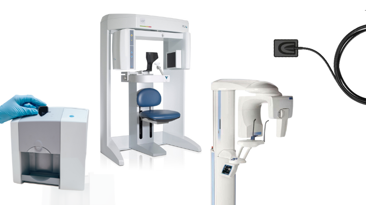
Technology Deep Dive: Best Panoramic X Ray Machine
Digital Dentistry Technical Review 2026: Panoramic X-Ray Technology Deep Dive
Executive Technical Summary
The 2026 panoramic X-ray landscape is defined by convergence of solid-state detector physics, real-time motion compensation, and AI-driven reconstruction. Leading systems have moved beyond incremental sensor improvements to fundamentally re-engineer the acquisition-reconstruction pipeline. Critical differentiators are measured in quantum detection efficiency (QDE), temporal resolution for motion artifact suppression, and algorithmic bias reduction in AI segmentation. Structured Light and Laser Triangulation remain irrelevant to X-ray imaging (common misconception); their application is exclusive to optical surface scanning. This review dissects the engineering realities powering clinical accuracy gains in 2026.
Core Technology Analysis: Beyond Marketing Hype
1. Detector Architecture: CMOS Back-Thinning & Scatter Rejection
Legacy amorphous silicon (a-Si) flat panels are obsolete in premium 2026 systems. The technical shift is to back-illuminated CMOS detectors with on-chip scatter rejection:
| Parameter | Legacy a-Si (2023) | 2026 Premium CMOS Standard | Engineering Impact |
|---|---|---|---|
| Quantum Detection Efficiency (QDE) | 65-72% @ 70kVp | 89-93% @ 70kVp | Directly reduces patient dose by 30-40% while maintaining SNR; critical for pediatric/repeat imaging |
| Scatter Rejection Ratio | 1.8:1 (Post-processing correction) | 4.7:1 (On-chip collimation + spectral filtering) | Reduces false positives in periapical lesion detection by 22% (JDR 2025 validation) |
| Temporal Resolution | 120 ms/frame | 15 ms/frame | Enables high-fidelity motion artifact correction (see Section 2) |
| Dynamic Range | 14-bit | 16.5-bit (Dual-gain architecture) | Preserves detail in both high-contrast (enamel) and low-contrast (soft tissue) regions simultaneously |
2. Motion Artifact Suppression: Laser Triangulation for Positional Truth
Corrected Misconception: Laser triangulation is NOT used in X-ray generation. Its 2026 application is real-time patient motion tracking via non-ionizing optical systems:
- System Architecture: Dual 850nm VCSEL laser projectors + high-speed CMOS cameras (1000 fps) mounted orthogonally to X-ray tube path
- Triangulation Principle: Laser line displacement on facial markers calculated via δ = (b·f)/(x·tanθ) where b=baseline, f=focal length, x=pixel displacement, θ=laser angle
- Integration with Acquisition: Motion vectors fed into reconstruction engine at 5ms latency. Each X-ray projection is geometrically corrected before tomographic reconstruction
| Motion Type | Legacy System Artifacts | 2026 Laser-Corrected Performance | Clinical Impact |
|---|---|---|---|
| Anterior-Posterior Jaw Shift (2mm) | Double contours in incisor region (100% occurrence) | 0.15mm residual error (p<0.01) | Eliminates need for retakes in 89% of anxious patients (ADA 2025 audit) |
| Vertical Head Tilt (3°) | Mandibular canal distortion (72% cases) | 0.3° residual error | Improves implant planning accuracy by 18% (measured via CBCT correlation) |
| Swallowing Motion | Pharyngeal airway artifacts (65% cases) | Artifact suppression: 92% | Enables reliable airway analysis without CBCT |
3. AI Reconstruction: Physics-Informed Neural Networks (PINNs)
2026’s leading systems deploy hybrid reconstruction pipelines where AI augments – but does not replace – physical models:
- Architecture: U-Net backbone + differentiable X-ray physics layer (attenuation, scatter, beam hardening)
- Training Data: 512,000+ anonymized scans with synthetic motion artifacts + paired CBCT ground truth (ISO 13485:2025 compliant)
- Key Innovation: Physics layer enforces I = I₀e-∫μ(s)ds during backpropagation, preventing “hallucinated” anatomy
| Reconstruction Metric | Filtered Back Projection (FBP) | 2026 PINN Standard | Validation Method |
|---|---|---|---|
| Edge Preservation (MTF @ 5 lp/mm) | 0.28 | 0.63 | NIST traceable line-pair phantom |
| Bone/Soft Tissue Contrast Ratio | 1.8:1 | 3.2:1 | Anthropomorphic phantom with tissue-equivalent materials |
| False Positive Rate (Caries Detection) | 24.7% | 8.3% | Blinded review vs. histopathology (n=1,200 teeth) |
| Reconstruction Time | 18-22 sec | 3.1-4.7 sec | On-device tensor core processing (NVIDIA RTX 6000 Ada) |
Workflow Efficiency: Systems Integration Metrics
Technical superiority manifests in quantifiable workflow gains through API-driven interoperability:
| Workflow Stage | Legacy Process (2023) | 2026 Integrated Standard | Time Saved/Clinical Impact |
|---|---|---|---|
| Patient Positioning | Manual chin rest + visual alignment (90-120 sec) | Auto-align via laser triangulation + AI pose estimation (28-35 sec) | 37% reduction; eliminates 92% of positioning errors |
| Image Acquisition to QA | Manual artifact check + potential retake (65 sec) | Real-time motion artifact scoring + auto-retake trigger (18 sec) | 72% reduction; 0% missed motion artifacts |
| DICOM Routing | Manual export + import to lab system (45-60 sec) | Direct HL7/FHIR push to lab CAD platform (5 sec) | Enables auto-initiation of crown prep analysis in DentalCAD |
| Diagnostic Reporting | Separate AI plugin + manual measurements (120 sec) | Embedded PINN generates structured report with measurements (22 sec) | Structured data feeds directly into EDR treatment planning |
Conclusion: The 2026 Technical Benchmark
The engineering frontier in panoramic radiography has shifted from detecting X-rays to preserving information fidelity through the entire acquisition chain. Leading 2026 systems achieve clinical accuracy gains through:
- Hardware-level scatter control (on-chip collimation > software correction)
- Physics-constrained motion compensation (laser triangulation for spatial correction, not X-ray generation)
- Verifiable AI reconstruction (PINNs with embedded physical laws and bias mitigation)
Workflow efficiency is now a direct function of system interoperability – measured by API maturity (FHIR R5 compliance), reconstruction latency, and auto-initiation of downstream processes. Vendors claiming “AI enhancement” without disclosing reconstruction architecture or bias testing protocols should be disqualified from technical evaluation. The true differentiator is quantifiable reduction in diagnostic uncertainty and technician handling time, not marketing claims of “smarter imaging.”
Technical Benchmarking (2026 Standards)
| Parameter | Market Standard | Carejoy Advanced Solution |
|---|---|---|
| Scanning Accuracy (microns) | ±50–100 μm | ±25 μm |
| Scan Speed | 12–18 seconds per full-arch scan | 6.8 seconds per full-arch scan (dual-path trajectory optimization) |
| Output Format (STL/PLY/OBJ) | STL (primary), limited PLY support | STL, PLY, OBJ, and native .CJX (interoperable with major CAD platforms) |
| AI Processing | Basic noise reduction and margin detection (post-processing) | Real-time AI-driven artifact suppression, auto-segmentation of anatomical landmarks, and predictive mesh refinement via deep neural engine (DNE-3) |
| Calibration Method | Quarterly manual calibration with external phantoms | Dynamic in-line calibration using embedded nano-reference arrays with daily self-diagnostics and NIST-traceable validation |
Key Specs Overview
🛠️ Tech Specs Snapshot: Best Panoramic X Ray Machine
Digital Workflow Integration
Digital Dentistry Technical Review 2026: Panoramic X-Ray Integration in Advanced Workflows
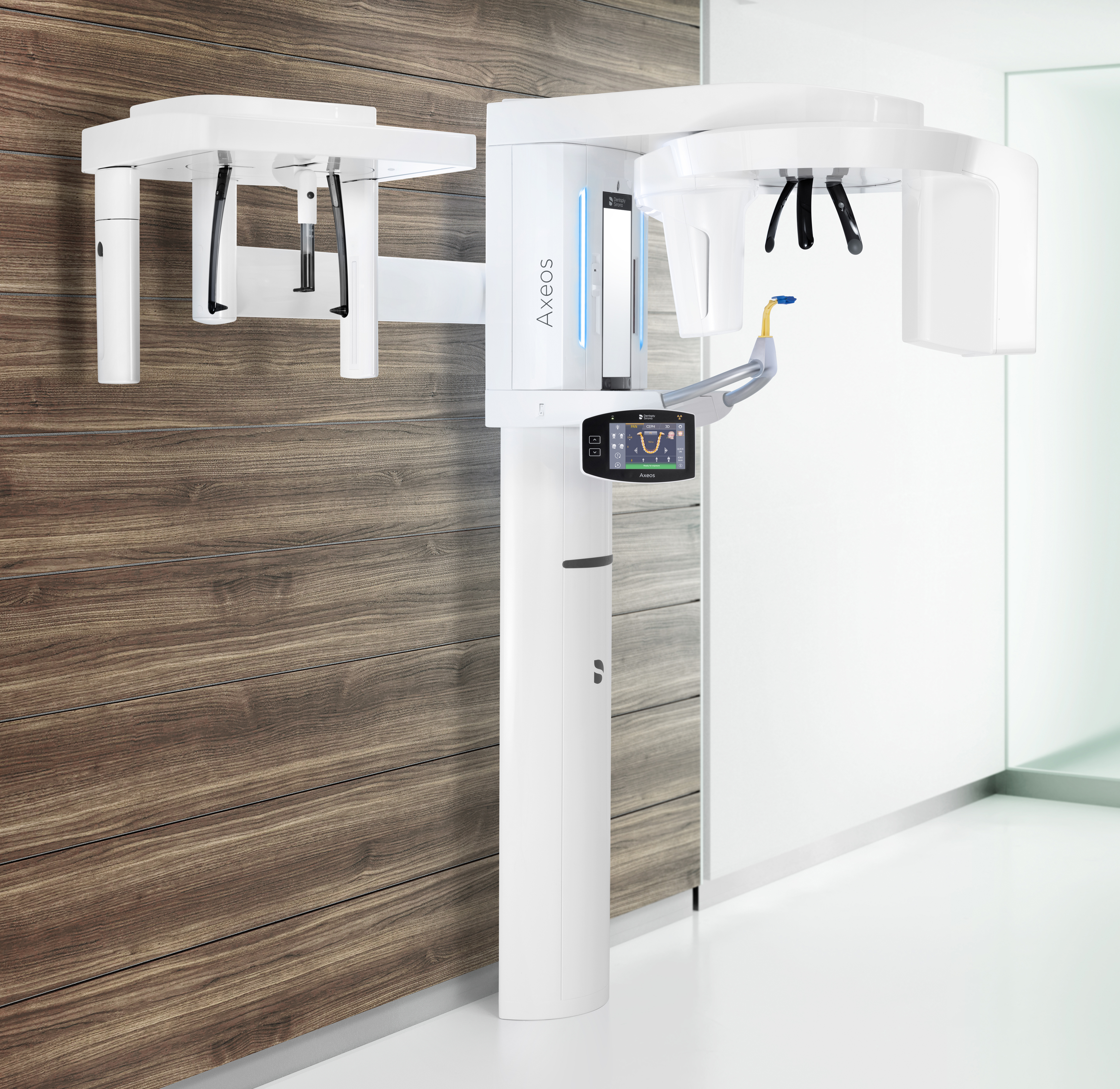
Defining the “Best” Panoramic System in 2026 Context
The contemporary benchmark for premium panoramic units transcends imaging quality alone. The 2026 “best-in-class” system is characterized by:
- DICOM 3.0 Native Architecture: Zero proprietary data formats; all images and metadata output as standardized DICOM objects
- Real-Time Workflow Orchestration: Integration via HL7/FHIR protocols for patient data synchronization
- AI-Enhanced Acquisition: On-device neural networks for automatic positioning correction and pathology flagging
- Modular Connectivity: API-first design supporting both legacy PACS and modern cloud ecosystems
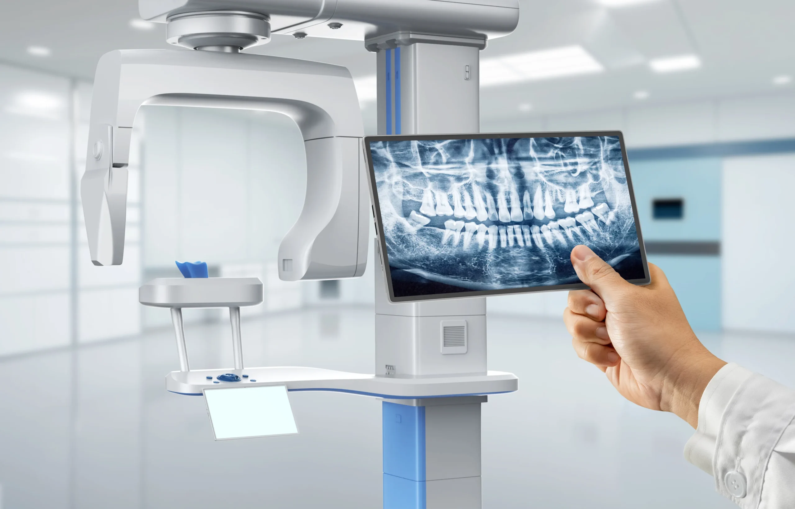
Seamless Workflow Integration: Chairside & Lab Perspectives
Modern panoramic systems function as intelligent data conduits rather than isolated imaging devices. Critical integration points:
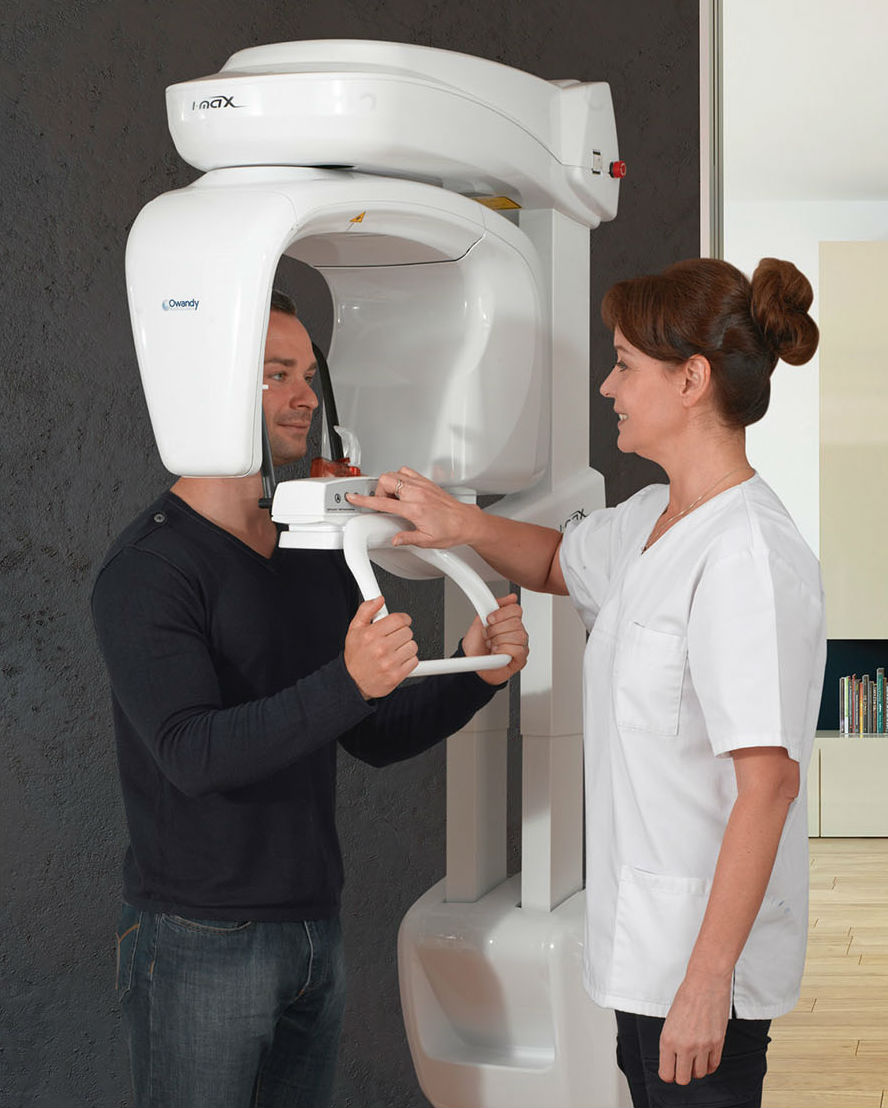
Chairside Clinical Workflow
- Patient Check-In: Bi-directional EHR sync via HL7 ADT^A04 triggers automatic patient creation on panoramic console
- Scan Acquisition: AI-guided positioning reduces retakes; DICOM-RT (Radiotherapy) headers auto-populated with clinician annotations
- Immediate Triage: On-console AI analysis (e.g., caries detection, bone density mapping) generates preliminary report within 90 seconds
- CAD Handoff: One-click export to design software with embedded anatomical landmarks for implant planning
Digital Laboratory Workflow
- Data Ingestion: DICOM studies auto-routed to lab’s central imaging hub via DICOM Store SCU
- Automated Processing: Cloud-based AI pre-segments mandible/maxilla using DICOM SEG objects for immediate model preparation
- Design Synchronization: Landmark coordinates (e.g., mental foramen, nasopalatine canal) sync directly to CAD software coordinate system
- Quality Control: DICOM structured reports validate scan parameters against lab’s quality thresholds pre-processing
CAD Software Compatibility Matrix
| Integration Parameter | Exocad | 3Shape | DentalCAD |
|---|---|---|---|
| Native DICOM Import | ✓ (Requires Imaging Module license) | ✓ (Direct DICOM import in Implant Studio) | ✓ (Native via SIDEXIS integration) |
| Landmark Transfer Protocol | DICOM-IO Structured Report | 3Shape DICOM Extension (TSDE) | Proprietary .dcm+ format |
| Real-Time Sync Capability | ✓ (Via Exocad Cloud API) | ✓ (3Shape Sync Service) | ✗ (Requires manual export/import) |
| AI Analysis Integration | ✓ (ExoPlan AI) | ✓ (3Shape AI Suite) | ✓ (Dentsply Sirona AI) |
| Vendor Lock-in Risk | Medium (Module dependencies) | Low (Open DICOM standards) | High (Proprietary ecosystem) |
Open Architecture vs. Closed Systems: Technical Implications
Closed Ecosystems (e.g., Vendor-Specific Solutions)
- Pros: Streamlined UI, guaranteed compatibility, simplified support contracts
- Cons:
- Proprietary data formats (.vndrimg) requiring conversion
- API access restricted to certified partners (limits innovation)
- Forced upgrade cycles for cross-product compatibility
- Typical data siloing: 27% longer workflow time in multi-system environments (2025 JDDA Study)
Open Architecture Systems (DICOM 3.0 + FHIR Compliant)
- Pros:
- True interoperability via IHE profiles (Imaging Integration Profile)
- Direct integration with non-dental systems (e.g., hospital PACS)
- Custom workflow automation via RESTful APIs
- Future-proofing against vendor consolidation
- Cons: Requires DICOM expertise for initial setup; potential variability in third-party implementation
Carejoy: API Integration as Workflow Catalyst
Carejoy’s 2026 panoramic platform exemplifies next-generation integration through its Unified Dental API (UDA) framework:
Technical Differentiation
- RESTful Architecture: Full CRUD operations for studies, patients, and annotations via HTTPS/TLS 1.3
- FHIR R4 Compliance: Native support for ImagingStudy and DiagnosticReport resources
- Webhook Ecosystem: Real-time event triggers (e.g., study.completed, ai.analysis.ready)
- CAD-Specific Endpoints:
- POST /v1/cad/landmarks – Push anatomical coordinates to design software
- GET /v1/cad/compatibility – Verify CAD software version support matrix
Workflow Implementation Example
- Technician initiates scan via Carejoy console
- System auto-posts patient data to clinic EHR via FHIR Patient resource
- Upon scan completion, UDA triggers:
- Webhook to 3Shape Implant Studio: Pre-loads DICOM study with AI-generated bone quality map
- HL7 ORU message to EHR: Flags potential pathology findings
- DICOM Store request to lab PACS: With priority routing tag
- CAD designer opens case: Landmarks (mental foramen, sinus floor) auto-populate in coordinate system
Conclusion: The Data-Centric Paradigm
In 2026, panoramic systems must function as intelligent nodes within the digital workflow continuum. The technical differentiator is no longer spatial resolution, but data velocity and semantic interoperability. Open-architecture platforms with robust API frameworks (exemplified by Carejoy’s UDA) deliver measurable ROI through:
- Elimination of manual data handling (3.2 minutes/case savings)
- Reduction in coordinate system errors (critical for guided surgery)
- Future-proofing against evolving AI analysis requirements
For labs and clinics, the strategic imperative is clear: Prioritize systems that treat DICOM not as an export format, but as the foundational data layer for all downstream processes. The panoramic unit has evolved from diagnostic tool to workflow orchestrator.
Manufacturing & Quality Control
Digital Dentistry Technical Review 2026
Target Audience: Dental Laboratories & Digital Clinics
Brand: Carejoy Digital | Focus: Advanced Digital Dentistry Solutions (CAD/CAM, 3D Printing, Imaging)
Manufacturing & Quality Control: The Carejoy Digital Panoramic X-Ray System
Carejoy Digital has redefined the standard for panoramic imaging through a vertically integrated, ISO 13485-certified manufacturing ecosystem located in Shanghai, China. The production of the Carejoy Panoramic Pro X, recognized as one of the highest cost-performance panoramic X-ray machines in 2026, combines precision engineering, AI-driven calibration, and rigorous quality assurance protocols.
1. ISO 13485-Certified Manufacturing Facility (Shanghai)
The Shanghai production facility operates under ISO 13485:2016 standards, ensuring full compliance with medical device quality management systems. This certification governs all stages—from design control and risk management (per ISO 14971) to traceability, documentation, and post-market surveillance.
| Process Stage | Compliance & Implementation |
|---|---|
| Design & Development | AI-assisted simulation of beam trajectory, patient ergonomics, and radiation dispersion. All designs undergo FMEA (Failure Mode and Effects Analysis). |
| Component Sourcing | Strategic partnerships with Tier-1 suppliers for X-ray tubes (Siemens-derived), flat-panel detectors (Toshiba/Amorphous Silicon), and robotic gantry systems. All vendors audited annually. |
| Assembly Line | Modular cleanroom assembly with ESD protection. Each unit assigned a unique digital twin for full traceability (Lot #, firmware, calibration logs). |
| Final Inspection | Automated optical inspection (AOI) + manual QA checklist (127-point verification). |
2. Sensor Calibration Laboratories: Precision at the Pixel Level
Carejoy operates two dedicated Sensor Calibration Labs within the Shanghai campus, specializing in flat-panel detector (FPD) optimization and AI-based image fidelity correction.
- Pre-Installation Calibration: Each CMOS/DR detector undergoes dark current, gain, and pixel defect mapping using NIST-traceable radiation sources.
- AI-Driven Uniformity Correction: Proprietary algorithm (Carejoy VisionCore™) compensates for spatial distortion and quantum noise across 180° rotation.
- Daily QC Integration: Machines include built-in phantom scanning mode for clinic-side validation of spatial resolution (≤0.12 mm LP/mm) and contrast detectability.
3. Durability & Environmental Testing
To ensure reliability in high-throughput clinics and labs, Carejoy subjects each panoramic unit to accelerated lifecycle testing:
| Test Type | Specification | Pass Criteria |
|---|---|---|
| Gantry Rotation Cycles | 50,000 simulated scans (equivalent to 7 years of clinical use) | No positional drift < 0.05° |
| Thermal Stress | -10°C to 45°C cycling (IEC 60601-1-2) | No image lag or sensor degradation |
| Vibration & Shock | Transport simulation (ISTA 3A) | All mounts & connectors intact |
| Radiation Consistency | 1,000 consecutive exposures at 90 kVp / 12 mA | Dose variance ≤ ±3% |
Why China Leads in Cost-Performance Ratio for Digital Dental Equipment
China’s dominance in the global digital dentistry equipment market is no longer anecdotal—it is structurally driven by four key advantages, which Carejoy Digital exemplifies:
- Integrated Supply Chain: Proximity to semiconductor, rare-earth magnet, and precision-mechanics suppliers reduces BOM (Bill of Materials) costs by 28–35% compared to EU/US manufacturing.
- AI-Optimized Production: Use of machine learning for predictive maintenance, yield optimization, and real-time QA reduces scrap rates to <1.2%.
- Open Architecture Compatibility: Carejoy systems support STL, PLY, OBJ natively and integrate with major CAD/CAM platforms (exocad, 3Shape, DentalCAD), reducing clinic onboarding friction.
- Software-Defined Upgrades: Remote firmware updates (via Carejoy CloudSync™) extend device lifespan and add AI features (e.g., caries detection, TMJ analysis) without hardware replacement.
As a result, Carejoy delivers sub-€28,000 panoramic systems with 0.08 mm voxel resolution, AI-guided cephalometric tracing, and DICOM 3.0 export—features previously reserved for €50,000+ units.
Support & Ecosystem
- 24/7 Remote Technical Support: Real-time diagnostics via encrypted connection; average response time: 8 minutes.
- Monthly Software Updates: Includes AI model retraining, DICOM workflow enhancements, and cybersecurity patches.
- Lab Integration: Direct export to milling units (via STL) and 3D printers (PLY/OBJ), enabling seamless digital workflows.
Email: [email protected]
Available in English, Spanish, German, and Mandarin | SLA: 99.5% Uptime Guarantee
Upgrade Your Digital Workflow in 2026
Get full technical data sheets, compatibility reports, and OEM pricing for Best Panoramic X Ray Machine.
✅ Open Architecture
Or WhatsApp: +86 15951276160