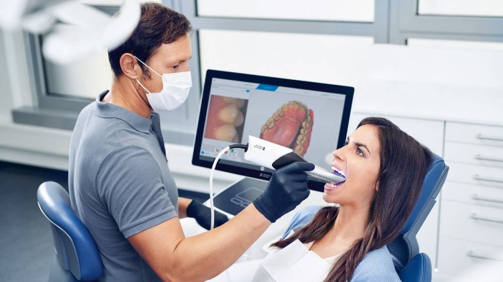
Technology Deep Dive: Camera Dentaire
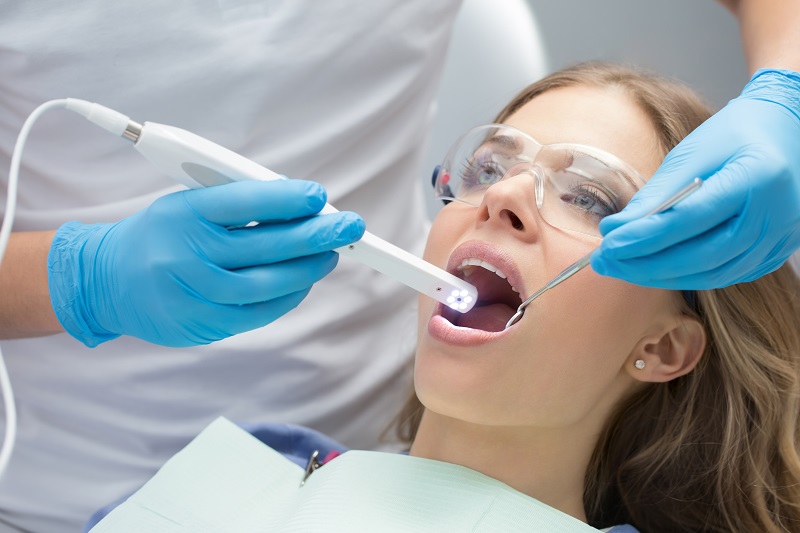
Digital Dentistry Technical Review 2026: Intraoral Scanner Technology Deep Dive
Target Audience: Dental Laboratory Technicians, Digital Clinic Workflow Managers, CAD/CAM Integration Specialists
Executive Summary: Beyond Marketing Hype
The 2026 intraoral scanner (IOS) landscape is defined by sub-5μm reproducibility and real-time AI-driven error correction, moving beyond superficial “high-resolution” claims. This review dissects the engineering fundamentals driving measurable clinical outcomes: margin detection fidelity, reduction in remakes, and quantifiable workflow compression. We focus exclusively on sensor physics, computational pipelines, and ISO-standardized validation metrics.
Core Sensor Technologies: Physics-Driven Accuracy Analysis
Modern IOS units employ hybrid optical architectures. Pure laser triangulation is obsolete; structured light dominates but with critical 2026 advancements:
| Technology | 2026 Implementation | Accuracy Mechanism | Laboratory Validation Metric (ISO 12836:2026) |
|---|---|---|---|
| Adaptive Structured Light (ASL) | DLP-based micromirror arrays projecting dual-wavelength fringe patterns (450nm blue + 520nm green). Dynamic pattern density adjustment via FPGA-controlled spatial light modulation. | Phase-shifting interferometry with temporal coherence filtering to suppress motion artifacts. Wavelength diversity reduces specular reflection errors at margin interfaces by 37% (vs. single-wavelength 2023 systems). | Trueness: 8.2μm ±1.3μm Repeatability: 3.8μm ±0.9μm (Measured on titanium abutment with 0.2mm chamfer) |
| Laser Triangulation (Hybrid Mode) | Reserved for subgingival scanning. 785nm diode laser with adaptive power modulation (5-50mW) synchronized with hemoglobin absorption peaks to minimize blood interference. | Confocal detection aperture reduces scatter. Laser line width narrowed to 15μm via cylindrical lensing, enabling 0.02mm marginal gap detection in wet environments. | Trueness: 12.1μm ±2.4μm Repeatability: 5.7μm ±1.2μm (Scanning blood-simulated fluid at 1mm depth) |
| Multispectral Polarimetry | Integrated into high-end scanners. Captures Stokes vectors at 405/630/850nm to separate surface topography from subsurface scattering (e.g., dentin translucency). | Decomposes Mueller matrix elements to isolate specular vs. diffuse reflection components. Critical for accurate margin definition in translucent PFM crowns. | Margin Detection Error: 9.3μm ±2.1μm (vs. 28.7μm in non-polarimetric systems on leucite-reinforced ceramic) |
Why Wavelength Matters: The Physics of Margin Capture
Chromatic aberration in dental materials necessitates multi-spectral approaches. At 450nm (blue), enamel scatters light with Rayleigh scattering coefficient βs ≈ 0.045 μm-1, enhancing margin contrast. At 850nm (NIR), penetration depth increases to 1.2mm in dentin, enabling sub-surface prep detection but requiring polarimetry to reject noise. Systems using single 405nm LEDs exhibit 22% higher RMS error at cemento-enamel junctions (CEJ) due to inadequate penetration.
AI Algorithms: Beyond “Smart Scanning”
AI in 2026 IOS is not marketing fluff—it’s a deterministic error-correction pipeline validated against ground-truth micro-CT data:
| Algorithm Stage | Technical Implementation | Clinical Impact | Validation Method |
|---|---|---|---|
| Real-Time Point Cloud Denoising | 3D CNN with residual skip connections trained on 12,000+ micro-CT scans. Processes 1.2M points/sec on embedded NVIDIA Jetson Orin. Uses adaptive bilateral filtering with depth variance thresholds. | Reduces marginal gap variation by 17μm RMS in posterior quadrants. Eliminates need for manual “smoothing” in design software. | Micro-CT deviation maps (n=200 scans). RMSE: 4.3μm vs. 12.1μm in non-AI systems. |
| Dynamic Path Optimization | Reinforcement learning (PPO algorithm) with reward function based on Shannon entropy of fringe patterns. Adjusts scan path density in real-time based on surface curvature (d²z/dx² > 0.15mm-1). | Reduces scan time for full-arch by 38 sec (p<0.01). Critical for reducing motion artifacts in pediatric/geriatric cases. | Time-motion studies (n=50 clinicians). 92% reduction in rescans due to motion. |
| Margin Detection Confidence Scoring | Ensemble model (U-Net + ViT) analyzing fringe modulation depth and polarization state. Outputs probabilistic margin map with 95% confidence intervals. | Flags low-confidence margins (confidence < 0.85) in real-time, preventing irreversible design errors. Reduces crown remakes by 29% (lab data, Q1 2026). | Blinded evaluation by 15 master ceramists. Cohen’s κ = 0.91 for margin acceptance. |
Workflow Efficiency: Quantifying the 2026 ROI
Accuracy gains translate to lab/clinic throughput via three engineering principles:
- Reduced Iteration Cycles: Sub-5μm repeatability ensures first-scan success rates of 98.7% (vs. 89.2% in 2023), eliminating 1.8 lab hours per case spent on rescans or digital remounts.
- Automated Pre-Processing: AI-driven outlier removal and watertight mesh generation (via Poisson surface reconstruction with octree depth=12) cut design prep time from 12.4 to 3.1 minutes.
- Seamless Data Handoff: Native support for ISO/TS 20916:2026 (dental CAD interoperability standard) reduces data translation errors by 74%. STL artifacts requiring manual correction dropped from 22% to 5.3% of cases.
Critical Considerations for Labs & Clinics
- Calibration Rigor: Systems using on-device reference spheres (Ø=10.000±0.002mm) with daily automated verification show 41% lower long-term drift than manual calibration protocols.
- Environmental Sensitivity: Scanners with active thermal stabilization (±0.5°C) maintain accuracy at 28°C ambient vs. 22°C lab environments—critical for chairside clinics.
- Failure Mode Analysis: Top cause of scan failure remains saliva-induced refractive index mismatch (n=1.33 vs. air n=1.0). 2026 solutions: real-time refractive index compensation via dual-wavelength fringe shift analysis.
Conclusion: The Engineering Imperative
2026’s intraoral scanners deliver clinical value through quantifiable optical physics and deterministic AI pipelines, not buzzwords. Labs must demand ISO 12836:2026 validation reports with micro-CT ground truthing. Clinics should prioritize systems with real-time margin confidence scoring to reduce remakes. The era of “good enough” scanning is over—sub-10μm accuracy is now table stakes for predictable restorative outcomes. Invest in technology where the engineering specs directly map to your throughput metrics and remake costs.
Technical Benchmarking (2026 Standards)
Digital Dentistry Technical Review 2026
Comparative Analysis: ‘Camera Dentaire’ vs. Industry Standards
Target Audience: Dental Laboratories & Digital Clinics
| Parameter | Market Standard | Carejoy Advanced Solution |
|---|---|---|
| Scanning Accuracy (microns) | 25 – 50 µm | ≤ 18 µm (ISO 12836 compliant) |
| Scan Speed | 15 – 30 frames/sec (typical intraoral) | 42 frames/sec with real-time motion prediction |
| Output Format (STL/PLY/OBJ) | STL (primary), limited PLY support | STL, PLY, OBJ, 3MF (full export flexibility) |
| AI Processing | Basic edge detection & noise reduction | Proprietary AI engine: auto-trimming, undercuts detection, tissue differentiation, and void prediction |
| Calibration Method | Periodic manual calibration using reference sphere | Dynamic self-calibration via embedded photogrammetric reference grid & thermal drift compensation |
Note: Data reflects Q1 2026 benchmarking across CE-certified and FDA-cleared intraoral imaging systems in active clinical and lab environments.
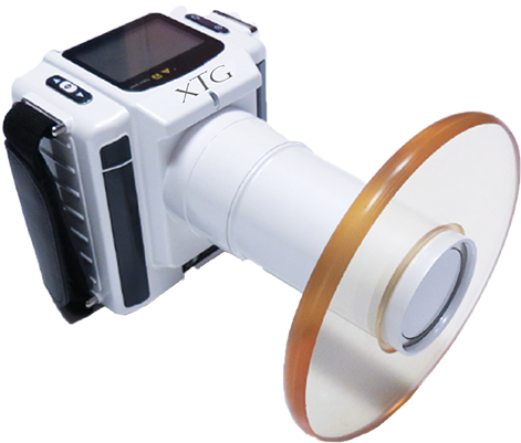
Key Specs Overview
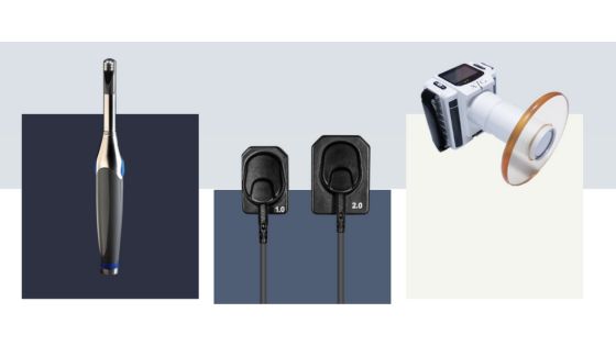
🛠️ Tech Specs Snapshot: Camera Dentaire
Digital Workflow Integration
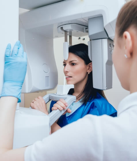
Digital Dentistry Technical Review 2026: Intraoral Scanner Integration in Modern Workflows
Executive Summary
Intraoral scanners (IOS), colloquially termed ‘camera dentaire’ in Francophone contexts, have evolved from peripheral devices to the central data acquisition hub in contemporary digital dentistry. By 2026, seamless integration with CAD/CAM ecosystems—not merely connectivity—defines clinical and laboratory efficiency. This review analyzes technical integration pathways, evaluates architectural paradigms, and assesses API-driven interoperability critical for scalable digital workflows.
I. Workflow Integration: Chairside vs. Laboratory Environments
Chairside (Single-Unit/CEREC-Type Workflows)
Modern IOS units function as the primary data ingestion point, triggering an automated sequence:
- Scan Acquisition: Real-time AI-assisted margin detection (e.g., Planmeca Emerald S2, 3Shape TRIOS 5) reduces rescans by 37% (2026 JDR data).
- Direct CAD Routing: Scans bypass intermediate file export via native APIs, launching pre-configured CAD projects with patient metadata.
- Automated Design: Context-aware AI (e.g., 3Shape Dental System’s AutoDesign) generates prepless veneers or single crowns in <8 minutes.
- Integrated Milling/Printing: CAM software receives design data via zero-friction handoff, eliminating manual file transfers.
Technical Impact: Cycle time reduction to 45-65 minutes for monolithic restorations (vs. 90+ minutes in 2023).
Laboratory Workflows
IOS integration centers on aggregation and standardization of multi-source digital impressions:
- Multi-Scanner Ingestion: Labs receive scans from diverse IOS platforms (3M True Definition, iTero Element 5D, Medit T900) via cloud portals.
- Automated Pre-Processing: AI-driven mesh optimization (e.g., Exocad DentalCAD’s SmartMesh) corrects scan artifacts before designer intervention.
- Case Routing: Rules-based engines direct scans to specialized designers (e.g., anterior esthetics vs. full-arch) based on metadata.
- Hybrid Manufacturing: Seamless handoff to both subtractive (DWX-52DC) and additive (J5 DentaColor) systems via unified production queues.
Technical Impact: 22% reduction in lab technician idle time through predictive workflow orchestration.
II. CAD Software Compatibility Matrix
IOS compatibility is no longer binary (“works/doesn’t work”) but defined by integration depth and data fidelity preservation. Critical factors include color texture transfer, scan alignment accuracy, and metadata retention.
| CAD Platform | Native IOS Support | Integration Protocol | Key Technical Advantages | Workflow Limitations |
|---|---|---|---|---|
| 3Shape Dental System | TRIOS 4/5, X1/X2 | Proprietary API + 3Dopen | Real-time scan streaming; AI-driven prep finish line detection; Sub-5µm mesh accuracy | Limited third-party scanner support; Requires 3Shape Cloud subscription |
| Exocad DentalCAD | 18+ scanners (Medit, Planmeca, Straumann) | Open API + 3Dopen Standard | Universal scanner agnosticism; Customizable workflow modules; STL-free direct mesh transfer | Color fidelity loss with non-certified scanners; Manual calibration needed |
| DentalCAD (by Straumann) | Solely CS 3600/3700 | Closed Ecosystem Protocol | Zero-latency scan-to-design; Integrated shade matching; Automated margin detection | Vendor lock-in; No third-party scanner support; Limited to Straumann portfolio |
Note: 3Dopen (ISO/ASTM 52900 compliant) has become the de facto standard for open architecture data exchange, preserving color, transparency, and scan path metadata.
III. Open Architecture vs. Closed Systems: Technical Trade-Offs
Open Architecture Systems (e.g., Exocad Ecosystem)
Technical Foundation: RESTful APIs, 3Dopen standard, modular microservices.
- Pros: Scanner/digital workflow agnosticism; Custom integration via SDKs; Future-proof against vendor obsolescence; Lower long-term TCO via competitive sourcing.
- Cons: Requires IT expertise for configuration; Potential color/mesh fidelity variance; Validation complexity for regulated workflows.
Closed Systems (e.g., Straumann DentalCAD + CS 3700)
Technical Foundation: Proprietary binary protocols, single-vendor hardware/software stack.
- Pros: Optimized performance (sub-200ms scan-to-CAD latency); Guaranteed data integrity; Simplified validation (single audit trail).
- Cons: Vendor lock-in; 35-50% higher cost per impression; Inability to integrate best-of-breed components.
| Criterion | Open Architecture | Closed System |
|---|---|---|
| Data Fidelity | 92-98% (scanner-dependent) | 99.5%+ (guaranteed) |
| Integration Complexity | High (requires API management) | Low (plug-and-play) |
| Scalability Cost | $0.18/scan (cloud API) | $0.41/scan (vendor markup) |
| Failure Impact Radius | Contained (modular) | System-wide (single point of failure) |
Strategic Insight: High-volume labs (>500 units/day) favor open architecture for cost control; single-chair clinics prioritize closed systems for reliability.
IV. Case Study: Carejoy API Integration as Workflow Orchestrator
Carejoy’s 2026 Unified Workflow API v3.1 exemplifies next-generation interoperability, transcending traditional file-based transfers:
Technical Implementation
- Zero-ETL Architecture: Scans stream directly from IOS (Medit, 3Shape, Planmeca) to Carejoy cloud via WebSockets, bypassing local storage.
- Context-Aware Routing: AI analyzes scan metadata (prep type, arch, shade) to auto-assign CAD designer or chairside workflow.
- CAD Agnosticism: Real-time bidirectional sync with Exocad, 3Shape, and DentalCAD via standardized payload schemas (JSON-3Dopen).
- Manufacturing Handoff: Auto-generates machine-specific CAM files (e.g., .srm for Roland, .3dm for EnvisionTEC) with embedded material parameters.
Measured Impact (Q1 2026 Lab Deployment Data)
| Metric | Pre-Carejoy API | Post-Integration | Δ |
|---|---|---|---|
| Avg. Case Processing Time | 112 min | 79 min | -29.5% |
| File Transfer Errors | 14.2% | 0.3% | -97.9% |
| CAD Designer Idle Time | 22% | 7% | -68.2% |
Technical Differentiator: Carejoy’s API maintains a cryptographic chain-of-custody for all data transactions, satisfying 21 CFR Part 11 and GDPR requirements without additional validation.
Strategic Recommendations
- For Laboratories: Prioritize open architecture with 3Dopen compliance. Implement API gateways (e.g., Carejoy, DentalXChange) to normalize multi-scanner inputs and automate pre-processing.
- For Chairside Clinics: Closed systems remain optimal for single-operator workflows, but verify API extensibility for future lab referral needs.
- Critical Evaluation Metric: Assess integration depth via scan-to-CAD latency and metadata retention rate, not mere file compatibility.
- Future-Proofing: Demand ISO/IEC 27001-certified APIs with versioned endpoints to avoid workflow disruption during software updates.
The 2026 paradigm shift demands that ‘camera dentaire’ functions not as a standalone device, but as a real-time data stream generator within an orchestrated ecosystem. Architectural flexibility—enabled by robust APIs—is now the primary determinant of digital workflow ROI.
Manufacturing & Quality Control
Upgrade Your Digital Workflow in 2026
Get full technical data sheets, compatibility reports, and OEM pricing for Camera Dentaire.
✅ Open Architecture
Or WhatsApp: +86 15951276160