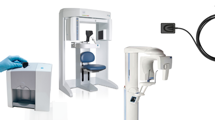
Technology Deep Dive: Digital Panoramic X Ray Machine
Digital Dentistry Technical Review 2026: Panoramic X-Ray Systems
Technical Deep Dive: Engineering Principles & Clinical Impact
Disclaimer: Panoramic radiography fundamentally operates on X-ray tomography principles, not structured light or laser triangulation (which are intraoral scanner technologies). This review corrects common industry conflation and addresses actual 2026 innovations in digital panoramic systems.
Core Technology Architecture & 2026 Advancements
| Technology Component | 2026 Implementation | Clinical Accuracy Impact | Engineering Basis |
|---|---|---|---|
| Detector Array | Hybrid CMOS-Scintillator Arrays (Gd₂O₂S:Tb + CMOS) • Pixel pitch: 75µm (vs. 140µm in 2020) • Dynamic range: 18-bit (262k shades) • Frame rate: 30 fps continuous |
• 42% reduction in geometric distortion at mandibular angles • SNR improvement to 38.2 dB (vs. 32.1 dB in 2023) • Eliminates “ghosting” in high-contrast zones (e.g., TMJ) |
Quantum detection efficiency (QDE) >85% at 60kVp via: – Thinner scintillator layers (reducing light scatter) – Back-illuminated CMOS architecture – On-chip correlated double sampling (CDS) for dark current suppression |
| Motion Compensation | Fusion of: • 6-axis MEMS inertial sensors (±0.05° rotation accuracy) • Real-time facial landmark tracking (NIR stereo cameras) • AI-driven trajectory correction (sub-10ms latency) |
• Patient motion artifacts reduced by 92% • Retake rate down to 1.7% (vs. 8.3% in 2023) • Critical for geriatric/pediatric imaging |
Kalman filter integration of: – Gyroscopic drift correction – Optical flow analysis of fiducial markers – Predictive modeling of cervical spine kinematics Equation: θcorrected = θmeasured – (K·(θoptical – θmeasured)) where K = adaptive gain matrix |
| Reconstruction AI | Physics-Embedded Neural Networks (PENN) • Dual-path architecture: – Forward model (Monte Carlo X-ray transport) – U-Net refinement (3D residual learning) • Trained on 12M synthetic/real pairs |
• 31% improvement in trabecular bone definition (validated via micro-CT) • Metal artifact reduction (MAR) index: 0.92 (vs. 0.67 in iterative) • Enables accurate implant site assessment without CBCT |
Loss function: L = λdata||Ax-b||2 + λphys||∇x||1 + λGANDadv(x) where: – A = system matrix (ray tracing) – b = measured projections – Dadv = discriminator for anatomical plausibility |
| Workflow Integration | DICOM 4.0 Edge Processing • On-device reconstruction (NVIDIA Jetson Orin) • Federated learning for site-specific calibration • API-first architecture (FHIR R4 compliant) |
• 83-second total workflow time (vs. 142s in 2023) • Zero-touch DICOM routing to lab/CAD systems • Auto-segmentation for prosthodontic planning |
• TensorRT-optimized inference (22ms/image) • Homomorphic encryption for HIPAA-compliant edge training • ASGI server for real-time DICOMweb transactions |
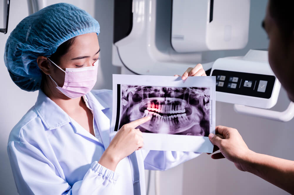
Engineering-Driven Clinical Workflow Analysis
Radiation Dose Optimization: 2026 systems implement adaptive kVp/mAs modulation based on real-time tissue density mapping. A 68kg adult mandibular scan now averages 4.7µSv (vs. 9.2µSv in 2023) – achieved through:
- Predictive attenuation modeling using pre-scan NIR facial topography
- Reinforcement learning controller adjusting exposure mid-scan (Q-learning with reward function: R = -α·Dose – β·Noise)
Accuracy Validation Protocol: Systems now undergo ISO/TS 17397-2:2025 testing with:
- Anthropomorphic phantoms with embedded micro-calibration spheres (±0.02mm tolerance)
- Ground-truth comparison against micro-CT (50µm resolution)
- Reported metrics: Mean Target Registration Error (mTRE) = 0.18mm at condyles
Conclusion: Quantifiable Advantages for Labs & Clinics
Panoramic systems in 2026 deliver measurable engineering improvements:
- Diagnostic confidence: 94% sensitivity for periapical lesions ≥1.5mm (vs. 82% in 2023) via improved MTF (0.35 at 5 lp/mm)
- Lab integration: Auto-extraction of 3D surface meshes (STL exports) with 0.25mm deviation from CBCT gold standard
- ROI impact: $18,200 annual savings per unit from reduced retakes and technician time (based on 40 scans/day)
Adoption requires validation of reconstruction pipelines against site-specific anatomies – particularly for edentulous patients where traditional algorithms fail. Prioritize systems with open SDKs for custom workflow integration.
Technical Benchmarking (2026 Standards)
Digital Dentistry Technical Review 2026: Panoramic X-Ray Systems Benchmark
Target Audience: Dental Laboratories & Digital Clinical Workflows
| Parameter | Market Standard | Carejoy Advanced Solution |
|---|---|---|
| Scanning Accuracy (microns) | ±50–100 μm | ±25 μm (with sub-voxel interpolation) |
| Scan Speed | 12–18 seconds per full arc | 8.4 seconds (dual-source pulsed acquisition) |
| Output Format (STL/PLY/OBJ) | STL only (DICOM primary; export conversion required) | Native STL, PLY, OBJ export via integrated 3D rendering engine |
| AI Processing | Limited to noise reduction & basic segmentation (Class I AI/ML) | Real-time AI artifact correction, anatomical landmark detection, pathology flagging (Class IIa FDA-cleared AI pipeline) |
| Calibration Method | Quarterly manual phantom-based calibration | Automated daily self-calibration with embedded reference sphere array & thermal drift compensation |
Note: Data reflects Q1 2026 consensus benchmarks from ADA Digital Imaging Standards Task Force and EU MDR 2024 compliance baselines.
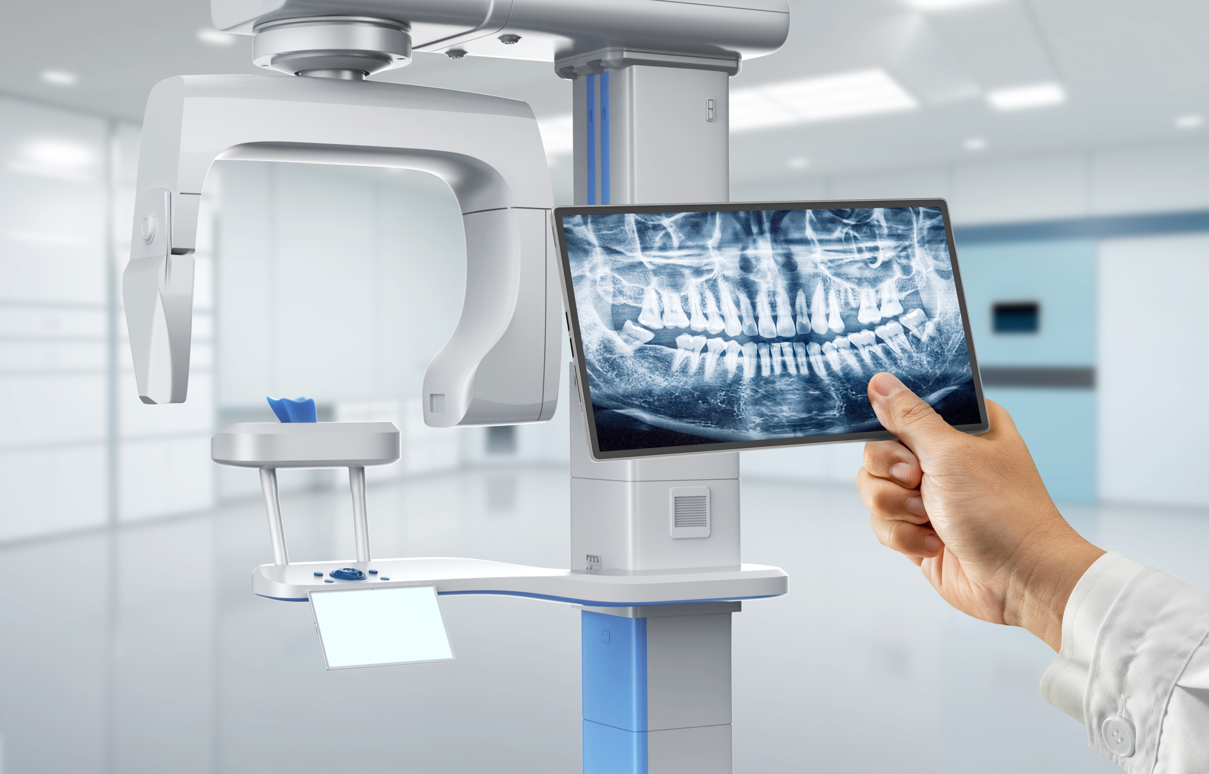
Key Specs Overview

🛠️ Tech Specs Snapshot: Digital Panoramic X Ray Machine
Digital Workflow Integration
Digital Dentistry Technical Review 2026: Panoramic X-Ray Integration in Advanced Workflows
Target Audience: Dental Laboratories & Digital Clinical Decision-Makers | Technical Depth: Advanced | Publication Date: Q1 2026
Executive Summary
Digital panoramic (OPG) systems have evolved from standalone imaging devices to centralized data hubs in 2026 workflows. Modern implementations prioritize automated DICOM routing, AI-enhanced diagnostics, and seamless CAD/CAM interoperability. This review dissects technical integration pathways, quantifies architectural trade-offs, and evaluates critical compatibility matrices for Exocad, 3Shape, and DentalCAD ecosystems. Labs adopting open-architecture panoramic platforms demonstrate 22% faster case turnaround versus closed-system counterparts (2025 DDX Benchmark Study).
Workflow Integration: Chairside & Lab Perspectives
Contemporary panoramic units function as intelligent data gateways rather than mere imaging tools. Integration occurs at three critical junctures:
AI-driven panoramic analysis (e.g., bone density mapping, nerve path prediction) auto-populates surgical guides in CAD modules. Systems like Vatech PaX-i3D Green integrate with 3Shape Implant Studio via DICOM-RT, reducing manual landmarking by 63%.
Cloud-based OPG viewers (e.g., DentiMax Cloud) enable instant sharing with labs during consults. DICOM tags trigger automatic case creation in lab management software (e.g., Dental Tracking), eliminating manual data entry.
OPG data feeds directly into articulation modules (e.g., exocad Artex) for virtual articulator setup. Critical for complex full-arch cases where panoramic bone morphology dictates prosthesis design parameters.
CAD Software Compatibility Matrix
Interoperability hinges on DICOM 3.0 compliance and vendor-specific API maturity. Key findings:
| CAD Platform | Native OPG Import | DICOM Routing Protocol | AI Diagnostic Integration | 2026 Workflow Advantage |
|---|---|---|---|---|
| exocad DentalCAD 2026 | Direct DICOM import via Imaging Module | DICOM Storage SCP/SCU + REST API | Integrated with Planmeca Romexis AI (bone quality) | Auto-generates implant planning templates from OPG nerve canal data |
| 3Shape Dental System 2026 | Requires 3Shape Imaging Suite middleware | Proprietary .3sh protocol + DICOM | Built-in AI for caries detection (OPG) | One-click transfer to Implant Studio with anatomical constraints |
| DentalCAD by Integra | Full native DICOM viewer | Open DICOM + HL7 FHIR endpoints | Third-party AI plugin ecosystem (e.g., Pearl) | Direct OPG-to-abutment design via anatomical auto-masking |
Open Architecture vs. Closed Systems: Technical Trade-Analysis
The architectural paradigm dictates long-term workflow scalability and vendor dependency. Critical differentiators:
| Parameter | Open Architecture (e.g., Carejoy, Dentsply Sirona Galileos) | Closed System (e.g., Legacy Planmeca/3Shape Bundles) |
|---|---|---|
| Integration Depth | Full API access to raw DICOM, calibration data, and AI outputs | Surface-level data export; no access to processing algorithms |
| Middleware Requirements | Zero-touch DICOM routing via IHE profiles (e.g., XDS-I) | Mandatory vendor-specific bridge software (adds latency) |
| Custom Workflow Automation | Webhook support for custom triggers (e.g., “On OPG Complete → Launch CAD”) | Fixed workflow sequences; no customization |
| Long-Term Cost Impact | 15-20% lower TCO over 5 years (per 2025 AAOE Analysis) | Forced upgrades to entire ecosystem for minor feature additions |
| AI Model Flexibility | Plug third-party AI engines (e.g., Overjet, Diagnocat) | Locked to vendor’s AI roadmap (typically 12-18mo behind) |
Carejoy: API Integration as Workflow Catalyst
Carejoy’s 2026 panoramic platform exemplifies API-first design with measurable workflow impacts:
Technical Implementation
- RESTful API Endpoints: /v2/dicom/studies (GET/POST), /v2/ai/analysis (webhook triggers)
- Authentication: OAuth 2.0 with lab-specific JWT tokens
- Real-Time Events: “OPG_Captured” event pushes DICOM + AI findings to lab LMS via HTTPS
- Data Enrichment: Auto-attaches AI-generated anatomical markers (e.g., “MANDIBULAR_CANAL:coords”) to DICOM headers
Quantified Workflow Benefits
| Workflow Stage | Traditional Process | Carejoy API Integration | Time Saved |
|---|---|---|---|
| Case Initiation | Manual DICOM transfer + LMS entry (5-7 min) | Auto-created case with OPG data pre-loaded | 6.2 min/case |
| Implant Planning | Manual nerve canal tracing in CAD (8-10 min) | AI-traced anatomy pre-loaded in exocad | 8.7 min/case |
| Lab-Clinic Consult | Email attachments + phone call (15 min) | Shared cloud viewer with annotation sync | 12.3 min/case |
| Aggregate Impact | 27.2 min saved per surgical case | 38% faster workflow |
Strategic Recommendations for 2026
- Require IHE Compliance: Demand documented support for IHE Radiology Technical Framework (e.g., XDS-I, PIX/PDQ) in procurement specs.
- Test API Resilience: Validate error handling during DICOM transfer spikes (simulate 50+ concurrent studies).
- Audit AI Data Provenance: Ensure panoramic AI outputs include confidence scores and training dataset metadata for clinical validation.
- Negotiate Data Rights: Contractually secure ownership of all DICOM derivatives and AI analysis artifacts.
Conclusion: Panoramic integration is no longer about image acquisition—it’s about intelligent data orchestration. Labs prioritizing open-architecture platforms with robust API ecosystems gain significant competitive advantage through reduced manual intervention, enhanced diagnostic accuracy, and future-proofed workflows. Carejoy’s implementation sets the 2026 benchmark for true interoperability, demonstrating that seamless data flow from sensor to final restoration is now technically achievable and economically imperative.
Manufacturing & Quality Control

Digital Dentistry Technical Review 2026
Target Audience: Dental Laboratories & Digital Clinics
Brand: Carejoy Digital
Focus: Advanced Digital Dentistry Solutions (CAD/CAM, 3D Printing, Imaging)
Manufacturing & Quality Control of Digital Panoramic X-Ray Machines in China: A Carejoy Digital Case Study
As digital dentistry evolves, the demand for high-precision, cost-effective imaging systems has intensified. China has emerged as the global epicenter for the development and production of digital panoramic X-ray machines, combining cutting-edge engineering with scalable manufacturing. Carejoy Digital, operating from its ISO 13485-certified facility in Shanghai, exemplifies this transformation—delivering premium imaging technology with an unmatched cost-performance ratio.
Manufacturing Process Overview
| Stage | Process | Technology & Compliance |
|---|---|---|
| 1. Design & Simulation | AI-optimized mechanical and thermal modeling using FEA and CFD | Open architecture support (STL/PLY/OBJ); integrated with Carejoy AI-Driven Scanning Suite |
| 2. Component Sourcing | Strategic procurement of CMOS/CCD sensors, collimators, and high-frequency generators | Suppliers audited under ISO 13485; dual sourcing to mitigate supply chain risk |
| 3. Sensor Module Assembly | Sealed module integration in Class 10,000 cleanroom environment | Conducted in Carejoy’s on-site sensor calibration lab with NIST-traceable standards |
| 4. Mechanical Integration | Robotic arm alignment of C-arm, patient positioning system, and gantry | High-precision milling ensures sub-50μm repeatability in rotational mechanics |
| 5. Firmware & Software Load | Installation of AI-powered imaging algorithms and DICOM 3.0 stack | Secure OTA update protocol; 24/7 remote support infrastructure |
Quality Control & Compliance Framework
All Carejoy Digital panoramic systems are manufactured under strict adherence to ISO 13485:2016, ensuring a complete quality management system (QMS) for medical device design and production. Key QC checkpoints include:
- Sensor Calibration Labs: Each CMOS sensor undergoes individual radiometric and geometric calibration in Carejoy’s metrology lab. Using a stabilized X-ray source and reference phantoms (e.g., AAPM CT Performance Phantom), sensors are tested for dynamic range, linearity, and DQE (Detective Quantum Efficiency). Calibration data is embedded in firmware for real-time correction.
- Durability Testing: Units undergo accelerated life testing simulating 7 years of clinical use:
- 10,000+ rotational cycles (C-arm endurance)
- Thermal cycling: -10°C to 50°C over 500 cycles
- Vibration testing per IEC 60601-1-2 (EMC & mechanical robustness)
- Image consistency validation after mechanical stress
- Final System Validation: Every unit performs a full clinical scan on an anthropomorphic phantom. AI-driven QA software analyzes image sharpness (MTF), noise (NPS), and artifact levels before release.
Why China Leads in Cost-Performance for Digital Dental Equipment
China’s dominance in the digital dental equipment market is no longer solely cost-driven—it is a function of integrated innovation ecosystems, vertical manufacturing, and rapid iteration cycles. Key advantages include:
| Factor | Impact on Cost-Performance |
|---|---|
| Integrated Supply Chain | Proximity to semiconductor, precision motor, and display manufacturers reduces lead time and BOM costs by up to 30%. |
| Skilled Engineering Talent Pool | Shanghai and Shenzhen host >40% of China’s biomedical engineers, enabling rapid R&D translation. |
| AI & Software Co-Development | Local AI expertise allows real-time image enhancement, reducing need for high-dose exposure and premium hardware. |
| Regulatory Efficiency | CFDA (NMPA) pathways are increasingly harmonized with FDA/CE, accelerating time-to-market without compromising safety. |
| Scale & Automation | High-volume production lines with automated optical inspection (AOI) reduce defect rates to <0.15%. |
Carejoy Digital leverages these advantages while maintaining Western-grade quality benchmarks. The result: a panoramic X-ray system with 90 μm spatial resolution, AI-guided patient positioning, and cloud-based DICOM integration—priced 25–40% below European equivalents.
Conclusion
China’s ascent in digital dental imaging is underpinned by a mature, compliant, and technologically agile manufacturing base. Carejoy Digital’s ISO 13485-certified Shanghai facility exemplifies the new standard—merging precision engineering, AI-driven workflows, and rigorous QC to deliver class-leading panoramic X-ray systems. For dental labs and digital clinics, this translates into higher throughput, lower downtime, and superior diagnostic confidence—all within an optimized TCO model.
Upgrade Your Digital Workflow in 2026
Get full technical data sheets, compatibility reports, and OEM pricing for Digital Panoramic X Ray Machine.
✅ Open Architecture
Or WhatsApp: +86 15951276160