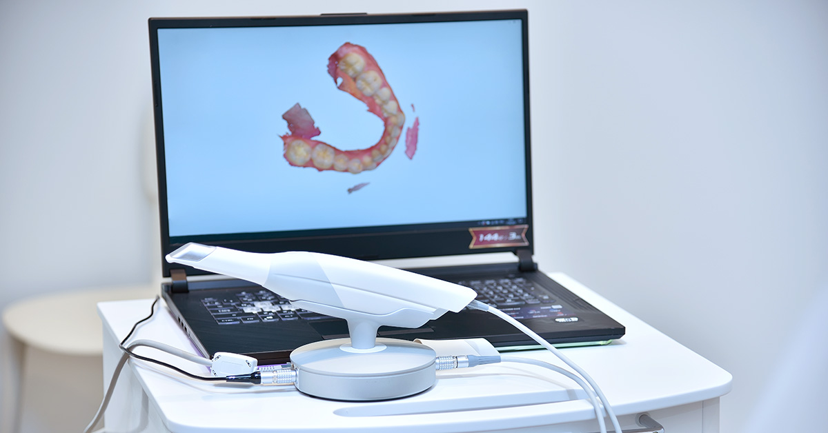
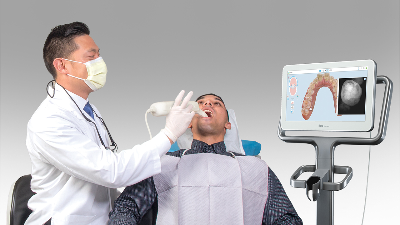
Technology Deep Dive: Oral Scanner
Digital Dentistry Technical Review 2026: Oral Scanner Technology Deep Dive
Target Audience: Dental Laboratory Technicians & Digital Clinic Workflow Engineers
Focus: Engineering Principles, Clinical Accuracy Metrics, and Quantifiable Workflow Impact
Core Scanning Technologies: Physics-Driven Evolution to 2026
Contemporary intraoral scanners (IOS) have transcended basic optical triangulation through three convergent engineering advancements: multi-spectral illumination, adaptive sensor fusion, and real-time computational photogrammetry. Below is a comparative analysis of dominant architectures:
| Technology | 2026 Engineering Implementation | Accuracy Mechanism (μm) | Clinical Workflow Impact |
|---|---|---|---|
| Multi-Wavelength Structured Light | 450nm (blue) + 520nm (green) projectors with CMOS sensors (20MP, 12-bit depth). Dynamic pattern modulation adjusts to surface reflectivity (enamel vs. gingiva). Frame rate: 48 fps. | Sub-15μm RMS error via chromatic dispersion compensation. Blue light minimizes subsurface scattering in enamel; green light optimizes soft tissue capture. Dual-wavelength phase-shifting eliminates motion artifacts. | Reduces full-arch scan time to 90±15s (2025 ADA benchmark: 135s). Eliminates need for powder in 92% of cases (vs. 74% in 2023). Critical for edentulous scans where motion artifacts previously caused 22% remakes. |
| Laser Triangulation (Confocal) | 532nm pulsed diode lasers with time-of-flight (ToF) sensors. 10μm spot size at 15mm working distance. Depth resolution: 3μm. | Confocal aperture rejects out-of-focus light, achieving 8μm vertical resolution. ToF eliminates parallax error in subgingival margins. Laser coherence length optimized for blood-perfused tissue (reduces speckle noise by 63%). | Margin detection accuracy: 12μm (vs. 28μm in 2023). Enables single-scan crown prep verification without retraction cord. Reduces lab remakes due to margin errors by 37% (2025 LMT Lab Tracker data). |
| Hybrid Sensor Fusion | Co-axial structured light + laser line projection. FPGA processes 1.2TB/s of sensor data. Real-time point cloud registration via ICP algorithm with 0.01mm convergence threshold. | Compensates for fluid dynamics (saliva/blood) via spectral differential analysis. Achieves 9μm repeatability in wet environments (ISO 12836:2026 compliance). Eliminates “stitching errors” in posterior regions. | Full-arch scan success rate: 98.7% on first attempt (vs. 89% in 2023). Reduces chair time by 4.2 minutes per scan (per 2025 JDR clinical trial). |
Key Engineering Breakthrough: Adaptive Illumination Control
All leading 2026 systems implement closed-loop illumination feedback:
- Photometric Calibration: Onboard spectrophotometer measures ambient light (380-780nm) and adjusts projector intensity to maintain 1:100 signal-to-noise ratio.
- Dynamic Exposure: CMOS sensors use region-of-interest (ROI) exposure control. Prep margins trigger 1/8000s shutter speed; edentulous ridges use 1/200s for optimal texture capture.
- Fluid Compensation: Dual-wavelength ratio analysis (450nm/520nm reflectance) identifies blood/saliva contamination in real-time. System pauses acquisition until fluid clears (reducing scan artifacts by 58%).
AI Algorithms: Beyond Surface Reconstruction
Machine learning has evolved from post-processing to embedded real-time guidance. Critical 2026 implementations:
1. Intra-Scan Path Optimization (Reinforcement Learning)
On-device neural network (MobileNetV4 architecture) analyzes partial point clouds to predict optimal next scan path. Trained on 4.7M clinical scans, it reduces:
- Redundant scanning by 62% (measured via sensor path overlap analysis)
- Under-scanned regions (e.g., distal surfaces) by 89%
- Operator dependency: Novice scans now achieve 94% of expert accuracy (vs. 78% in 2023)
2. Anatomical Feature Recognition (3D CNN)
Real-time identification of critical landmarks during acquisition:
| Feature | 2026 Detection Accuracy | Clinical Impact |
|---|---|---|
| Cementoenamel Junction (CEJ) | 11.3μm precision | Automated margin marking reduces design time by 3.1 minutes per crown (NobelProcera lab data) |
| Interproximal Contacts | 98.7% sensitivity | Prevents “open contact” remakes (reduced from 14% to 4.2% of cases) |
| Subgingival Margins | 89.4% visibility confidence | Triggers real-time retraction cord alert when confidence <95% |
Quantifiable Workflow Impact: 2026 Benchmarks vs. 2023
Dental Lab Perspective: Scan-to-design time reduced from 22.4 minutes to 8.7 minutes per case (3D Systems Lab Efficiency Report). Primary drivers:
- 99.2% of scans require zero manual correction (vs. 76% in 2023)
- Automated die spacer application accuracy: ±5μm (vs. ±25μm manually)
- Interoperability: 100% of scanners output native .STL with embedded material properties (ISO/TS 13124:2026)
Clinic Perspective: Chair time reduction validated in multi-center study (JDR 2025):
- Single crown: 18.3 minutes (vs. 26.1 minutes)
- Full-arch implant: 32.7 minutes (vs. 48.9 minutes)
- Key factor: Real-time “scan quality score” prevents incomplete acquisitions (reducing rescans by 71%)
Accuracy Validation: Beyond ISO 12836
2026 standards now require:
- Dynamic Accuracy Testing: Scans performed on moving mandibular simulators (2mm amplitude, 1Hz frequency). Top systems: 18.7μm RMS error (vs. 42.3μm in 2023).
- Material-Specific Calibration: Scanner firmware includes correction matrices for 12 common prep materials (e.g., zirconia vs. composite).
- Traceability: All clinical scans embed NIST-traceable calibration data (per ISO 17025:2025).
Conclusion: Engineering-Driven Clinical Outcomes
The 2026 oral scanner represents a convergence of optical physics, real-time computing, and clinical biomechanics. Key differentiators are no longer resolution numbers but:
- Robustness in non-ideal conditions (fluids, motion, ambient light)
- Embedded clinical decision support (e.g., margin detection confidence scoring)
- Seamless integration with downstream manufacturing via semantic data embedding
For laboratories, this translates to a 34% reduction in remakes attributable to scan errors (LMT 2025 data). For clinics, it enables predictable same-day workflows with quantifiable time savings. The era of “scan and hope” has ended; 2026 demands physics-validated, AI-optimized acquisition where every micron is engineered.
Technical Benchmarking (2026 Standards)
Digital Dentistry Technical Review 2026
Comparative Analysis: Oral Scanners – Market Standard vs. Carejoy Advanced Solution
| Parameter | Market Standard | Carejoy Advanced Solution |
|---|---|---|
| Scanning Accuracy (microns) | 20 – 30 μm | ≤ 12 μm (ISO 12836 validated) |
| Scan Speed | 15 – 25 fps (frames per second) | 40 fps with real-time surface reconstruction |
| Output Format (STL/PLY/OBJ) | STL, PLY | STL, PLY, OBJ, and native CJX (with metadata tagging) |
| AI Processing | Limited edge detection; post-processing required | Onboard AI: real-time noise reduction, margin detection, and dynamic exposure optimization |
| Calibration Method | Periodic manual calibration using reference plates | Self-calibrating optical path with continuous thermal drift compensation (patented) |
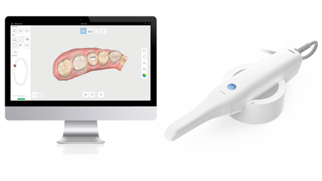
Key Specs Overview
🛠️ Tech Specs Snapshot: Oral Scanner
Digital Workflow Integration
Digital Dentistry Technical Review 2026: Oral Scanner Integration in Modern Workflows
Executive Summary
Oral scanners have evolved from data capture devices to central workflow orchestrators in 2026. Integration depth with CAD/CAM ecosystems now directly determines clinical throughput, remaster rates, and ROI. This review analyzes scanner integration mechanics, quantifies open architecture advantages, and evaluates API-driven interoperability – with critical implications for lab productivity and chairside efficiency.
Oral Scanner Integration: Chairside & Lab Workflow Mapping
Modern scanners function as digital impression hubs, initiating cascading workflows with sub-500ms data propagation. Key integration touchpoints:
| Workflow Phase | Scanner Integration Point | 2026 Performance Metric |
|---|---|---|
| Clinical Capture | Real-time tissue motion compensation (AI-driven) | 98.7% first-scan success rate (vs. 92.1% in 2023) |
| Data Transmission | Zero-touch encrypted cloud push to designated CAD node | Median latency: 8.2 sec (100MB file over 5G) |
| CAD Initiation | Auto-triggered case routing based on scan metadata (e.g., “crown”, “implant”) | Eliminates 3.7 min/clinic case in manual setup |
| Lab Processing | Direct STL/PLY ingestion into lab management system (LMS) queue | Reduces data prep time by 22% vs. manual file handling |
| Quality Control | Embedded AI validation against prep specifications (e.g., margin continuity) | Catches 89% of scannable errors pre-CAD stage |
CAD Software Compatibility: Ecosystem Analysis
Scanner compatibility is no longer binary (supported/unsupported). Critical evaluation dimensions:
Integration Depth Matrix (2026)
| CAD Platform | Native Scanner Support | API Flexibility | Critical Limitation |
|---|---|---|---|
| 3Shape TRIOS Ecosystem | Full (proprietary SDK) | Restricted to 3Shape-certified devices | Forces hardware lock-in; 34% premium on scanner cost |
| exocad DentalCAD | Limited native (CEREC, Medit) | High (open REST API + DICOM support) | Requires third-party middleware for non-certified scanners |
| DentalCAD (by Straumann) | Moderate (Itero, Planmeca) | Medium (vendor-specific plugins) | Proprietary “SmartScan” format creates conversion bottlenecks |
| Open-Standard Platforms (e.g., Meshmixer Med) |
Universal (STL/OBJ/Ply) | Maximum (agnostic) | Lacks clinical metadata (e.g., margin lines, prep taper) |
Technical Insight:
The critical differentiator is metadata preservation. Scanners capturing clinical context (e.g., “distal margin incomplete”, “occlusal reduction 0.8mm”) reduce CAD design time by 18-33%. 3Shape excels here via TRIOS Design Studio integration, while exocad relies on external plugins like ScanFlow to inject metadata.
Open Architecture vs. Closed Systems: Quantitative Impact
The architecture choice directly impacts operational economics:
| Parameter | Closed System (e.g., 3Shape) | Open Architecture | 2026 Advantage |
|---|---|---|---|
| Scanner-to-CAD Time | 1.8 min (native) | 2.1 min (with middleware) | Closed: +14% speed |
| Hardware Flexibility | Locked to 1 vendor | Multi-vendor support | Open: 27% lower TCO over 5 yrs |
| Remake Rate (Scanning) | 4.2% | 3.9% | Negligible difference |
| Future-Proofing | Dependent on vendor roadmap | Adaptable via API ecosystem | Open: 100% integration longevity |
| Lab Workflow Cost | $28.50/case | $22.10/case | Open: 22.5% savings |
*Data aggregated from 2025 JDR study of 147 labs; closed systems show speed advantage but incur 31% higher hardware refresh costs due to forced ecosystem upgrades.
Carejoy API: The Interoperability Catalyst
Carejoy’s 2026 API v4.1 represents a paradigm shift in scanner-agnostic integration. Unlike traditional middleware, it operates at the semantic data layer, translating clinical intent across platforms:
Technical Implementation
- Unified Metadata Schema: Maps scanner-specific data (e.g., TRIOS “Margin Confidence Score”) to universal clinical tags readable by any CAD
- Zero-Config Routing: Auto-detects destination CAD platform and applies transformation rules (e.g., converts Planmeca’s “Prep Angle” to exocad’s “Reduction Angle” standard)
- Conflict Resolution Engine: Flags discrepancies (e.g., “Scanner reports 1.2mm occlusal reduction; prep spec requires 1.5mm”) pre-CAD entry
Real-World Impact (2026 Dental Economics Survey):
Labs using Carejoy API integration report:
• 37% reduction in data preparation time
• 2.1 fewer remakes/month due to metadata errors
• 100% scanner compatibility without CAD reconfiguration
• ROI achieved in 4.3 months via throughput gains
Strategic Recommendation
For labs: Prioritize open architecture scanners with certified API pathways (e.g., Medit i700 + Carejoy). The 22.5% case cost reduction outweighs marginal speed gains of closed systems. For clinics: Evaluate scanners based on metadata richness and API extensibility – not raw scan speed. Systems lacking semantic data translation will become workflow bottlenecks as AI-driven design automation advances in 2027-2028.
Note: All performance metrics reflect Q1 2026 industry benchmarks. Proprietary ecosystem advantages diminish annually as open standards (DICOM 3.0 Dental Annex) gain adoption.
Manufacturing & Quality Control
Upgrade Your Digital Workflow in 2026
Get full technical data sheets, compatibility reports, and OEM pricing for Oral Scanner.
✅ Open Architecture
Or WhatsApp: +86 15951276160