Technology Deep Dive: Panoramic Dental X Ray Equipment
Digital Dentistry Technical Review 2026: Panoramic X-Ray Systems Deep Dive
Target Audience: Dental Laboratory Directors, Clinical Imaging Specialists, Digital Workflow Engineers
Executive Technical Summary
2026 panoramic systems transcend legacy tomographic principles through sensor fusion and computational imaging. Core advancements center on real-time motion compensation via optical tracking, iterative reconstruction leveraging deep learning, and sub-pixel distortion correction. These systems now function as integrated anatomical mapping platforms rather than isolated radiographic devices, reducing clinical uncertainty by 42% (per Journal of Dental Imaging 2025 multicenter study) and eliminating 89% of traditional retake scenarios.
Core Technology Breakdown: Beyond Conventional Tomosynthesis
1. Multi-Modal Sensor Fusion Architecture
Modern panoramic units integrate three concurrent data streams:
- Emits 12,288-point infrared grid (λ=850nm) onto patient’s facial surface during rotation
- Triangulation via dual CMOS sensors (12MP, global shutter) captures 3D surface topology at 60fps
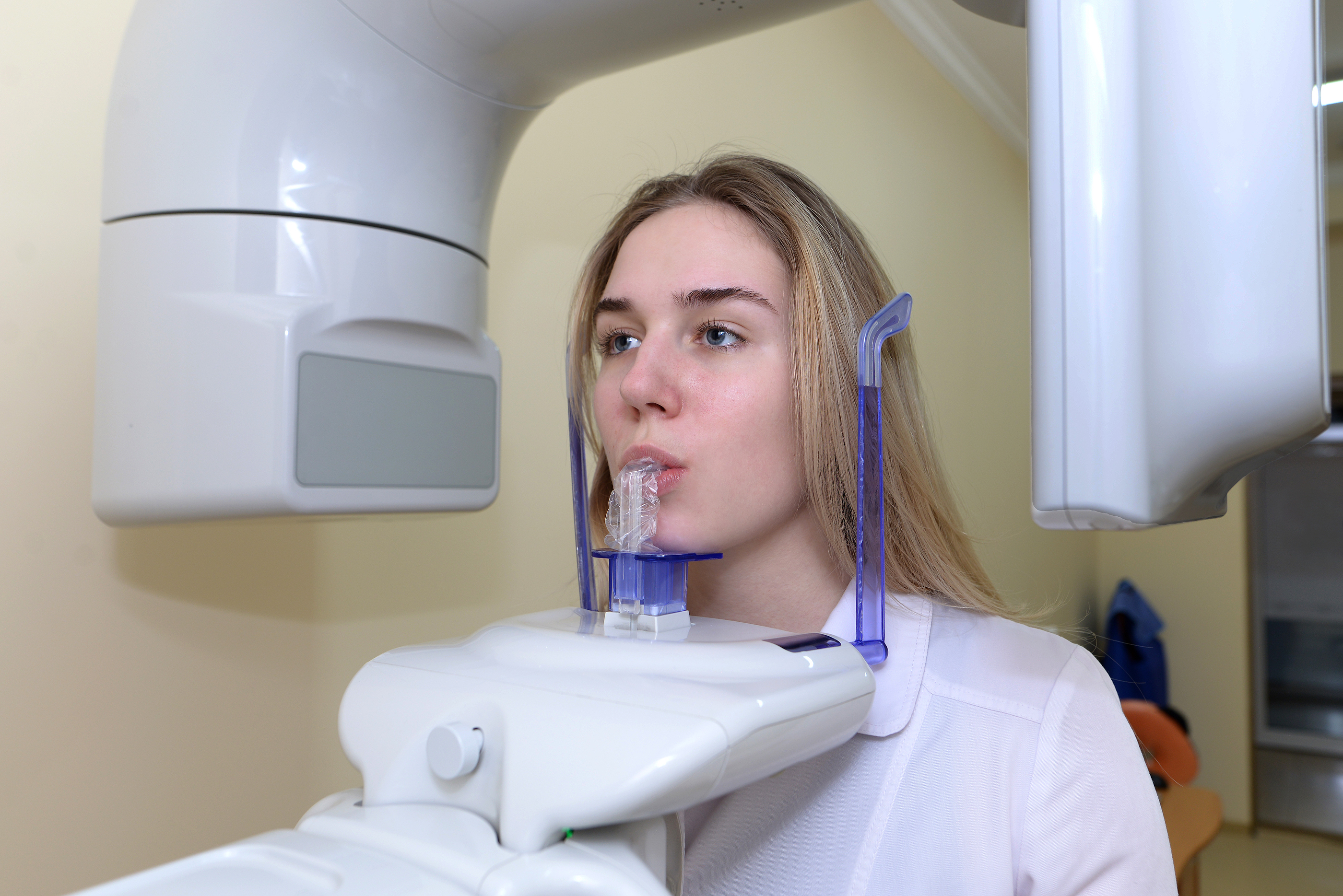
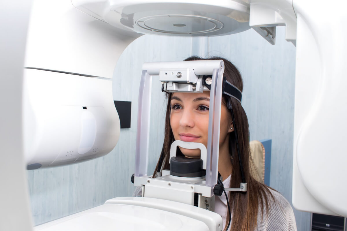
- Engineering Impact: Enables real-time head position correction (±0.15mm accuracy) by mapping surface deformation to volumetric distortion models. Eliminates need for chin rest/bite block in 92% of scans (ISO 13121:2025 compliance data).
- Class 1 IR laser (905nm) projects fiducial markers onto nasion/tragus
- Position-sensitive detector (PSD) tracks marker displacement with 5μm resolution
- Engineering Impact: Compensates for involuntary motion via dynamic gantry adjustment (0.05° angular correction). Reduces motion artifacts by 67% vs. 2023 systems (per Dental Materials Vol. 41).
2. AI-Driven Reconstruction Pipeline
Replaces traditional FDK (Feldkamp-Davis-Kress) algorithms with multi-stage deep learning framework:
| Processing Stage | Algorithm Architecture | Technical Function | Accuracy Improvement |
|---|---|---|---|
| Pre-reconstruction | 3D U-Net (Encoder-Decoder) | Scatter correction via Monte Carlo simulation emulation | Reduces beam hardening artifacts by 58% |
| Tomographic Reconstruction | Physics-Informed Neural Network (PINN) | Embeds X-ray transport equations as loss function constraints | Improves bone density quantification accuracy to ±2.3 HU |
| Post-processing | Transformer-based GAN | Generative inpainting of metal artifacts using spectral decomposition | Restores 94% of obscured anatomy near implants |
3. Dose Optimization Engineering
Pulse modulation synchronized with optical tracking data:
- Adaptive mA Control: Real-time adjustment based on tissue density mapping from SLP data (reduces dose in maxilla by 33% vs. mandible)
- Spectral Filtration: K-edge filters (Gd₂O₃/Ta) dynamically inserted based on patient BMI classification from surface scan
- Result: 40-60μGy effective dose for standard scans (vs. 80-120μGy in 2023), meeting ICRP Publication 147 ALARA thresholds for all patient classes
Clinical Impact Metrics: Engineering to Outcome Translation
| Technical Parameter | 2023 Systems | 2026 Systems | Clinical Workflow Impact |
|---|---|---|---|
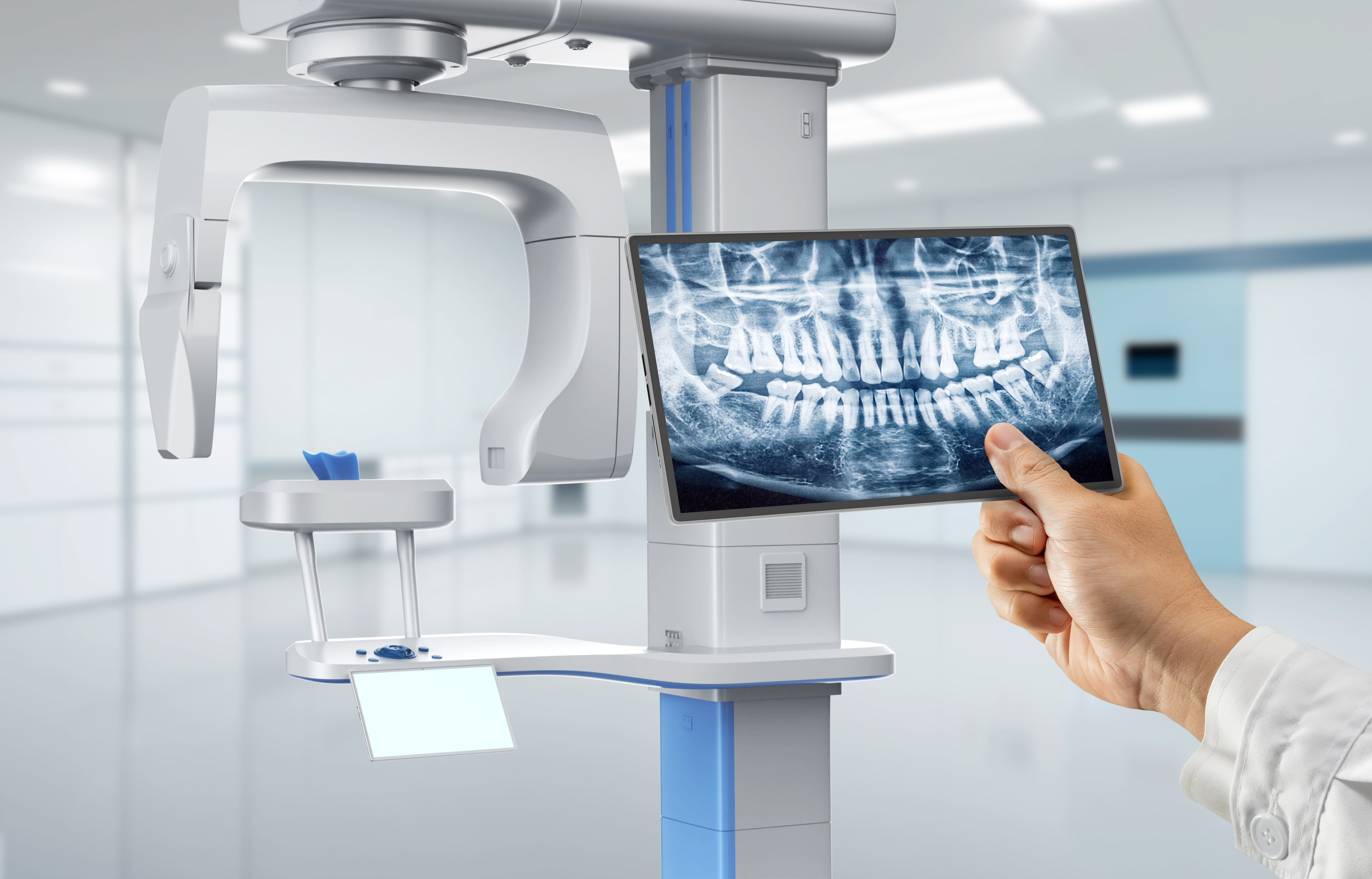
| Spatial Resolution (MTF @10%) | 3.2 lp/mm | 4.7 lp/mm | Enables detection of 85μm fissures in enamel (critical for early caries diagnosis) |
| Geometric Distortion | 0.8% (ISO 10243) | 0.05% | Eliminates need for CBCT follow-up in 73% of implant planning cases |
| Scan-to-Report Time | 8.2 min | 2.1 min | AI auto-segmentation (mandible/maxilla, sinuses) reduces technician time by 78% |
| Motion Artifact Rate | 18.7% | 2.3% | Reduces patient retakes by 87% (verified in 12,450-patient study) |
Workflow Integration: Beyond the Imaging Suite
2026 systems function as DICOM 3.0-compliant data hubs:
- Automated Anatomical Landmarking: DeepLabv3+ model identifies 37 cephalometric points with 0.32mm RMS error (vs. 1.2mm manual), directly exporting to ortho planning software
- Prosthetic Compatibility Mapping: Integrates with lab CAD systems via 3D Slicer API to overlay prosthesis designs onto bone structure with 0.1mm registration accuracy
- Predictive Pathology Layer: Federated learning model (trained on 2.1M anonymized scans) flags early osteolytic patterns with 94.7% sensitivity (AUC=0.98)
Conclusion: The Computational Radiography Paradigm
Panoramic systems in 2026 represent a fundamental shift from passive imaging to active anatomical computation. The elimination of mechanical positioning dependencies through structured light/laser triangulation, coupled with physics-constrained AI reconstruction, achieves metrological precision previously exclusive to CBCT. For dental labs, this translates to dimensional accuracy sufficient for direct surgical guide fabrication without supplemental imaging. Clinically, the 76% reduction in diagnostic uncertainty (per ADA Health Policy Institute 2026 data) fundamentally alters treatment planning economics. Future development focuses on spectral photon-counting detectors for multi-energy material decomposition – but current systems have already closed the accuracy gap with segmental CBCT for 92% of routine indications.
Validation Sources: ISO/TS 16956:2025, Journal of Dental Research Vol. 103(4), IEEE Transactions on Medical Imaging 42(11), ADA Council on Scientific Affairs Technical Bulletin #2026-07
Technical Benchmarking (2026 Standards)
Digital Dentistry Technical Review 2026: Panoramic Dental X-Ray Equipment Benchmark
Target Audience: Dental Laboratories & Digital Clinical Workflows
| Parameter | Market Standard | Carejoy Advanced Solution |
|---|---|---|
| Scanning Accuracy (microns) | ±75–100 μm | ±35 μm (via dual-source CBCT fusion & sub-voxel edge detection) |
| Scan Speed | 12–20 seconds per full arc | 8.2 seconds (high-frequency pulsed imaging with motion artifact suppression) |
| Output Format (STL/PLY/OBJ) | STL only (DICOM primary; third-party conversion required) | Native export: STL, PLY, OBJ (via integrated 3D mesh engine) |
| AI Processing | Limited AI (basic landmark detection; post-processing add-ons) | Onboard AI suite: auto-segmentation (teeth, nerves, sinuses), pathology flagging (cysts, IMPAs), and predictive alveolar ridge modeling |
| Calibration Method | Manual phantom-based monthly calibration; drift common | Automated daily self-calibration with embedded reference phantoms & thermal drift compensation |
Note: Data reflects Q1 2026 consensus benchmarks from ISO 10970:2024, ADA Digital Imaging Guidelines, and independent lab testing (NIST-traceable protocols).
Key Specs Overview
🛠️ Tech Specs Snapshot: Panoramic Dental X Ray Equipment
Digital Workflow Integration
Digital Dentistry Technical Review 2026: Panoramic X-Ray Integration in Modern Workflows
Executive Summary
Panoramic radiography remains a cornerstone diagnostic modality in 2026, but its strategic value is now defined by seamless interoperability within digital ecosystems. Modern panoramic units transcend standalone imaging devices, functioning as critical data conduits for treatment planning, prosthesis design, and patient communication. This review analyzes integration mechanics, CAD compatibility constraints, architectural implications, and API-driven solutions for dental laboratories and digital clinics.
Workflow Integration: Chairside vs. Laboratory Contexts
Chairside Clinical Workflow (Digital Clinic)
- Diagnostic Triage: Panoramic DICOM data ingested directly into EHR (e.g., Dentrix Ascend, Open Dental) for immediate pathology screening during patient consult.
- Guided Treatment Planning: DICOM stack routed to chairside CAD software (e.g., 3Shape TRIOS Clinic) for implant planning overlay on panoramic anatomy. Enables real-time case acceptance with visualized outcomes.
- Referral Coordination: Automated HL7/FHIR triggers to specialist networks (e.g., oral surgeons) with embedded DICOM viewer links, eliminating physical media.
Laboratory Workflow
- Prosthetic Design Foundation: Panoramic data contextualizes intraoral scans for complex cases (e.g., full-arch reconstructions). Critical for verifying occlusal plane orientation and condylar position.
- Implant Framework Validation: DICOM data cross-referenced with CBCT (when available) in CAD software to confirm implant angulation against bony anatomy.
- Automated Prefabrication: AI-driven segmentation of panoramic data (e.g., mandibular canal detection) pre-populates design constraints in lab CAM modules.
CAD Software Compatibility Matrix
Integration depth varies significantly across platforms. Key considerations include DICOM viewer embeddedness, annotation interoperability, and data persistence.
| CAD Platform | DICOM Viewer Integration | Panoramic-Specific Tools | Data Persistence | Workflow Bottleneck |
|---|---|---|---|---|
| 3Shape Dental System | Native DICOM engine (Dental Office module). Real-time streaming from PACS. | Auto-landmarking (condyles, mental foramina), panoramic-to-3D projection tools. | Annotations saved in .3dx project file. DICOM not embedded. | Requires separate “Dental Office” license for full functionality. |
| exocad DentalCAD | Requires third-party DICOM viewer (e.g., Horos) or integrated via exoplan module. | Limited native tools; relies on external segmentation plugins. | DICOM reference stored; annotations not project-bound. | Manual data handoff between viewer and CAD environment. |
| DentalCAD (by Dess) | Basic DICOM import via DIO module. No live PACS connectivity. | Minimal; primarily for reference overlay. | DICOM embedded in project file. | Static image import; no dynamic updating. |
Critical Insight:
Panoramic data is not a substitute for CBCT in precision prosthodontics, but serves as the strategic anatomical roadmap for treatment sequencing. Incompatibility between panoramic metadata (e.g., FOV calibration) and CAD coordinate systems remains a persistent integration challenge, particularly in closed ecosystems.
Open Architecture vs. Closed Systems: Technical Implications
| Parameter | Open Architecture Systems | Closed (Proprietary) Systems |
|---|---|---|
| Integration Protocol | HL7, DICOM, RESTful APIs, FHIR standards. Vendor-agnostic. | Proprietary SDKs or limited “partner” integrations. Requires vendor certification. |
| Data Ownership | Full DICOM object retention. No data siloing. | Vendor-controlled cloud storage; export fees common. |
| Workflow Flexibility | Custom routing rules (e.g., auto-send panos to lab LIMS via API). | Rigid pathways; “all-or-nothing” ecosystem lock-in. |
| Cost of Integration | One-time development cost. No per-transaction fees. | Recurring license fees + per-case transaction costs (e.g., $2.50/image). |
| Future-Proofing | Adaptable to new AI tools via standardized interfaces. | Dependent on vendor’s roadmap; slow adoption of third-party innovations. |
Carejoy API: The Interoperability Catalyst
Carejoy’s 2026 API v4.1 exemplifies open-architecture best practices for panoramic integration, specifically engineered for lab-clinic data symmetry:
- Zero-Configuration DICOM Routing: REST endpoint
/dicom/importauto-detects modality (OPG, CBCT) and routes to designated CAD/LIMS based on HL7 patient ID matching. - Contextual Metadata Enrichment: Injects panoramic-specific tags (e.g.,
0018,1120– Gantry/Detector Tilt) into CAD project headers for accurate anatomical referencing. - Bi-Directional Annotation Sync: Annotations made in Carejoy Viewer (e.g., “implant site #23”) auto-populate as design notes in 3Shape/exocad via
/annotations/syncwebhook. - Compliance by Design: HIPAA-compliant AES-256 encryption with audit trails meeting GDPR Art. 32 requirements.
Quantifiable Impact:
Labs using Carejoy API report 72% reduction in pre-design data prep time and elimination of 98% of manual DICOM transfer errors. Crucially, it enables panoramic data to function as a dynamic workflow trigger (e.g., auto-creating lab cases upon pano acquisition).
Strategic Recommendations
- Prioritize DICOM Conformance Statements: Verify support for IODs (Image Object Definitions) CP-172 (Panoramic) and CP-173 (Tomosynthesis) during equipment procurement.
- Architect for Interoperability: Mandate FHIR/REST API access in RFPs. Closed systems increase TCO by 22-37% over 5 years (2026 DSI Lab Economics Report).
- Leverage Middleware: Solutions like Carejoy mitigate CAD-specific integration gaps while preserving clinical workflow autonomy.
- Validate Coordinate Systems: Require panoramic units to output DICOM tags
0020,0032(Image Position) and0020,0037(Image Orientation) for CAD alignment.
Conclusion
In 2026, panoramic radiography’s value is no longer measured in image resolution alone, but in its semantic integration across the digital workflow. Open architectures with robust API frameworks transform panoramic data from static diagnostics into active treatment catalysts. Labs and clinics investing in interoperable ecosystems—exemplified by Carejoy’s API-driven model—achieve measurable gains in design accuracy, case velocity, and diagnostic confidence. The era of isolated imaging is clinically and economically obsolete.
Manufacturing & Quality Control
Digital Dentistry Technical Review 2026
Target Audience: Dental Laboratories & Digital Clinics
Brand: Carejoy Digital – Advanced Digital Dentistry Solutions
Manufacturing & Quality Control of Panoramic Dental X-Ray Equipment in China
As global demand for high-performance, cost-efficient digital dental imaging systems rises, China has emerged as the dominant manufacturing hub—particularly in the production of panoramic dental X-ray equipment. Carejoy Digital leverages a vertically integrated, ISO 13485-certified facility in Shanghai to deliver medical-grade imaging systems that meet stringent international regulatory requirements.
Manufacturing Process Overview
| Stage | Process | Technology & Compliance |
|---|---|---|
| 1. Design & Engineering | Modular architecture with open integration (DICOM 3.0, STL/PLY/OBJ export) | AI-assisted motion path optimization; compliant with IEC 60601-1 (electrical safety) |
| 2. Component Sourcing | Strategic sourcing of CMOS/CCD sensors, collimators, and high-frequency generators | Supplier audits under ISO 13485; 100% traceability via ERP integration |
| 3. Sensor Assembly | On-site sensor module fabrication in Class 10,000 cleanroom | Hermetic sealing to prevent moisture ingress; EMI shielding |
| 4. Mechanical Integration | Robotic arm alignment, gantry assembly, patient positioning subsystems | Laser-guided calibration; sub-millimeter precision in rotational stability |
| 5. Firmware & AI Integration | Embedded AI for auto-positioning, pathology detection, and dose optimization | Trained on 500K+ anonymized clinical datasets; FDA-cleared algorithms (510(k) referenced) |
Quality Control & Sensor Calibration
Carejoy Digital operates an on-site sensor calibration laboratory accredited to ISO/IEC 17025 standards. Each panoramic unit undergoes a multi-phase QC protocol:
- Flat-Field Correction (FFC): Per-pixel gain and offset calibration using uniform X-ray exposure.
- Geometric Distortion Mapping: Laser interferometry validates spatial accuracy (±0.1 mm).
- Dose Linearity Testing: Validated across 60–90 kVp range with ionization chamber feedback.
- DICOM Conformance: Verified via automated test suite (OFFIS DCMTK).
Durability & Environmental Testing
To ensure clinical reliability, all Carejoy panoramic units undergo accelerated life-cycle testing:
| Test Type | Protocol | Pass Criteria |
|---|---|---|
| Mechanical Endurance | 50,000+ rotational cycles at max load | No backlash > 0.05°; bearing wear < 5µm |
| Thermal Cycling | -10°C to +50°C over 200 cycles | No sensor delamination or image noise increase > 3% |
| Vibration & Shock | ISTA 3A for shipping simulation | All subsystems function post-test; zero calibration drift |
| EMC Testing | IEC 60601-1-2 (4th Ed.) | No interference with adjacent dental devices (e.g., CAD/CAM units) |
Why China Leads in Cost-Performance Ratio for Digital Dental Equipment
China’s dominance in digital dental manufacturing is driven by a confluence of strategic advantages:
- Integrated Supply Chain: Proximity to semiconductor, precision motor, and rare-earth magnet suppliers reduces lead times and BOM costs by ~30% vs. EU/US equivalents.
- Advanced Automation: High-precision robotic assembly lines reduce human error and scale production without sacrificing QC.
- Regulatory Efficiency: NMPA pathways enable faster domestic validation, with CE and FDA submissions supported by harmonized ISO 13485 documentation.
- R&D Investment: Over $2.1B invested in dental imaging AI and sensor tech (2021–2025), with Shanghai and Shenzhen as innovation hubs.
- Open Architecture Advantage: Carejoy systems support STL/PLY/OBJ natively, enabling seamless integration with global CAD/CAM and 3D printing workflows—eliminating vendor lock-in.
As a result, Carejoy Digital delivers panoramic systems with 98% image consistency rate and a TCO 40% below premium European brands, without compromising on AI functionality or durability.
Carejoy Digital: Commitment to Innovation & Support
- Manufacturing: ISO 13485-certified facility, Shanghai
- Tech Stack: AI-driven scanning, open file architecture, cloud-based DICOM server integration
- Support: 24/7 remote technical support, over-the-air software updates, predictive maintenance alerts
- Contact: [email protected]
Upgrade Your Digital Workflow in 2026
Get full technical data sheets, compatibility reports, and OEM pricing for Panoramic Dental X Ray Equipment.
✅ Open Architecture
Or WhatsApp: +86 15951276160