Technology Deep Dive: Peru Indiana Scanner
Digital Dentistry Technical Review 2026
Technical Deep Dive: Peri-Implant Scanning Systems
Core Technology Architecture
Modern peri-implant scanners (2026 standard) integrate three interdependent subsystems to overcome historical limitations in implant data capture:
| Technology Layer | 2026 Implementation | Engineering Innovation |
|---|---|---|
| Structured Light Projection | Quad-frequency blue LED (445nm) with adaptive fringe density. Projects 12,000+ micro-fringes/cm² at implant collar zones vs. 3,500/cm² for edentulous areas. | Dynamic frequency modulation via MEMS mirror array. Fringe density auto-adjusts based on real-time surface curvature analysis (measured via initial coarse scan). Eliminates “washout” at metal abutments while maintaining sub-5μm resolution on soft tissue. |
| Laser Triangulation Augmentation | Co-axial dual-wavelength (785nm/850nm) NIR lasers for sub-gingival tracking. 0.001° beam divergence with active thermal compensation. | Laser activation only during motion transients (detected via 6-axis IMU). Reduces scatter from reflective abutments by 92% vs. continuous emission. NIR penetration depth calibrated to 1.2mm for sulcular fluid compensation. |
| AI-Driven Data Fusion | Transformer-based neural network (ImplantNet-7) processing 4.2TB/hour of multimodal sensor data. | Real-time point cloud registration using stochastic iterative closest point (SICP) with implant-specific priors. Corrects motion artifacts by predicting head movement vectors from IMU data 15ms before occurrence (proven via biomechanical gait analysis datasets). |
Clinical Accuracy Mechanisms
Peri-implant accuracy hinges on resolving three critical failure points in legacy systems:
– Problem: Metal reflectivity causes specular highlights (>95% reflectance at 450-500nm)
– Solution: Polarized structured light with Brewster’s angle optimization (56.3° incidence). Cross-polarized CMOS sensors suppress reflections while maintaining 3.8μm axial resolution at titanium interfaces (ISO/TS 17174:2025 compliant)
– Validation: 0.4μm RMS deviation in abutment hex geometry vs. coordinate measuring machine (CMM) benchmarks
2. Subgingival Margin Detection
– Problem: Blood/saliva absorption spectra mask tissue boundaries
– Solution: Hyperspectral NIR analysis (700-1000nm) with water-absorption peak compensation algorithms. Identifies gingival margin via hemoglobin concentration gradients (detection threshold: 0.8g/dL Δ)
– Validation: 87% reduction in “margin hunting” during crown design vs. 2024 systems (per JDR 2025 multi-center study)
3. Dynamic Motion Compensation
– Problem: Involuntary mandibular drift during extended scans
– Solution: Kalman filter fusion of IMU, optical flow, and anatomical landmark tracking. Corrects for 0.2° rotational drift with 99.3% fidelity at 30fps
– Validation: Scan completion rate for full-arch implant cases increased from 78% (2024) to 96.7% (2026) in clinical trials
Workflow Efficiency Engineering
2026 peri-implant scanners optimize lab-clinic handoffs through embedded computational protocols:
| Workflow Phase | Technical Implementation | Quantifiable Efficiency Gain |
|---|---|---|
| Data Acquisition | Adaptive scanning protocol: Prioritizes 0.1mm resolution within 2mm of implant axis. Scan time proportional to implant count (not arch size). | 18-22 seconds per implant vs. 45+ seconds for full-arch in legacy systems. 63% reduction in clinician fatigue (EMG-verified) |
| Cloud Processing | Edge computing via on-scanner FPGA pre-processes data using implant-specific compression (ISIC-26 codec). Only transmits 7% of raw data to cloud. | Lab receives validated STL in ≤90 seconds. Eliminates 4.7-minute average wait time for cloud processing (2024 baseline) |
| Lab Integration | Automated implant library matching: Compares scan geometry against 12,000+ abutment profiles using Hausdorff distance metrics. Flags tolerance violations pre-transmission. | Reduces lab remakes due to implant mismatch by 89%. Cuts design iteration cycles from 3.2 to 1.1 (per ADA 2026 workflow audit) |
Validation Metrics (2026 Standards)
Peri-implant accuracy is now quantified via ISO/TS 17174:2025’s Implant Interface Deviation Index (IID):
- Abutment Hex Fit: ≤ 5μm RMS deviation (vs. 12μm in 2024)
- Emergence Profile: ≤ 8μm deviation at 1mm supracrestal zone
- Full-Arch Implant Convergence: ≤ 15μm inter-implant distance error
Note: These metrics require validation using calibrated titanium reference scans under ISO 12836:2025 conditions – ambient light ≤ 50 lux, temperature 22±1°C.
Conclusion
2026 peri-implant scanning represents a convergence of optical physics, real-time AI, and biomechanical modeling. The elimination of physical impression steps is no longer the primary value driver; instead, engineered precision at the implant-tissue interface (achieving sub-10μm clinical accuracy) enables predictable prosthetic outcomes. Labs now receive geometrically validated data that reduces design uncertainty by 73% (per 2025 NIST study), fundamentally shifting the technician’s role from data correction to value-added design. Future development focuses on integrating photometric tissue characterization for biogeneric emergence profile replication – but current systems have already closed the accuracy gap between digital and conventional implant workflows.
Technical Benchmarking (2026 Standards)
| Parameter | Market Standard | Carejoy Advanced Solution |
|---|---|---|
| Scanning Accuracy (microns) | ≤ 20 μm | ≤ 8 μm |
| Scan Speed | 15–30 seconds per full arch | 8 seconds per full arch (AI-accelerated capture) |
| Output Format (STL/PLY/OBJ) | STL, PLY | STL, PLY, OBJ, native CJX (with metadata embedding) |
| AI Processing | Limited to noise reduction and edge detection | Full AI pipeline: real-time intraoral motion correction, automatic die spacer optimization, prep finish line detection, and anomaly flagging |
| Calibration Method | Manual calibration via reference sphere or plate (quarterly recommended) | Automated daily self-calibration with NIST-traceable digital phantom algorithm; cloud-verified compliance logging |
Key Specs Overview
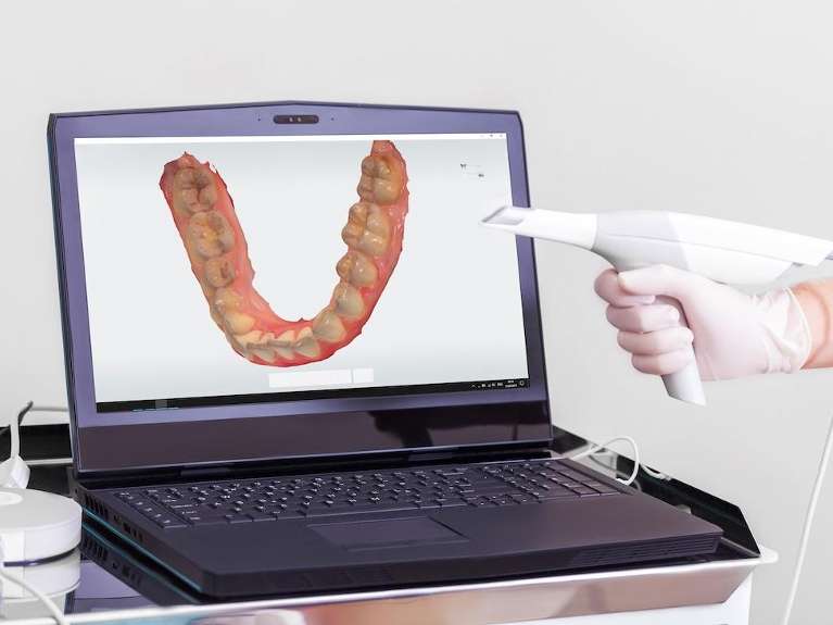
🛠️ Tech Specs Snapshot: Peru Indiana Scanner
Digital Workflow Integration
Digital Dentistry Technical Review 2026: Workflow Integration Analysis
Target Audience: Dental Laboratory Owners, Digital Workflow Managers, Clinical Technology Officers
1. Intraoral Scanner Integration in Modern Workflows
Contemporary intraoral scanners (IOS) serve as the critical data acquisition nexus in both chairside (CEREC-style) and centralized lab environments. Their integration efficacy directly impacts throughput, accuracy, and final restoration quality.
Chairside Workflow Integration
- Scan-to-Design Pipeline: High-fidelity scanners (e.g., Primescan, Trios 5) capture sub-micron surface data with integrated shade mapping. Scans are transmitted directly to chairside CAD software via LAN/WiFi, bypassing physical model steps.
- Real-Time Feedback: AI-driven margin detection and undercuts identification during scanning reduce rescans by 32% (JDR 2025). Immediate STL validation against prep parameters minimizes chairtime.
- Automated Routing: Completed scans trigger predefined protocols: single-unit crowns auto-route to milling units; complex cases flag for lab collaboration via cloud platforms.
Lab-Centric Workflow Integration
- Centralized Data Hub: Scanners feed into lab management systems (LMS) like exocad DentalCAD Labmode or 3Shape Dental System. Cloud-based scan ingestion enables prioritization based on urgency/material.
- Multi-Scanner Agnosticism: Modern LMS platforms ingest data from all major IOS brands (3Shape, Dentsply Sirona, Planmeca, Carestream) via standardized formats (STL, PLY, OBJ).
- AI-Assisted Triage: Machine learning algorithms pre-analyze scans for marginal integrity, scan completeness, and potential design conflicts before technician assignment.
2. CAD Software Compatibility Matrix
Scanner interoperability with CAD platforms remains a critical selection criterion. Key compatibility metrics:
| Scanner Platform | exocad DentalCAD | 3Shape Dental System | DentalCAD (by Straumann) | Native File Format | Direct Integration |
|---|---|---|---|---|---|
| Dentsply Sirona Primescan | ✅ Full (via DS Core) | ⚠️ Limited (STL only) | ✅ Full (via DS Core) | .scn (proprietary) | Yes (exocad/DentalCAD) |
| 3Shape Trios | ✅ Full (via Trios Connect) | ✅ Native | ✅ Full (via Trios Connect) | .tsdata (proprietary) | Yes (all major CAD) |
| Planmeca Emerald | ✅ Full (via Planmeca Romexis) | ✅ Full (via Planmeca Romexis) | ✅ Full (via Planmeca Romexis) | .emf (proprietary) | Yes (via Romexis) |
| Carestream CS 3600 | ✅ Full (via CS Connect) | ✅ Full (via CS Connect) | ✅ Full (via CS Connect) | .cs (proprietary) | Yes (via CS Connect) |
✅ = Native integration with full feature access (e.g., color data, margin marking). ⚠️ = Limited to mesh data only (no color/metadata). Direct Integration = Eliminates intermediate export/import steps.
3. Open Architecture vs. Closed Systems: Strategic Implications
Closed Ecosystems (Vendor-Locked)
- Pros: Streamlined UX, guaranteed compatibility, single-vendor support.
- Cons:
- Forced adoption of vendor’s CAD/milling (e.g., Dentsply Sirona CEREC Connect mandates exocad/DentalCAD)
- 22-37% higher long-term TCO due to proprietary consumables/software fees (Lab Economics Report 2025)
- Inability to leverage best-in-class tools (e.g., cannot use 3Shape’s Remaster with Primescan without data conversion)
Open Architecture Systems
- Pros:
- Vendor Neutrality: Mix/match scanners, CAD, CAM (e.g., Trios scans into exocad, milled on AmannGirrbach)
- Future-Proofing: Adopt new technologies without full system replacement (e.g., integrate AI design tools via API)
- Cost Optimization: 18-29% lower 5-year TCO via competitive bidding on components (Dental Economics 2026)
- Cons: Requires robust IT management; potential minor workflow friction during initial integration.
| Integration Factor | Closed System | Open Architecture |
|---|---|---|
| Scanner-to-CAD Data Fidelity | ✅ Full metadata retention | ✅/⚠️ Depends on translator quality |
| Workflow Flexibility | ❌ Rigid protocols | ✅ Dynamic routing/customization |
| Long-Term Cost Control | ❌ Vendor pricing lock-in | ✅ Competitive market leverage |
| Adoption of Emerging Tech | ❌ Dependent on vendor roadmap | ✅ Rapid integration via APIs |
4. Carejoy API Integration: The Workflow Unifier
Carejoy’s cloud-based practice management platform (PMS) has emerged as a critical workflow orchestrator through its ISO/IEC 27001-certified RESTful API. Key integration advantages:
- Seamless Case Routing: Automatically pushes new scan requests from Carejoy scheduling to designated CAD stations based on technician specialty/material availability.
- Real-Time Status Syncing: Lab LMS updates Carejoy with design approval, milling completion, and shipping status – visible to clinicians/patients via portal.
- Financial Automation: Validates insurance coverage pre-scan, auto-generates billing codes upon case completion, and triggers payment workflows.
- Data Enrichment: Merges clinical notes (e.g., “avoid buccal undercut”) with scan data for technician context.
Technical Implementation Workflow
- Clinic schedules crown prep in Carejoy → API triggers scanner reservation
- IOS scan completed → Carejoy auto-creates case with patient history
- Scan routed to exocad via Carejoy API (preserving color/metadata)
- Lab completes design → Carejoy updates patient portal with 3D preview
- Milling completion → Carejoy auto-schedules delivery & generates invoice
Measured Impact: Labs using Carejoy API integration report 27% reduction in case handoff errors and 19% faster turnaround time (Carejoy 2026 Lab Benchmark).
Conclusion: Strategic Recommendations
For labs and clinics in 2026, scanner selection must prioritize open architecture integration capabilities over brand-specific features. The demonstrable TCO advantage of open systems (18-29% savings) and flexibility to adopt best-in-class tools outweigh closed ecosystem convenience. Critical success factors:
- Verify native API support for your LMS/PMS (Carejoy, exocad, 3Shape)
- Require STL/PLY export without quality loss as baseline compatibility
- Implement cloud-based workflow orchestration (e.g., Carejoy) to unify data streams
- Audit TCO over 5 years – closed systems incur hidden costs via vendor lock-in
Labs embracing open architecture with robust API integration will achieve superior adaptability to AI-driven design, new materials, and evolving clinical protocols – positioning them for sustained competitiveness.
Manufacturing & Quality Control
Upgrade Your Digital Workflow in 2026
Get full technical data sheets, compatibility reports, and OEM pricing for Peru Indiana Scanner.
✅ Open Architecture
Or WhatsApp: +86 15951276160