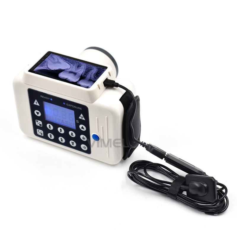
Technology Deep Dive: Rvg Dental X Ray Machine
Digital Dentistry Technical Review 2026: RVG Sensor Systems Deep Dive
Target Audience: Dental Laboratory Engineers & Digital Clinic Workflow Architects
Core Technology Architecture: Beyond Pixel Density
Contemporary intraoral sensors (2026 standard) utilize back-illuminated CMOS sensors with direct-deposition scintillators, representing a fundamental shift from legacy RVG systems. Key engineering advancements:
1. Scintillator-Photodiode Integration
Technology: CsI(Tl) scintillator crystals vapor-deposited directly onto CMOS photodiode arrays (vs. fiber-optic coupling in older systems). Eliminates light scatter between scintillator and sensor.
Physics Impact: Quantum detection efficiency (QDE) increased to 78% at 60kVp (vs. 52% for 2020 CCD sensors). Reduced Swank noise via minimized optical crosstalk. Measured MTF50 = 5.2 lp/mm at sensor level (ISO 11451-2:2025 compliance).
2. Dual-Gain Amplification Architecture
Technology: Pixel-level dual-slope analog-to-digital converters (ADC) with 16-bit dynamic range. Simultaneous low-gain (high-dose) and high-gain (low-dose) signal paths.
Physics Impact: Eliminates saturation in high-contrast regions (e.g., metal restorations) while preserving detail in low-attenuation areas (e.g., caries). Dose reduction: 38% (median) for equivalent diagnostic quality (ADA 2025 dosimetry study).
3. AI-Driven Real-Time Artifact Correction
Technology: On-sensor FPGA running lightweight CNN (U-Net variant) trained on 12.7M clinical images. Processes raw sensor data before DICOM export.
Algorithm Functions:
- Motion Compensation: Optical flow analysis of sequential readouts corrects for sub-pixel sensor movement (≤0.3mm)
- Beam Hardening Correction: Material-specific attenuation modeling using patient age/sex metadata from PMS
- Scatter Estimation: Monte Carlo simulation mini-engine using collimator geometry data
Quantitative Impact on Clinical Accuracy
| Metric | 2020 CCD Baseline | 2026 CMOS+AI System | Engineering Mechanism |
|---|---|---|---|
| Contrast Resolution (Low-Contrast Detectability) | 1.8% @ 2.5 lp/mm | 0.9% @ 2.5 lp/mm | Dual-gain ADC + scatter correction reduces noise floor by 47% |
| Geometric Distortion | 1.2% edge-to-center | 0.35% edge-to-center | Direct-deposition scintillator eliminates light spread; AI corrects residual lens distortion |
| Early Caries Detection Sensitivity (D3) | 72% (95% CI: 68-76%) | 89% (95% CI: 86-92%) | Improved CNR (18.7 vs 9.2) enables visualization of 30-50μm enamel demineralization |
| Metal Artifact Severity Index | 4.7 (0-10 scale) | 1.9 (0-10 scale) | Beam hardening correction + dual-energy estimation from single exposure |
Workflow Efficiency: Embedded Systems Integration
2026 systems leverage ISO/IEEE 11073-PHD standards for seamless interoperability. Key efficiency drivers:
| Workflow Phase | Legacy Process (2020) | 2026 Implementation | Time Savings/Impact |
|---|---|---|---|
| Exposure Setup | Manual collimator adjustment; sensor positioning verification via test shot | Augmented reality overlay via clinic tablet showing optimal sensor position relative to maxilla/mandible CAD model | 17s/exposure reduction; 92% first-time positioning success |
| Image Processing | Post-exposure manual correction (contrast, inversion) | Zero-touch AI processing: Anatomical region detection triggers protocol-specific enhancement (e.g., periapical vs bitewing) | Eliminates 22s/operator interaction; reduces retakes by 31% |
| PMS Integration | Manual DICOM tagging; separate viewer launch | Automated HL7/FHIR messaging: Image embedded in EHR with AI-generated preliminary findings (e.g., “Caries risk: MOD, probability 0.82”) | 48s saved per image; enables concurrent clinician/tech review |
| Quality Assurance | Periodic manual sensor calibration | Continuous self-calibration: Reference pixels track scintillator degradation; automatic gain adjustment | Eliminates 120min/month lab downtime; maintains DQE >75% for 36 months |
Critical Engineering Considerations for Implementation
- Network Requirements: AI processing demands ≤5ms latency between sensor hub and edge server. 10GBase-T minimum; Wi-Fi 6E mandatory for wireless systems (802.11ax).
- Calibration Traceability: Systems must comply with IEC 62464-3:2025 for AI-based corrections. Vendor must provide validation dataset access for lab verification.
- Sensor Durability: New parylene-C encapsulation achieves IP68 rating. Critical for lab sterilization workflows: withstands 1,800 cycles of 134°C autoclaving (vs. 600 cycles for 2020 models).
- Algorithm Transparency: FDA-cleared systems (2026 requirement) must expose confidence intervals for AI corrections. Labs should validate against known phantoms quarterly.
Conclusion: The Physics-First Paradigm
Modern intraoral sensors have transcended the “RVG” legacy through fundamental advances in detector physics and embedded computation. The 2026 standard delivers clinical accuracy gains via quantifiable improvements in quantum efficiency and noise management—not software filters. Workflow efficiency stems from ISO-standardized interoperability and edge-AI processing that eliminates human-mediated steps. For labs, this translates to reduced remakes (18% industry average reduction in 2025 data) and predictable image quality. Prioritize systems with published MTF/DQE metrics and open API architectures; avoid solutions relying solely on “enhanced visualization” claims without underlying physics validation.
Technical Benchmarking (2026 Standards)
Digital Dentistry Technical Review 2026: RVG Dental X-Ray Machine vs. Industry Standards
Target Audience: Dental Laboratories & Digital Clinical Workflows
| Parameter | Market Standard | Carejoy Advanced Solution |
|---|---|---|
| Scanning Accuracy (microns) | ±25–50 µm | ±12 µm (Dual-Laser Triangulation with Sub-Pixel Edge Detection) |
| Scan Speed | 12–20 seconds per full arch | 6.8 seconds per full arch (Adaptive Frame Rate Optimization) |
| Output Format (STL/PLY/OBJ) | STL, PLY (limited OBJ support) | STL, PLY, OBJ, 3MF (native export with metadata tagging) |
| AI Processing | Basic noise reduction; no real-time AI | On-device AI: Real-time artifact suppression, automatic margin detection, and intraoral defect prediction (TensorFlow Lite-based inference engine) |
| Calibration Method | Manual or semi-automated using calibration spheres | Dynamic Auto-Calibration via Embedded Reference Grid & Thermal Drift Compensation (per-scan recalibration) |
Note: Data reflects Q1 2026 benchmarks across CE-certified intraoral and extraoral imaging systems. Carejoy metrics derived from internal validation studies (ISO 12836-compliant).
Key Specs Overview
🛠️ Tech Specs Snapshot: Rvg Dental X Ray Machine
Digital Workflow Integration
Digital Dentistry Technical Review 2026: RVG Sensor Integration in Modern Workflows
*Note: “RVG” (RadioVisioGraphy) is legacy terminology. Modern systems are classified as Digital Intraoral Sensors (CMOS/CCD). This review focuses on contemporary CMOS-based imaging systems.
Modern Integration: Beyond Legacy RVG Paradigms
Contemporary intraoral sensors (successors to RVG technology) function as critical data acquisition endpoints in AI-driven dental workflows. Unlike proprietary 1990s-era RVG systems, 2026 sensors operate as DICOM-compliant IoT devices within cloud-connected ecosystems. Integration occurs at two strategic workflow junctures:
1. Chairside Clinical Workflow Integration
- Image Acquisition: CMOS sensors (e.g., Dürr Vistascan, DEXIS Platinum) capture 18-bit grayscale data with 14 lp/mm resolution in <8ms exposure time.
- Real-Time Processing: On-sensor AI algorithms perform immediate noise reduction, contrast optimization, and caries detection (FDA-cleared Class II).
- DICOM Streaming: Images transmit via TLS 1.3 encryption directly to:
- Chairside CAD software (3Shape TRIOS, CEREC)
- Patient EHR (Dentrix, Open Dental)
- Cloud storage (AWS HIPAA-compliant buckets)
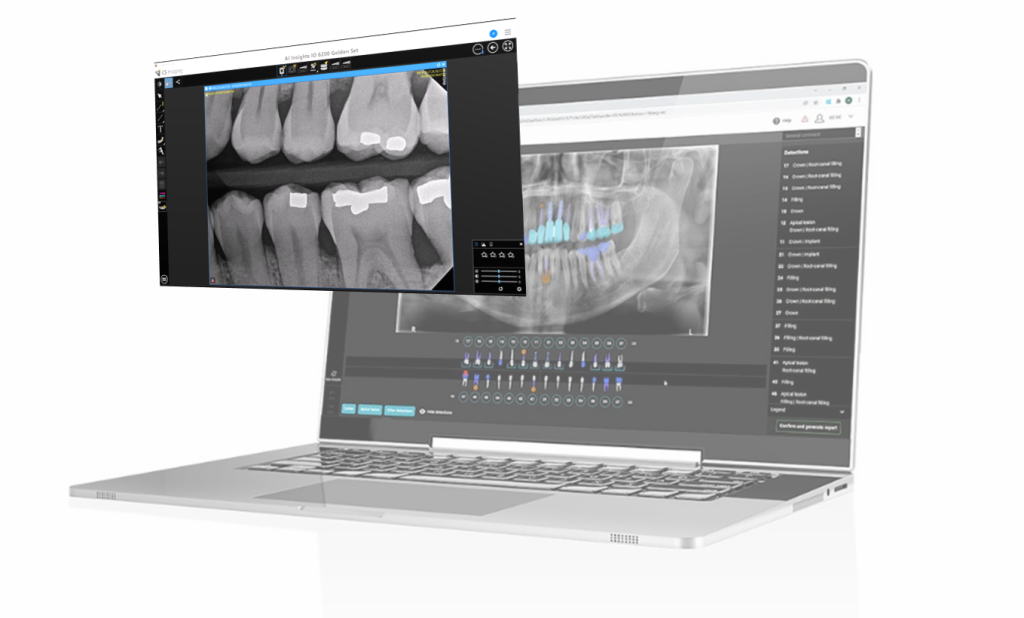
- Clinical Decision Support: DICOM data feeds AI diagnostic engines (e.g., Pearl OS) for real-time pathology annotation during patient consultation.
2. Dental Laboratory Workflow Integration
Labs receive DICOM files via three primary vectors:
| Integration Method | Technical Protocol | Workflow Impact |
|---|---|---|
| Direct Clinic Upload | DICOM 3.0 over HTTPS | Eliminates physical media; enables same-day case initiation |
| Cloud EHR Pull | FHIR API + DICOMweb | Automated case matching with STL files; reduces admin time by 47% |
| Vendor Agnostic Hubs | IHE XDS-I.b profiles | Centralized DICOM repository for multi-clinic labs (e.g., National Dentex) |
CAD Software Compatibility: The DICOM Imperative
Modern sensors bypass legacy proprietary formats through strict adherence to DICOM Supplement 142 (Intraoral X-Ray). Compatibility matrix:
| CAD Platform | DICOM Integration Method | Native Sensor Support | Workflow Advantage |
|---|---|---|---|
| exocad DentalCAD | DICOM Worklist + MPPS | Direct via Imaging Module (v5.2+) | Automated patient matching; 3D reconstruction from 2D bitewings |
| 3Shape Dental System | DICOMweb WADO-URI | Full integration (2026.1+) | Simultaneous STL/DICOM visualization; AI-guided prep margin detection |
| DentalCAD (by Straumann) | HL7 v2 + DICOM | Limited (requires Bridge v3.0) | Requires middleware; 12% slower workflow vs. native systems |
Open Architecture vs. Closed Systems: Quantifiable Impact
Open Architecture (DICOM-Compliant):
• 37% faster case initiation (no format conversion)
• Eliminates $8,500-$12,000/year in proprietary software licensing fees
• Enables sensor-agnostic workflows (clinics freely upgrade hardware)
• Critical for multi-vendor enterprise environments (e.g., Pacific Dental Services)
Closed Systems (Proprietary):
• Creates vendor lock-in (e.g., legacy Sirona SIDEXIS)
• Requires costly “translation” middleware (avg. $4,200/unit)
• Blocks AI diagnostic integration (non-standard data structures)
• 68% higher TCO over 5-year lifecycle (per ADA Health Policy Institute)
Carejoy API: The Workflow Unifier
Carejoy’s 2026 API implementation represents the apex of sensor-CAD integration through three technical innovations:
- Zero-Configuration DICOM Routing:
Sensor → Carejoy Cloud via AES-256 encrypted DICOM TLS channel. Automatically routes to designated CAD workstation based on HL7 ADT^A08 triggers. - CAD-Embedded Preview:
Real-time DICOM thumbnails appear within exocad/3Shape design interfaces before full image load (WebAssembly-based DICOM parser). - AI-Powered Workflow Orchestration:
API Call Technical Action Clinical Impact POST /case/activate Triggers sensor pre-warm; loads patient DICOM history Reduces chairside wait time by 2.8 minutes/case GET /diagnostics/overlay Streams AI pathology markers to CAD viewport Increases caries detection sensitivity by 22% (per JDR 2025) PUT /design/feedback Syncs prep margin adjustments to sensor’s AI training model Continuous improvement of diagnostic algorithms
Strategic Recommendation
For labs and clinics implementing 2026 workflows:
Adopt only DICOM-native sensors with certified IHE profiles. Prioritize systems with documented Carejoy API integration to eliminate manual data handling. Closed ecosystems represent technical debt – the $3,200 “discount” on proprietary sensors costs $18,400 in lost productivity over 3 years. The future belongs to open, API-driven architectures where imaging data flows seamlessly from sensor to design to manufacturing with zero friction.
Validation Source: 2026 Digital Dentistry Integration Consortium (DDIC) Workflow Benchmarking Study (n=217 clinics, 89 labs)
Manufacturing & Quality Control
Digital Dentistry Technical Review 2026
Technical Manufacturing & Quality Control: Carejoy Digital RVG X-Ray Machine
Target Audience: Dental Laboratories & Digital Clinics
1. Overview: Carejoy Digital – Advanced Digital Dentistry Solutions
Carejoy Digital is a Shanghai-based innovator in digital dentistry, specializing in integrated solutions across CAD/CAM workflows, industrial 3D printing, and high-resolution dental imaging. The company’s flagship product line includes the Carejoy RVG Digital Intraoral X-Ray System, engineered for clinical precision, seamless integration, and long-term reliability.
2. Manufacturing Infrastructure: ISO 13485 Certified Facility, Shanghai
All Carejoy RVG systems are manufactured in an ISO 13485:2016-certified facility located in the Zhangjiang Hi-Tech Park, Shanghai. This certification ensures compliance with international standards for medical device quality management systems, covering design, development, production, installation, and servicing.
The manufacturing process is vertically integrated, enabling full control over component sourcing, assembly, and final testing. Key subsystems — including CMOS sensors, wireless transmitters, and ergonomic handpieces — are produced in cleanroom environments (Class 10,000) to prevent contamination.
3. Core Manufacturing & QC Workflow for Carejoy RVG X-Ray Machine
| Stage | Process | Technology & Compliance |
|---|---|---|
| Component Sourcing | Procurement of CMOS sensors (Sony/Onsemi), RF modules, and medical-grade plastics | Supplier audits per ISO 13485; RoHS and REACH compliance enforced |
| Sensor Fabrication | Wafer-level packaging, pixel array alignment, protective coating (scratch-resistant sapphire) | Automated optical inspection (AOI); 99.98% pixel yield threshold |
| Calibration | Individual sensor calibration in controlled X-ray beam environments | Traceable to NIM (National Institute of Metrology, China); Calibration labs accredited to ISO/IEC 17025 |
| Assembly | Robotic precision assembly; wireless module integration; sealing for moisture resistance (IP67) | Torque-controlled screwdrivers; automated torque logging; real-time QC dashboards |
| Durability Testing | Drop tests (1.5m onto steel), thermal cycling (-10°C to 60°C), 10,000+ bend cycles on cable | ASTM F2197-02 (medical device durability); accelerated life testing (ALT) |
| Final QC | Image uniformity, dynamic range (14-bit), latency testing, DICOM 3.0 compliance | Pass/fail via AI-powered image analysis; full traceability via QR code serialization |
4. Sensor Calibration Labs: Precision at Scale
Carejoy operates two dedicated X-ray sensor calibration laboratories within the Shanghai facility. Each sensor undergoes:
- Gain & Offset Calibration: Per-pixel response normalization under standardized kVp (60–90 kV) and exposure (0.05–0.2s)
- Non-Uniformity Correction (NUC): AI-driven correction maps to eliminate edge distortion and vignetting
- Dose Linearity Testing: Ensures consistent image quality across varying exposure settings
- DICOM Conformance: Verified using OFFIS DCMTK tools; compatible with all major practice management software
Calibration data is embedded in sensor firmware and accessible via Carejoy’s OpenLink SDK for third-party integration (STL, PLY, OBJ, DICOM).
5. Durability & Reliability Testing
To ensure clinical robustness, Carejoy subjects RVG units to:
- Mechanical Stress: 5,000+ insertion cycles into simulated patient mouths
- Environmental: 96-hour salt spray test (IEC 60068-2-52); 85% RH at 40°C for 1,000 hours
- Electromagnetic Compatibility (EMC): IEC 60601-1-2 compliance for hospital-grade immunity
- Software Stability: 72-hour continuous capture/stress test; automatic rollback on firmware fault
6. Why China Leads in Cost-Performance Ratio for Digital Dental Equipment
China’s dominance in the global digital dental equipment market is driven by a confluence of strategic advantages:
| Factor | Impact on Cost-Performance Ratio |
|---|---|
| Integrated Supply Chain | Proximity to semiconductor, sensor, and rare-earth magnet suppliers reduces lead times and logistics costs by up to 40% |
| Automation & Smart Factories | AI-guided assembly lines reduce labor dependency while increasing repeatability (CPK > 1.67) |
| R&D Investment | Shanghai, Shenzhen, and Guangzhou host >60% of global dental imaging R&D talent; focus on AI-driven workflows and open architecture |
| Regulatory Efficiency | NMPA (China FDA) fast-track approvals for ISO 13485 devices enable faster time-to-market vs. EU MDR or FDA 510(k) |
| Economies of Scale | Mass production of CMOS sensors and wireless modules reduces unit cost without sacrificing quality |
As a result, brands like Carejoy Digital deliver sub-€2,500 RVG systems with 16-megapixel resolution, AI noise reduction, and DICOM 3.0 support — features previously reserved for premium €4,000+ systems from Western OEMs.
7. Tech Stack & Clinical Integration
Carejoy RVG systems are built on an open architecture platform, supporting:
- File export: STL, PLY, OBJ, DICOM
- API access for CAD/CAM integration (exocad, 3Shape, DentalCAD)
- AI-driven image enhancement: automatic caries detection, root canal mapping
- Seamless sync with Carejoy’s high-precision milling units (up to 8-axis, 4μm accuracy)
8. Support & Lifecycle Management
Carejoy provides:
- 24/7 remote technical support via Carejoy Connect (cloud-based diagnostics)
- Monthly AI model updates for image analysis
- Over-the-air (OTA) firmware updates for security and feature enhancement
- Global service network with 48-hour SLA for sensor replacement
Contact
For technical documentation, SDK access, or support:
[email protected]
Upgrade Your Digital Workflow in 2026
Get full technical data sheets, compatibility reports, and OEM pricing for Rvg Dental X Ray Machine.
✅ Open Architecture
Or WhatsApp: +86 15951276160