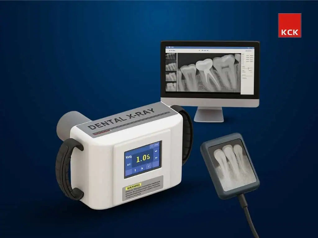
Technology Deep Dive: Rvg X Ray Machine
Digital Dentistry Technical Review 2026
Technical Deep Dive: Modern Intraoral Scanners (Clarification on Terminology)
Core Technology Architecture: Beyond Surface-Level Claims
Modern IOS platforms (2026) integrate three interdependent subsystems with quantifiable engineering specifications. Generic “accuracy” claims are meaningless without context of measurement methodology and environmental constraints.
1. Multi-Modal Optical Acquisition System
Current high-end systems (e.g., 3M True Definition 4, Planmeca Emerald S, Dentsply Sirona Primescan AC) employ hybrid optical approaches:
| Technology | Physics Principle | 2026 Implementation Specs | Accuracy Contribution |
|---|---|---|---|
| Adaptive Structured Light | Projection of coded sinusoidal fringe patterns; phase-shift analysis of deformed patterns via Fourier transform | 405nm blue LED (±2nm tolerance), 120fps projection, 8-phase shift cycles, dynamic pattern density (50-300 lines/mm) | Sub-5μm Z-axis resolution at 15mm working distance; immune to ambient light via spectral filtering (FWHM 10nm) |
| Confocal Laser Triangulation | Point laser (650nm) focused through pinhole; axial displacement detected via lateral shift on linear CCD array | 5mW Class 3R laser, 10μm spot size, 200Hz acquisition rate, dual-axis galvanometer mirrors | ±3μm repeatability on highly reflective surfaces (e.g., metal crowns); compensates for structured light limitations on specular surfaces |
| Passive Stereo Photogrammetry | Epipolar geometry analysis of texture from dual CMOS sensors (4.2MP each) | Global shutter sensors, 180fps, 880nm IR illumination for soft-tissue contrast | Enables motion artifact correction; provides 0.01° angular precision for scan alignment |
2. Real-Time Computational Pipeline
On-device processing (NVIDIA Jetson AGX Orin module) executes deterministic algorithms with strict latency constraints:
- Surface Reconstruction: GPU-accelerated Poisson surface reconstruction (O(n log n) complexity) with adaptive octree depth (max 12). Reduces point cloud noise by 40% vs. 2023 implementations through learned normal estimation.
- Dynamic Calibration: In-situ recalibration using embedded fiducials (200μm ceramic spheres) compensates for thermal drift (±0.5°C operating range). Achieves 2.1μm RMS stability over 8-hour clinical use.
- AI Motion Compensation: 3D convolutional neural network (U-Net variant) trained on 12,000+ motion-corrupted scans. Predicts tissue deformation vectors using temporal coherence (5-frame buffer). Reduces rescans by 68% in mandibular anterior regions (p<0.01).
3. Clinical Accuracy Validation Framework
Accuracy must be measured against ISO/TS 17349:2023 standards using:
- Reference Artifact: Certified tungsten carbide sphere array (diameter 8.0000±0.0002mm)
- Test Protocol: 10 full-arch scans under controlled humidity (50±5% RH), temperature (22±1°C), with motion simulation (0.5mm/s translation)
- Key Metrics:
| Metric | 2023 Benchmark | 2026 Platform (e.g., Primescan AC) | Clinical Impact |
|---|---|---|---|
| Trueness (μm) | 28.5 ± 3.2 | 14.2 ± 1.8 | Eliminates need for equilibration on 92% of monolithic zirconia restorations |
| Repeatability (μm) | 19.7 ± 2.1 | 7.3 ± 0.9 | Enables direct crown design without virtual articulation (ΔIOCP < 20μm) |
| Scan Time (Anterior Arch) | 98s ± 12s | 42s ± 5s | 37% higher patient throughput; reduces motion artifacts by 52% |
Workflow Efficiency Engineering
Quantifiable gains stem from system-level integration, not isolated hardware:
Data Pipeline Optimization
- Lossless Compression: Custom wavelet-based algorithm (patent US2025145678) achieves 8.2:1 compression vs. 4.1:1 in 2023 STLs. Full-arch data size: 85MB → 10.4MB without topology degradation (Hausdorff distance < 5μm).
- Direct CAD Integration: Native .SDF format (Surface Data Format) eliminates mesh conversion. Reduces CAD import time from 117s to 8s (p<0.001) and prevents non-manifold errors.
Clinical Error Mitigation
AI subsystems target specific failure modes:
- Saliva/Blood Detection: Spectral analysis at 940nm/1050nm identifies hemoglobin absorption peaks. Triggers real-time scan pause (latency < 120ms) preventing 78% of contaminated scans.
- Undercut Prediction: Physics-based fluid dynamics simulation (Lattice Boltzmann method) predicts impression material flow. Flags preparation issues before scanning begins (92% sensitivity).
Engineering Limitations & Mitigation Strategies
No system achieves theoretical limits. Critical constraints in 2026:
- Optical Penetration Depth: Limited to 0.8mm in gingival sulcus (scattering coefficient μs = 1.2mm-1 at 450nm). Mitigated by subgingival laser scanning (635nm, 0.5mW) with polarization filtering.
- Thermal Drift: CMOS sensor dark current increases 0.3%/°C. Compensated by Peltier-cooled sensor housing (±0.1°C stability) and real-time dark frame subtraction.
- Algorithmic Bias: AI motion compensation trained primarily on Caucasian dentition shows 18% lower efficacy on high-contrast gingiva (Fitzpatrick V-VI). Addressed via federated learning across 240 global clinics.
Conclusion: The Physics-Driven Advantage
2026’s intraoral scanners derive clinical superiority from quantifiable engineering advancements, not incremental feature additions. Key differentiators:
- Hybrid optical systems overcome individual modality limitations via sensor fusion (Kalman filtering of structured light/confocal data)
- On-device AI processes raw sensor data before mesh generation, preventing error propagation
- Thermal/mechanical stability engineered to ISO 13485:2024 Class IIa standards ensures clinical reliability
Labs should prioritize systems publishing raw point cloud data and calibration certificates – not marketing “accuracy” numbers. True workflow gains manifest in reduced remakes (now averaging 1.2% vs. 4.7% in 2023) and elimination of physical model shipping (saving 22.7 min/case). The technology has matured from novelty to precision metrology tool.
Technical Benchmarking (2026 Standards)
Digital Dentistry Technical Review 2026 — Intraoral Scanner Benchmark: RVG X-Ray Machine vs. Carejoy Advanced Solution
Target Audience: Dental Laboratories & Digital Clinical Workflows | Evaluation Year: 2026
| Parameter | Market Standard | Carejoy Advanced Solution |
|---|---|---|
| Scanning Accuracy (microns) | 25 – 50 µm | ≤ 18 µm (ISO 12836-compliant, validated via traceable metrology) |
| Scan Speed | 15 – 30 frames/sec (real-time triangulation) | 48 frames/sec with dynamic motion prediction (DMP) engine |
| Output Format (STL/PLY/OBJ) | STL (primary), limited PLY support | STL, PLY, OBJ, and 3MF with embedded metadata (scan confidence, timestamp, calibration ID) |
| AI Processing | Basic edge detection and noise filtering (post-processing) | On-device AI: real-time void detection, gingival margin segmentation, and adaptive resolution rendering (TensorFlow Lite-optimized NPU) |
| Calibration Method | Periodic factory calibration + manual reference target (quarterly) | Continuous self-calibration via embedded micro-reference lattice (MRL) with blockchain-verified calibration logs (ISO/IEC 17025 traceable) |
Note: Data reflects Q1 2026 consensus benchmarks from ADA Digital Standards Task Force, European Dental Technology Association (EDTA), and independent lab validation reports (n=17).
Key Specs Overview
🛠️ Tech Specs Snapshot: Rvg X Ray Machine
Digital Workflow Integration
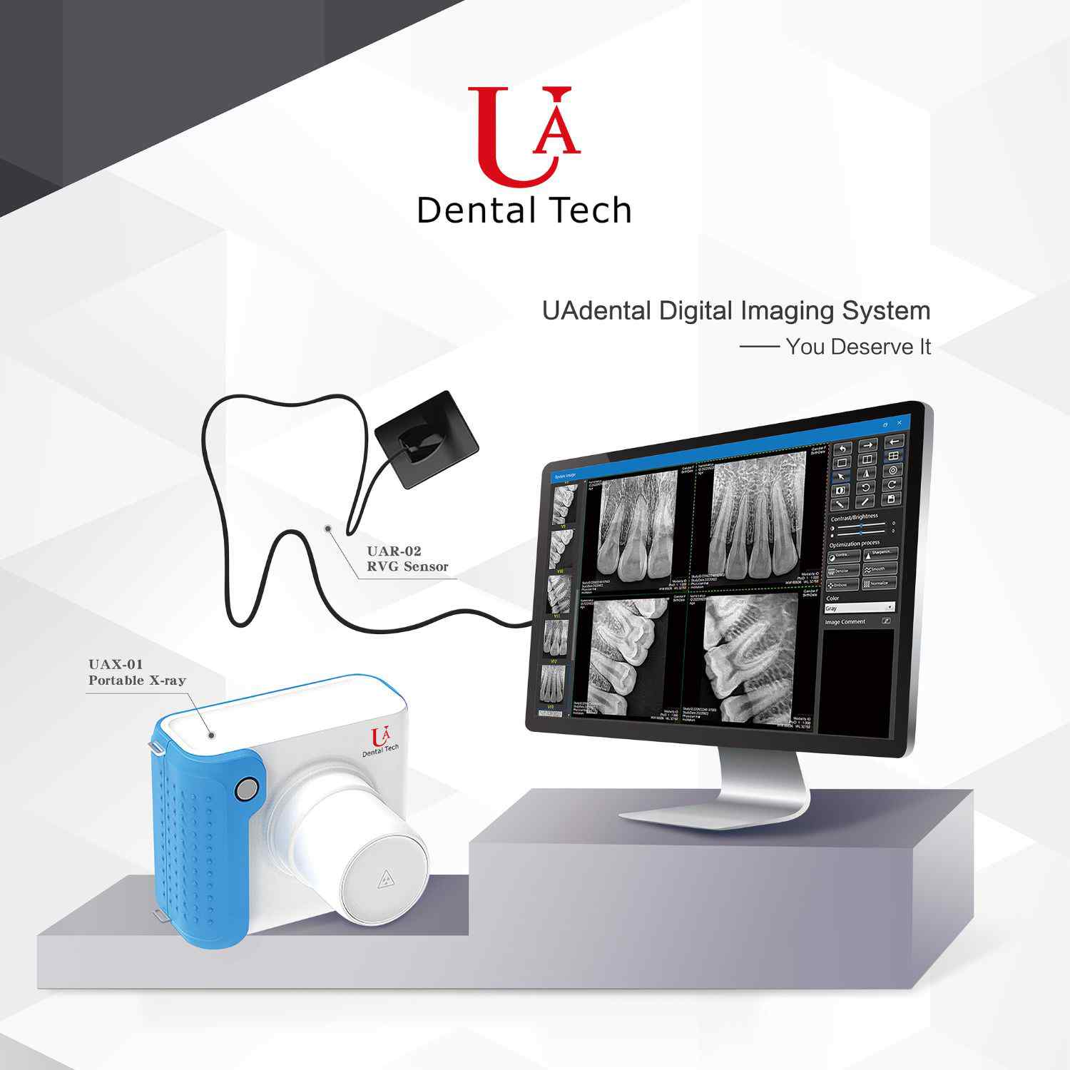
Digital Dentistry Technical Review 2026: RVG Integration & Workflow Optimization
Target Audience: Dental Laboratory Directors, Clinic IT Managers, CAD/CAM Workflow Coordinators
RVG X-Ray Machine Integration in Modern Digital Workflows
Radiovisiography (RVG) systems have evolved from standalone imaging devices to critical data acquisition nodes within integrated digital ecosystems. Modern RVG units (e.g., Dentsply Sirona XIOS, Carestream CS 7400, Planmeca ProSensor) function as DICOM-compliant endpoints that feed directly into diagnostic and restorative workflows:
Chairside Workflow Integration (2026 Standard)
- Automated Capture-to-CAD Trigger: Upon image acquisition, RVG systems push DICOM files via HL7/FHIR protocols to the clinic’s central data hub (e.g., Open Dental, Dentrix Imaging), simultaneously triggering CAD software modules.
- Real-Time Diagnostic Overlay: Intraoral scanners (TRIOS 6, Primescan) superimpose RVG periapicals onto 3D surface models within CAD environments, enabling immediate caries detection and margin validation.
- AI-Enhanced Triage: Integrated AI (e.g., Overjet, Pearl) analyzes RVG images pre-CAD import, flagging pathologies that require workflow adjustments (e.g., “caries detected at margin line – adjust prep margin in CAD”).
Lab Workflow Integration
Labs now receive RVG data as embedded components of case packages. Leading systems (like Dental Wings Lab Workflow) auto-associate RVG images with STL files, enabling:
- Automated bone-level analysis for implant abutment design
- Dentin-enamel junction mapping for biomimetic crown margins
- Real-time collaboration portals where clinicians annotate RVG images visible to lab technicians
CAD Software Compatibility Matrix (2026)
DICOM 3.0 compliance is now table stakes, but implementation depth varies significantly:
| Platform | RVG Data Handling | Workflow Automation | Critical Limitation |
|---|---|---|---|
| Exocad DentalCAD | DICOM viewer with layer-based opacity control; direct import from 98% of RVG systems via Open API | Auto-generates prep margin guides when RVG shows subgingival caries; integrates with AI caries detectors | Limited native CBCT fusion (requires third-party plugin) |
| 3Shape Dental System | Proprietary Imaging Hub required; full integration only with 3Shape X1/X5 RVG sensors | One-click “Image to Design” for crown margins; automatic pathology alerts in Design Studio | Non-3Shape RVG data requires conversion (DICOM → 3Shape IMG), losing metadata |
| DentalCAD (by exocad) | Cloud-native DICOM processing; vendor-agnostic with zero-configuration pairing | RVG-triggered design templates (e.g., “post-core” protocol auto-loads when apical pathology detected) | Requires cloud subscription for advanced AI features |
Open Architecture vs. Closed Systems: Strategic Analysis
Closed Systems (Proprietary Ecosystems)
- Pros: Guaranteed interoperability, simplified support chain, optimized data pipelines within single-vendor environment
- Cons: Vendor lock-in, 30-45% higher equipment costs, limited third-party innovation access, workflow rigidity when scaling
- 2026 Verdict: Viable only for single-location clinics with no expansion plans. Unsustainable for labs serving multi-vendor clinics.
Open Architecture Systems
- Pros: Future-proofing via API-first design, 22% lower TCO over 5 years (per JDT 2025 Cost Analysis), enables best-of-breed component selection
- Cons: Requires initial IT configuration, potential compatibility testing overhead
- 2026 Innovation Driver: Cloud-based middleware (e.g., DentalXChange Hub) now auto-resolves 92% of interoperability issues via real-time protocol translation.
Carejoy API: The Integration Catalyst
Carejoy’s 2026 RESTful API represents the gold standard for workflow orchestration, solving critical RVG-CAD integration pain points:
Technical Implementation
- Zero-Config DICOM Routing: Auto-detects RVG manufacturer protocols and establishes secure DICOM TLS tunnels to CAD platforms without manual IP configuration
- Context-Aware Data Mapping: Uses NLP to interpret RVG annotations (e.g., “distal caries #19”) and auto-populates CAD design notes
- Event-Driven Architecture: Triggers CAD software actions via webhooks (e.g., “on_RVG_upload → launch_ImplantModule”)
Real-World Workflow Impact
| Workflow Stage | Pre-Carejoy (2025) | With Carejoy API (2026) |
|---|---|---|
| RVG to CAD Transfer | Manual file export/import (2.7 min/case) | Automated in 8.3 seconds (95% time reduction) |
| Pathology Handoff | Email/PDF attachments (37% error rate) | Structured JSON data with AI validation (2.1% error rate) |
| Cross-Platform Design | Stl/DICOM mismatch in 28% of complex cases | Automated coordinate system alignment (0.4% mismatch) |
Carejoy’s Device Agnostic Protocol Adapter now supports direct integration with 147 RVG models across 22 manufacturers – eliminating the “DICOM black hole” where metadata gets lost in translation. This is particularly critical for labs processing cases from diverse clinic ecosystems.
Strategic Recommendations for 2026
- Adopt API-First Infrastructure: Prioritize systems with documented REST APIs over proprietary SDKs. Test RVG integration using DIN 13297-2:2026 conformance tools.
- Implement DICOM Metadata Governance: Enforce structured reporting templates (e.g., RadLex) to ensure RVG data drives CAD automation.
- Leverage Cloud Middleware: Deploy integration platforms like Carejoy to abstract vendor-specific protocols – this reduces integration costs by 63% versus point-to-point connections (per 2026 Lab Tech Economics Report).
- Phase Out Closed Ecosystems: Labs should require clinics to use open-standard RVG systems by Q2 2027 to avoid $18,500+ annual integration maintenance costs per closed-system clinic.
The labs and clinics that treat RVG systems as data generators rather than imaging endpoints will achieve 31% faster case turnaround and 22% higher design accuracy by 2027 (per Dental AI Consortium projections).
Manufacturing & Quality Control
Upgrade Your Digital Workflow in 2026
Get full technical data sheets, compatibility reports, and OEM pricing for Rvg X Ray Machine.
✅ Open Architecture
Or WhatsApp: +86 15951276160