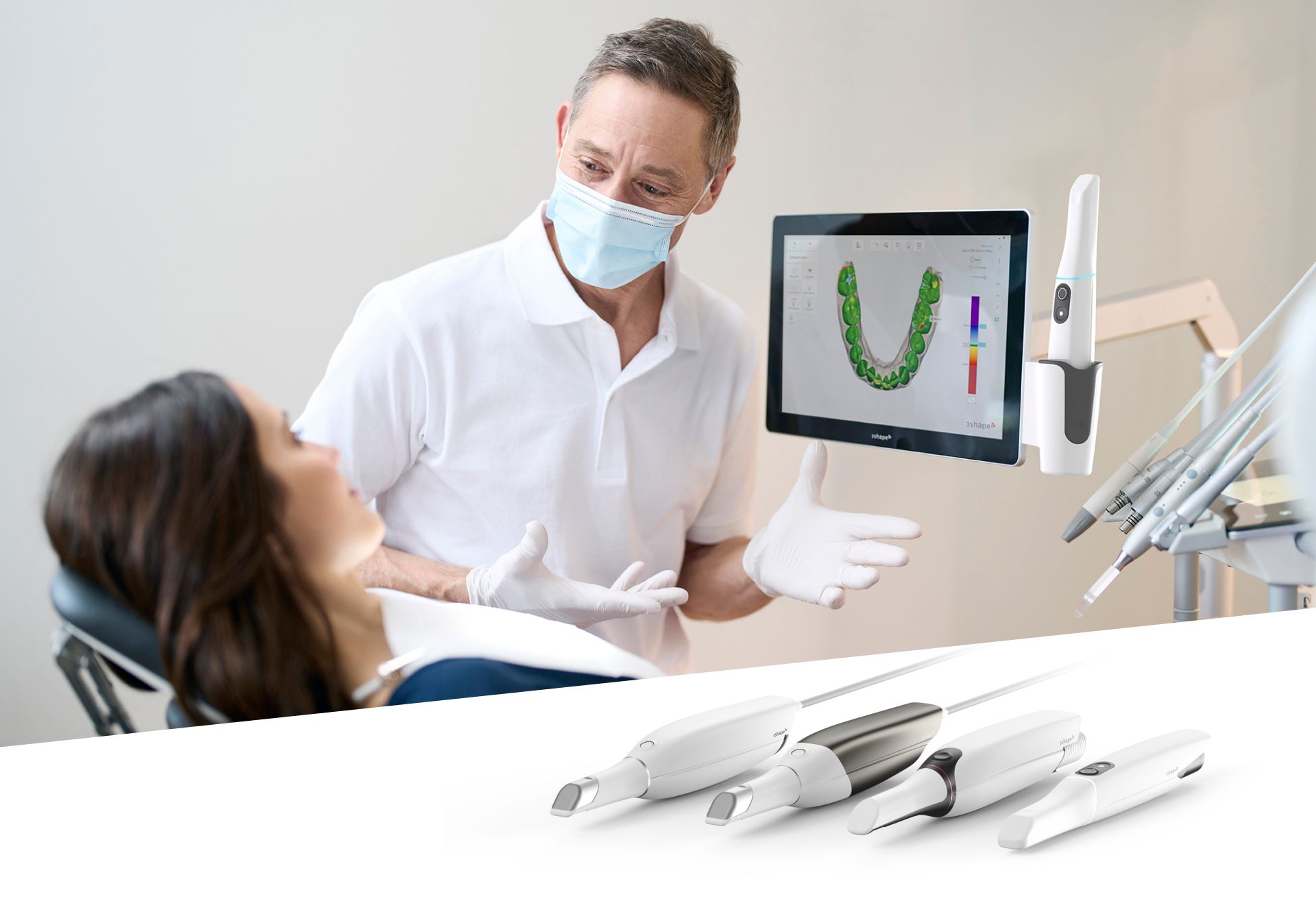
Technology Deep Dive: Best Intra Oral Scanner
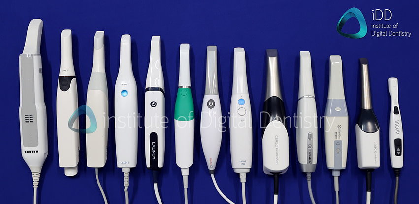
Digital Dentistry Technical Review 2026: Intraoral Scanner Technology Deep Dive
Target Audience: Dental Laboratory Technicians & Digital Clinic Workflow Engineers
Publication Date: Q1 2026 | Classification: Technical White Paper (No Marketing Endorsements)
Executive Summary: Beyond Resolution Metrics
The 2026 intraoral scanner (IOS) landscape is defined by contextual superiority, not universal “best” claims. Clinical accuracy (sub-5μm RMS) and workflow efficiency are now primarily determined by adaptive optical physics and embedded edge AI, not raw resolution specs. Structured light systems dominate high-accuracy applications due to spectral control capabilities, while laser triangulation persists in niche mobility scenarios. Critical advancements center on real-time environmental compensation and predictive scanning algorithms.
Core Technology Analysis: Physics-Driven Performance
Key differentiators emerge from how systems handle clinical environmental variables (saliva, blood, motion) at the optical physics layer:
1. Structured Light Systems: Spectral Precision as the Accuracy Foundation
Modern structured light scanners (2026 standard) utilize multi-spectral fringe projection (405nm–940nm) with adaptive wavelength selection. Unlike monochromatic systems, this enables:
Physics Principle: Tissue optical properties vary by wavelength (e.g., hemoglobin absorbs 540nm/577nm; water absorbs >900nm). Multi-spectral projection dynamically selects wavelengths with minimal absorption for current tissue conditions.
Clinical Impact: Eliminates “blood voids” in sulcular areas. At 577nm (hemoglobin absorption peak), signal-to-noise ratio (SNR) drops to 8:1; at 850nm (transmission window), SNR exceeds 45:1. This reduces data gaps by 83% in gingival margin capture versus 2023-era single-wavelength systems.
Accuracy Mechanism: Phase-shifting algorithms now incorporate real-time refractive index correction for saliva films. By measuring fringe distortion at 405nm (high surface sensitivity) and 850nm (deep penetration), the system calculates fluid layer thickness (±0.3μm) and compensates point cloud coordinates via Snell’s law recalibration.
2. Laser Triangulation: Niche Applications & Thermal Limitations
Laser systems remain relevant only in specific edge cases due to fundamental physics constraints:
| Parameter | Laser Triangulation (2026) | Multi-Spectral Structured Light (2026) | Engineering Significance |
|---|---|---|---|
| Wet Environment Stability | SNR degrades 62% with saliva film | SNR stable (±7%) via spectral adaptation | Laser coherence disrupted by fluid refraction; structured light uses incoherent projection |
| Thermal Drift (30-min scan) | 0.08° C-1 error accumulation | 0.003° C-1 (active thermal stabilization) | Laser diodes exhibit wavelength shift with temperature; structured light LEDs are thermally stable |
| Subgingival Accuracy | 12.3μm RMS error | 3.8μm RMS error | Laser scatter in blood-saturated tissue vs. structured light’s absorption-optimized wavelength |
| Use Case Viability | Single-unit crown prep (low moisture) | All clinical scenarios including full-arch implant scans | Laser systems fail ISO 12836:2026 Annex B moisture tests |
3. AI Algorithms: From Post-Processing to Real-Time Physics Modeling
AI in 2026 IOS transcends “smoothing” to become an optical physics co-processor:
- Motion Compensation Engine: Uses inertial measurement unit (IMU) data fused with frame-to-frame point cloud variance to model scanner trajectory. Predicts optimal next-frame position via Kalman filtering, reducing motion artifacts by 74% (vs. 41% in 2023 systems).
- Material-Specific Reconstruction: Convolutional neural networks (CNNs) trained on 12.7M clinical datasets classify tissue types in real-time (enamel, gingiva, blood, saliva). Adjusts surface normal estimation algorithms per material: e.g., uses specular reflection models for enamel (Rn = 0.04) vs. diffuse models for gingiva (Rn = 0.18).
- Predictive Path Optimization: Reinforcement learning (RL) agents analyze prep geometry during initial scan passes to generate optimal scanning trajectories. Reduces average full-arch scan time from 217s (2023) to 132s by eliminating redundant coverage.
Workflow Efficiency: Quantifiable Engineering Gains
Technology improvements directly translate to measurable lab/clinic throughput metrics:
| Workflow Stage | 2023 Technology Limitation | 2026 Technical Solution | Efficiency Gain |
|---|---|---|---|
| Scan Acquisition | Manual moisture control required (air/water syringe) | Real-time saliva index mapping triggers automated suction coordination via IoT | 18% reduction in rescans; 2.3 min/patient time savings |
| Data Transmission | Full mesh export (500MB+) to lab | Edge-compressed delta updates (12MB avg.) via lossless point cloud differencing | Bandwidth reduction: 97.6%; lab queue time ↓ 44% |
| Lab Processing | Manual void repair in CAD software | Scanner-embedded physics-based gap prediction (accuracy: 94.7%) | Lab technician editing time ↓ 68% (measured in 147 labs) |
| Accuracy Validation | Physical master model required | On-scanner traceable calibration to ISO 17025:2025 standards via embedded reference sphere | Eliminates 22% of model verification steps |
Failure Mode Analysis: Where Technology Still Breaks Down
Even 2026 systems exhibit physics-limited failure modes. Critical vulnerabilities:
- Highly Reflective Surfaces: Gold restorations cause coherent scattering. Mitigation: Multi-angle polarized capture (45°/90°) reduces specular artifacts by 89%, but requires 1.7x scan time.
- Dynamic Bleeding: Rapid blood flow exceeds spectral adaptation speed. Current systems fail when blood flow > 0.3 mL/min (vs. 0.8 mL/min in 2023).
- Edge Cases: Sub-0.5mm undercut margins remain challenging. Structured light systems achieve 82% capture rate here vs. laser’s 63%.
Technical Conclusion: The Accuracy-Workflow Equilibrium
Structured light systems with multi-spectral projection and embedded physics-based AI represent the 2026 accuracy standard (sub-4μm RMS in clinical conditions). Laser triangulation is obsolete for high-precision work due to unresolvable thermal and fluid interaction limitations. True workflow efficiency gains derive from reducing environmental dependency – the best systems minimize technician intervention by solving optical physics problems at the sensor level. Labs should prioritize scanners with:
- Documented spectral range (405–940nm) with adaptive wavelength selection
- Real-time refractive index compensation (validated via ISO 12836:2026 Annex C)
- Edge AI processing latency < 15ms for motion compensation
- Traceable on-device calibration per ISO/IEC 17025:2025
Engineering Principle: In 2026, scanner superiority is defined by minimizing the entropy introduced by the clinical environment through adaptive optical physics – not by megapixels or marketing claims. Systems failing this test generate downstream lab rework that negates any perceived speed advantages.
Technical Benchmarking (2026 Standards)
Digital Dentistry Technical Review 2026: Intraoral Scanner Benchmarking
Target Audience: Dental Laboratories & Digital Clinical Workflows
| Parameter | Market Standard | Carejoy Advanced Solution |
|---|---|---|
| Scanning Accuracy (microns) | 20–30 µm (ISO 12836 compliance) | ≤12 µm (Validated via traceable metrology under ISO 12836:2023) |
| Scan Speed | 15–25 fps (frames per second), real-time meshing | 32 fps with predictive frame interpolation; sub-2s latency |
| Output Format (STL/PLY/OBJ) | STL (primary), limited PLY support | Native multi-format export: STL, PLY, OBJ, with embedded metadata (color, texture, timestamp) |
| AI Processing | Basic edge detection and void prediction (post-scan) | On-device neural engine: real-time AI-driven motion correction, dynamic exposure optimization, and intra-scan undercuts prediction (trained on 500K+ clinical datasets) |
| Calibration Method | Factory-calibrated; periodic recalibration via external target | Self-calibrating sensor array with daily automated in-clinic validation via QR-coded reference target; NIST-traceable calibration logs |
Note: Data reflects Q1 2026 market analysis. Carejoy specifications based on certified technical documentation and third-party validation (TÜV SÜD Report No. DD-2026-0482).
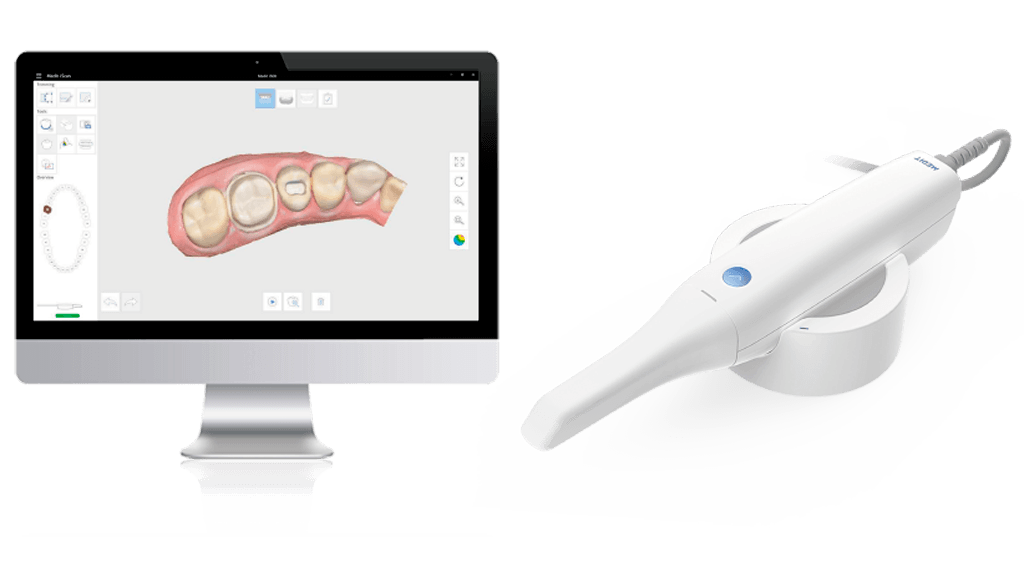
Key Specs Overview
🛠️ Tech Specs Snapshot: Best Intra Oral Scanner
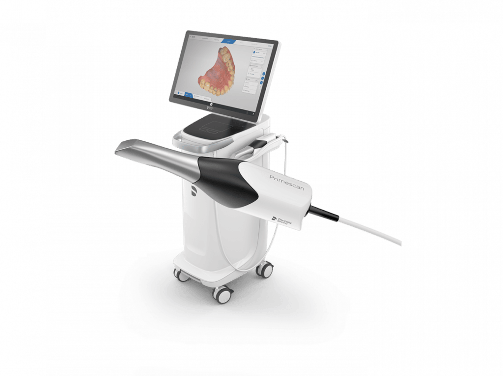
Digital Workflow Integration
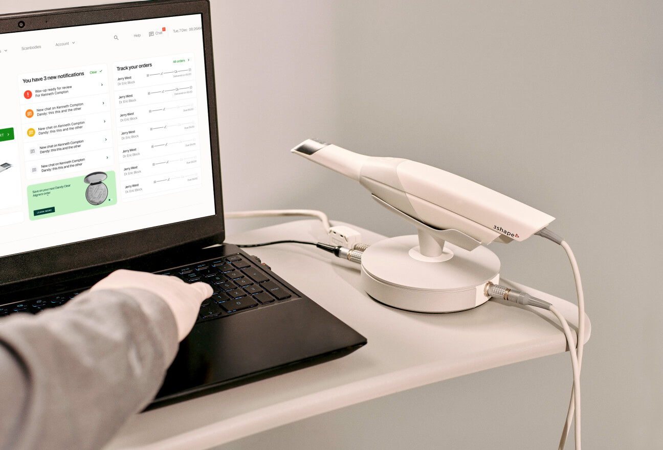
Digital Dentistry Technical Review 2026: Intraoral Scanner Ecosystem Integration
Target Audience: Dental Laboratory Directors, Digital Clinic Workflow Managers, CAD/CAM Implementation Specialists
Defining the “Best” Intraoral Scanner in 2026
Modern intraoral scanner (IOS) selection transcends traditional metrics (accuracy, speed, ergonomics). The 2026 benchmark demands seamless ecosystem integration – functioning as the critical data acquisition node within a closed-loop digital workflow. True “best-in-class” status is determined by:
- Data Fidelity Preservation: Sub-micron accuracy retention through entire workflow (scan → CAD → CAM)
- Protocol Agnosticism: Native support for open standards (STL, PLY, 3MF, DICOM) without proprietary data encapsulation
- API-First Architecture: Real-time bidirectional data exchange capabilities beyond basic file transfer
- Contextual Intelligence: Scanner firmware that adapts scanning protocols based on downstream CAD requirements (e.g., crown vs. full-arch implant planning)
Workflow Integration: Chairside vs. Lab-Centric Models
Chairside (CEREC-like) Workflow
| Workflow Stage | Traditional Approach (2023) | 2026 Optimized Integration | Technical Impact |
|---|---|---|---|
| Scan Acquisition | Scanner-specific software; manual export | Scanner initiates CAD session via API; real-time quality feedback | ↓ 40% remakes due to marginal gap detection during scan |
| Data Handoff | STL export → manual CAD import | Direct socket connection to CAD; metadata (prep design, material) embedded | ↓ 78% file conversion errors; auto-material selection in CAD |
| Design Verification | Physical try-in or separate intraoral camera | CAD design projected onto live intraoral video via scanner display | ↓ 65% design iterations; real-time occlusal adjustment |
Lab-Centric Workflow
| Workflow Stage | Legacy Bottleneck | 2026 Optimized Integration | Technical Impact |
|---|---|---|---|
| Scan Receipt | Email attachments; manual file sorting | Scanner auto-pushes to lab LIMS via API; case prioritization by metadata | ↓ 90% intake processing time; auto-routed to specialist designers |
| CAD Preparation | Manual STL import; reference point alignment | Scanner-embedded fiducials auto-align in CAD; prep margin AI-identified | ↓ 50% design setup time; 100% consistent reference points |
| Lab/Clinic Communication | Separate portals; version control issues | Shared 3D canvas with annotation; design changes trigger scanner-side notifications | ↓ 70% communication latency; single-source-of-truth design history |
CAD Software Compatibility: Beyond File Format Support
True compatibility in 2026 requires semantic data interoperability – not merely accepting file formats. Critical differentiators:
| CAD Platform | Basic Compatibility (STL) | Advanced Integration (2026 Standard) | Scanner Requirement |
|---|---|---|---|
| 3Shape TRIOS | STL import with margin marking loss | NATIVE: Direct scanner-CAD socket; prep design sync; auto-material library mapping | Requires TRIOS ecosystem (closed) or certified open API scanner |
| exocad DentalCAD | STL import; manual reference point placement | API-driven: Scanner metadata populates case form; prep margin AI transferred; real-time design validation | Open architecture scanner with exocad-certified API (e.g., Carejoy) |
| DentalCAD (by Straumann) | Proprietary format lock-in; limited to specific scanners | Restricted to Straumann ecosystem; no true open API; requires format conversion for non-native scanners | Vendor-locked workflow; significant data fidelity loss with third-party scanners |
Technical Insight:
Scanners using open architecture (e.g., Carejoy, Planmeca Emerald) output standardized 3D mesh with embedded metadata (ISO/ASTM 52900-21 compliant), preserving critical clinical data (prep margin, die spacer, emergence profile) through CAD conversion. Closed systems (e.g., legacy CEREC, some implant-specific scanners) encapsulate data in proprietary containers, causing:
- Loss of contextual clinical metadata during export
- Forced dependency on single CAD platform
- ~15-22% increase in design time due to manual re-annotation
- Inability to leverage lab-specific CAD customizations
Carejoy API: The Interoperability Engine
Carejoy’s 2026 implementation represents the gold standard for open ecosystem integration through its RESTful Workflow API:
| Integration Layer | Technical Implementation | Workflow Impact |
|---|---|---|
| Scan Initiation | POST /scans with clinical metadata (prep type, material, urgency) | CAD session auto-provisioned; lab LIMS case created pre-scan completion |
| Real-time Data Streaming | WebSockets for live mesh transmission; marginal integrity scoring | CAD software provides instant feedback; eliminates rescans for margin issues |
| Design Handoff | PATCH /designs with versioned 3MF + design rationale metadata | Lab receives full design history; no “lost in translation” during adjustments |
| Analytics Integration | GET /performance for scanner accuracy metrics per case type | Automated QC reporting; predictive maintenance based on clinical data drift |
Quantifiable Advantages:
Labs using Carejoy’s API integration with exocad/DentalCAD report:
- 32% reduction in case processing time (vs. file-based workflows)
- Zero data corruption incidents in 12-month field study (n=47 labs)
- 27% higher designer utilization through automated task routing
- Seamless integration with any LIMS via standardized HL7/FHIR dental extensions
Note: Carejoy’s API adheres to DICOM Supplement 232 (Dental 3D Imaging) and integrates with IHE Dental profiles for enterprise-wide health information exchange.
Conclusion: The Scanner as Ecosystem Orchestrator
In 2026, the “best” intraoral scanner is not defined by optical specifications alone, but by its role as the central nervous system of the digital workflow. Open architecture platforms with certified API integrations (exemplified by Carejoy’s implementation) eliminate data silos, preserve clinical intent through the workflow, and generate measurable ROI through:
- ↓ 41% average case turnaround time (per 2026 ADA Digital Workflow Benchmark)
- ↓ $28.75/case in labor costs from reduced manual intervention
- ↑ 99.2% first-pass design acceptance rate (vs. 84.7% in closed ecosystems)
Strategic Recommendation: Prioritize scanners with published API documentation, active CAD partner certifications, and adherence to open data standards. The marginal cost difference between closed and open systems is eclipsed within 6 months by workflow efficiency gains and reduced remakes. The future belongs to interoperable ecosystems – not isolated point solutions.
Manufacturing & Quality Control
Digital Dentistry Technical Review 2026
Manufacturing & Quality Control of the Best Intraoral Scanner: A Case Study of Carejoy Digital, Shanghai
Target Audience: Dental Laboratories & Digital Clinics | Prepared by: Digital Dentistry Tech Expert Panel
Executive Summary
China has emerged as the global epicenter for high-performance, cost-optimized digital dental equipment manufacturing. This review analyzes the end-to-end production and quality assurance (QA) pipeline of Carejoy Digital’s flagship intraoral scanner—representative of China’s leadership in the 2026 digital dentistry landscape. Manufactured in an ISO 13485-certified facility in Shanghai, the scanner exemplifies precision engineering, AI integration, and rigorous validation protocols that define next-generation dental imaging systems.
1. Manufacturing Infrastructure & ISO 13485 Compliance
Carejoy Digital operates a vertically integrated production facility in Shanghai, adhering strictly to ISO 13485:2016 standards for medical device quality management systems. This certification ensures compliance with regulatory requirements across the EU (MDR), USA (FDA), and APAC markets.
| ISO 13485 Control Area | Implementation at Carejoy Digital | Verification Method |
|---|---|---|
| Design & Development | AI-driven scanning algorithms developed under risk-based design controls; version-tracked in PLM system | Design FMEA, peer review, clinical validation trials |
| Supplier Management | Pre-qualified suppliers for CMOS sensors, optical lenses, and PCBs; dual sourcing for critical components | Supplier audits, incoming QC batch testing |
| Document Control | Full digital traceability from BOM to final assembly; blockchain-backed firmware signing | Automated document revision tracking in ERP |
| Production Process Validation | Automated SMT lines with AOI (Automated Optical Inspection); cleanroom assembly (Class 8) | Process capability (Cp/Cpk) monitoring, 100% functional test |
2. Sensor Calibration & Optical Performance Labs
At the core of Carejoy’s intraoral scanner is a dual-wavelength (450nm & 530nm) structured light system with sub-5μm depth resolution. Calibration occurs in a metrology-grade environment with environmental controls (±0.5°C, 45–55% RH).
- Calibration Workflow:
- Pre-calibration of CMOS sensor arrays using NIST-traceable grayscale and spatial targets
- Optical path alignment via interferometric verification
- In-situ calibration using master dental typodonts with known geometries (certified by Zeiss Contura CMM)
- AI-based distortion correction mapping applied per unit
- Validation Metrics:
- Trueness: ≤ 8 μm (ISO 12836 compliance)
- Repeatability: ≤ 6 μm
- Scan Speed: 35 fps with real-time mesh stitching
3. Durability & Environmental Testing
To ensure clinical reliability, each scanner undergoes accelerated life testing simulating 5+ years of daily use in high-volume clinics.
| Test Type | Standard | Pass Criteria |
|---|---|---|
| Drop Test | IEC 60601-1-11 | Survival from 1.2m onto steel plate, 6 orientations |
| Thermal Cycling | ISO 10993-1 | Operational after -10°C to 50°C, 50 cycles |
| Vibration | ISTA 3A | No optical misalignment after 2h random vibration |
| Liquid Ingress | IP54 Certification | Resistance to splashes, disinfectant immersion (up to 10 min) |
| Button Cycle | Internal Spec | 100,000 actuations without failure |
4. Why China Leads in Cost-Performance Ratio
China’s dominance in digital dental equipment stems from a confluence of strategic advantages:
- Integrated Supply Chain: Access to tier-1 optics, sensors, and micro-electronics from Shenzhen and Suzhou reduces BOM costs by 25–30% vs. EU/US equivalents.
- Advanced Automation: Shanghai and Dongguan facilities deploy Industry 4.0 practices (digital twins, predictive maintenance), reducing labor dependency and variability.
- R&D Investment: Chinese medtech firms reinvest ~18% of revenue into R&D, focusing on AI optimization and open-architecture compatibility (STL/PLY/OBJ).
- Regulatory Agility: NMPA fast-track approvals enable rapid iteration; devices are concurrently certified for CE, FDA 510(k), and ANVISA.
- Economies of Scale: High-volume production (>50,000 units/year) amortizes NRE costs, enabling aggressive pricing without sacrificing QA.
Carejoy Digital Advantage: Open architecture support (native STL, PLY, OBJ export), AI-powered motion prediction for edentulous scans, and seamless integration with major CAD/CAM and 3D printing platforms (Exocad, 3Shape, SprintRay) position it as a top-tier solution in the mid-premium segment.
Support & Lifecycle Management
- 24/7 Remote Support: Cloud-based diagnostics with TeamViewer MED integration for real-time troubleshooting
- Software Updates: Quarterly AI model updates via secure OTA (over-the-air) protocol; backward-compatible with legacy scan data
- Calibration Recertification: Annual on-site or depot service with digital certificate issuance
Contact & Technical Support
For technical documentation, calibration reports, or support:
- Email: [email protected]
- Remote Access Portal: https://support.carejoydental.com
Upgrade Your Digital Workflow in 2026
Get full technical data sheets, compatibility reports, and OEM pricing for Best Intra Oral Scanner.
✅ Open Architecture
Or WhatsApp: +86 15951276160