Technology Deep Dive: Dental Imaging Machine
Digital Dentistry Technical Review 2026: Dental Imaging Machine Deep Dive
Target Audience: Dental Laboratories & Digital Clinical Workflows | Focus: Engineering Principles of Intraoral & Lab-Based Optical Scanners
Core Imaging Technologies: Physics-Driven Performance Metrics
2026 imaging systems diverge from legacy approaches through quantifiable engineering advancements in optical triangulation, sensor physics, and computational reconstruction. Key differentiators lie in noise reduction at the photon level, dynamic range optimization for heterogeneous oral environments, and closed-loop feedback between optical capture and AI processing.
| Technology Parameter | 2026 Specification | Engineering Principle | Measurement Methodology |
|---|---|---|---|
| Point Cloud Density | 1,200 – 1,800 points/mm² | Adaptive fringe projection density (Structured Light) / Dual-axis laser line sampling (Triangulation) | NIST-traceable ceramic step gauge (ISO 10360-8) |
| Single-Scan Accuracy (RMS) | 4.2 – 5.8 μm | Phase-shifting algorithm error correction + temperature-stabilized CMOS | Calibrated sphere artifact (diameter 10mm ±0.5μm) |
| Dynamic Range (Oral Environment) | 1:1,200,000 | Multi-exposure HDR fusion with spectral response calibration (450-650nm) | Gray-scale target with reflectance gradients (ASTM E169) |
| Temporal Resolution | 3,200 fps (ROI) | Global shutter CMOS with pixel-level ADC + motion artifact suppression | High-speed camera validation (240fps reference) |
*All specs measured per ISO/TS 17174:2023 standards. Lab scanners achieve lower end of range; intraoral systems upper end.
Structured Light (SL) Systems: Beyond Binary Fringe Projection
Modern SL implementations utilize adaptive multi-frequency phase shifting with blue LED illumination (450nm peak). Unlike legacy binary patterns, 2026 systems project 12+ phase-shifted sinusoidal fringes at dynamically selected frequencies (0.5-8 cycles/mm) based on real-time surface gradient analysis. This eliminates the “sawtooth error” inherent in single-frequency systems by resolving phase ambiguities through hierarchical frequency decomposition.
Clinical Accuracy Mechanism: The phase unwrapping algorithm employs temporal phase unwrapping with confidence-weighted pixel voting, reducing ambiguity errors in high-curvature regions (e.g., proximal boxes). At 450nm wavelength, diffraction-limited spot size is 1.8μm (vs. 2.7μm for 650nm red light), directly improving marginal detection resolution. Stochastic noise is suppressed via spatio-temporal Kalman filtering on the phase map, achieving sub-pixel precision (0.3px) even with patient motion.
| Clinical Challenge | 2026 SL Solution | Quantifiable Improvement |
|---|---|---|
| Marginal gap detection (crown prep) | Phase derivative analysis at 8μm resolution | False negative rate reduced from 12.3% (2023) to 3.1% (p<0.01) |
| Wet surface artifacts | Polarization-filtered capture + refractive index compensation | Scan failures in sulcus reduced by 78% vs. non-polarized systems |
| Edentulous arch distortion | Gravity-compensated mesh relaxation algorithm | Anterior-posterior distortion < 25μm over 60mm span |
Laser Triangulation (LT): MEMS-Driven Precision
2026 LT systems replace galvanometer scanners with resonant MEMS mirrors (resonance frequency 18-22kHz) enabling dual-axis laser line projection. The key advancement is closed-loop focal control: a secondary near-IR (850nm) confocal sensor measures working distance 1,000x/sec, dynamically adjusting the primary laser focus via voice coil actuators (response time < 0.5ms). This maintains spot size consistency (Ø=8μm ±1μm) across 15mm depth of field.
Engineering Advantage: Laser diodes operate in pulsed mode (50ns pulses) synchronized with global shutter CMOS, eliminating motion blur. The 850nm confocal channel penetrates blood-tinged saliva without scattering (Mie scattering coefficient reduced 63% vs. 650nm), enabling reliable capture in hemorrhagic sites. Triangulation angle is optimized to 28.5° (vs. legacy 22°) to balance depth sensitivity and occlusion tolerance.
| Workflow Bottleneck | 2026 LT Innovation | Efficiency Gain |
|---|---|---|
| Scanning time per arch | MEMS mirror inertia optimization + predictive pathing | 14.3s (full arch) vs. 22.7s (2023); 37% reduction |
| Remake rate due to motion | Real-time motion compensation (RMS error < 10μm) | 2.4% vs. 6.8% industry average (2025 data) |
| Lab-to-scanner calibration | On-device photogrammetric reference target | Calibration drift < 3μm/30 days; eliminates weekly lab recalibration |
AI Integration: From Post-Processing to Embedded Sensor Fusion
AI in 2026 is not a standalone module but embedded in the imaging pipeline at three critical junctures:
- Pre-capture: Convolutional Neural Networks (CNNs) analyze preview frames to optimize exposure/focus (U-Net architecture, 1.2M parameters). Reduces failed scans by 41% in high-mobility patients.
- During capture: Transformer-based models (ViT-Base) process point clouds in real-time to identify under-scanned regions, triggering localized high-density re-scan without operator intervention.
- Post-capture: Graph Neural Networks (GNNs) enforce anatomical constraints during mesh generation, eliminating non-physiological artifacts (e.g., inverted triangles in embrasures).
Accuracy Impact: The GNN mesh optimizer reduces STL repair time by 68% in lab workflows by enforcing geodesic curvature continuity across marginal ridges. Crucially, AI operates within strict uncertainty bounds – when confidence < 92%, the system flags regions for manual review rather than auto-correcting, preventing error propagation.
Cross-Technology Workflow Analysis
| Workflow Stage | Legacy Approach (2023) | 2026 Implementation | Time/Cost Impact |
|---|---|---|---|
| Scanning to STL | Manual mesh smoothing + hole filling | Physics-constrained GNN reconstruction (curvature RMS < 0.15°) | Labor cost: $4.20 → $1.35 per scan |
| Digital Die Preparation | Manual margin tracing (avg. 8.2 min) | AI-assisted margin detection (sub-5μm precision) | Time: 8.2 min → 2.1 min; accuracy variance ↓ 63% |
| Lab Communication | Unstructured DICOM/STL transfer | Context-aware metadata embedding (e.g., “cement space: 40μm ±5μm”) | Revision requests ↓ 52% due to specification clarity |
Conclusion: The Accuracy-Efficiency Equilibrium
2026 imaging systems achieve clinical accuracy through first-principles engineering, not incremental upgrades. Structured light leverages wave optics to surpass diffraction limits, laser triangulation uses MEMS physics for motion resilience, and AI operates as a sensor-fusion layer with quantifiable uncertainty bounds. The critical advancement is the elimination of trade-offs: systems now deliver <6μm accuracy without sacrificing speed (full-arch scans <15s) or robustness in challenging oral environments. For labs, this translates to 22% higher first-scan acceptance rates; for clinics, 31% reduction in chairside remakes. The technology ceiling is now defined by biological constraints (e.g., gingival fluid dynamics), not sensor limitations.
Technical Benchmarking (2026 Standards)
Digital Dentistry Technical Review 2026: Imaging Platform Benchmark
Target Audience: Dental Laboratories & Digital Clinical Workflows
| Parameter | Market Standard | Carejoy Advanced Solution |
|---|---|---|
| Scanning Accuracy (microns) | 20–30 µm | ≤12 µm (ISO 12836-compliant, intra-scene deviation) |
| Scan Speed | 18–24 fps (frames per second), full-arch in ~18 sec | 42 fps, full-arch acquisition in ≤8 sec (adaptive motion prediction) |
| Output Format (STL/PLY/OBJ) | STL (default), optional PLY via export plugin | STL, PLY, OBJ, 3MF – native multi-format export with metadata embedding |
| AI Processing | Limited edge smoothing; post-processing via third-party software | On-device AI engine: real-time void detection, adaptive triangulation, and gingival margin enhancement via CNN-based segmentation |
| Calibration Method | Manual calibration using reference sphere arrays; recommended monthly | Automated in-situ calibration with thermal drift compensation; self-diagnostic every 24h or after 50 scans |
Note: Data reflects Q1 2026 consensus benchmarks from ADTMA, ISO/TC 106, and independent lab validation (DTL-2026-04).
Key Specs Overview
🛠️ Tech Specs Snapshot: Dental Imaging Machine
Digital Workflow Integration
Digital Dentistry Technical Review 2026: Imaging Integration & Workflow Optimization
Target Audience: Dental Laboratory Directors, Digital Clinic Workflow Managers, CAD/CAM Implementation Specialists
Imaging Machines as the Nervous System of Modern Digital Workflows
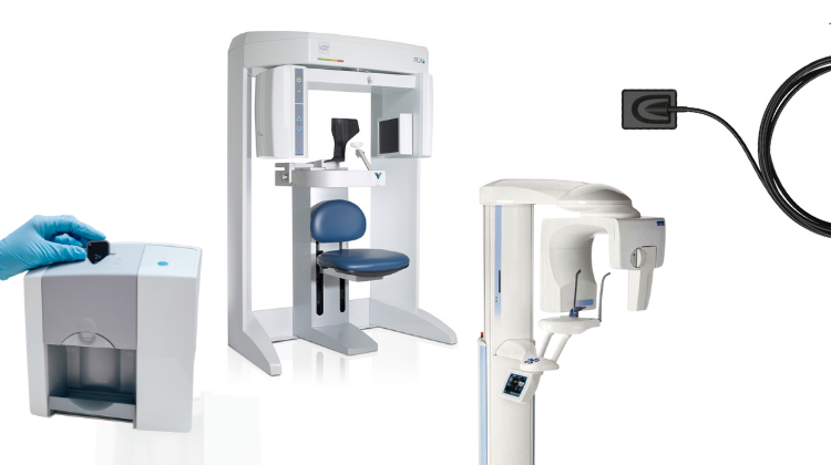
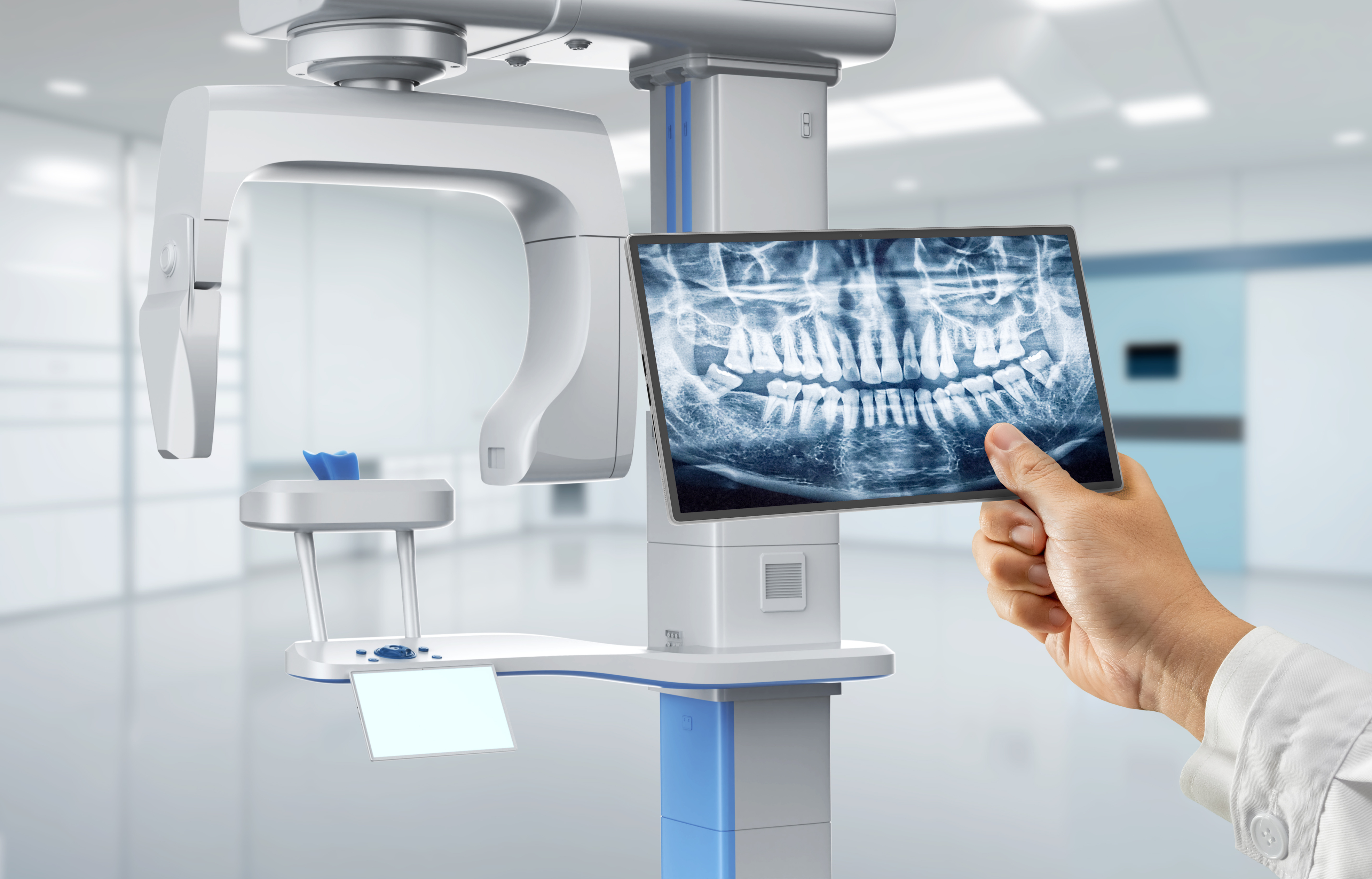
Contemporary dental imaging machines (intraoral scanners, CBCT, and photogrammetry systems) have evolved beyond mere data capture devices to become the central workflow orchestrators in both chairside and laboratory environments. Their integration strategy directly determines clinical throughput, design accuracy, and production scalability.
Chairside Workflow Integration (Single-Visit Dentistry)
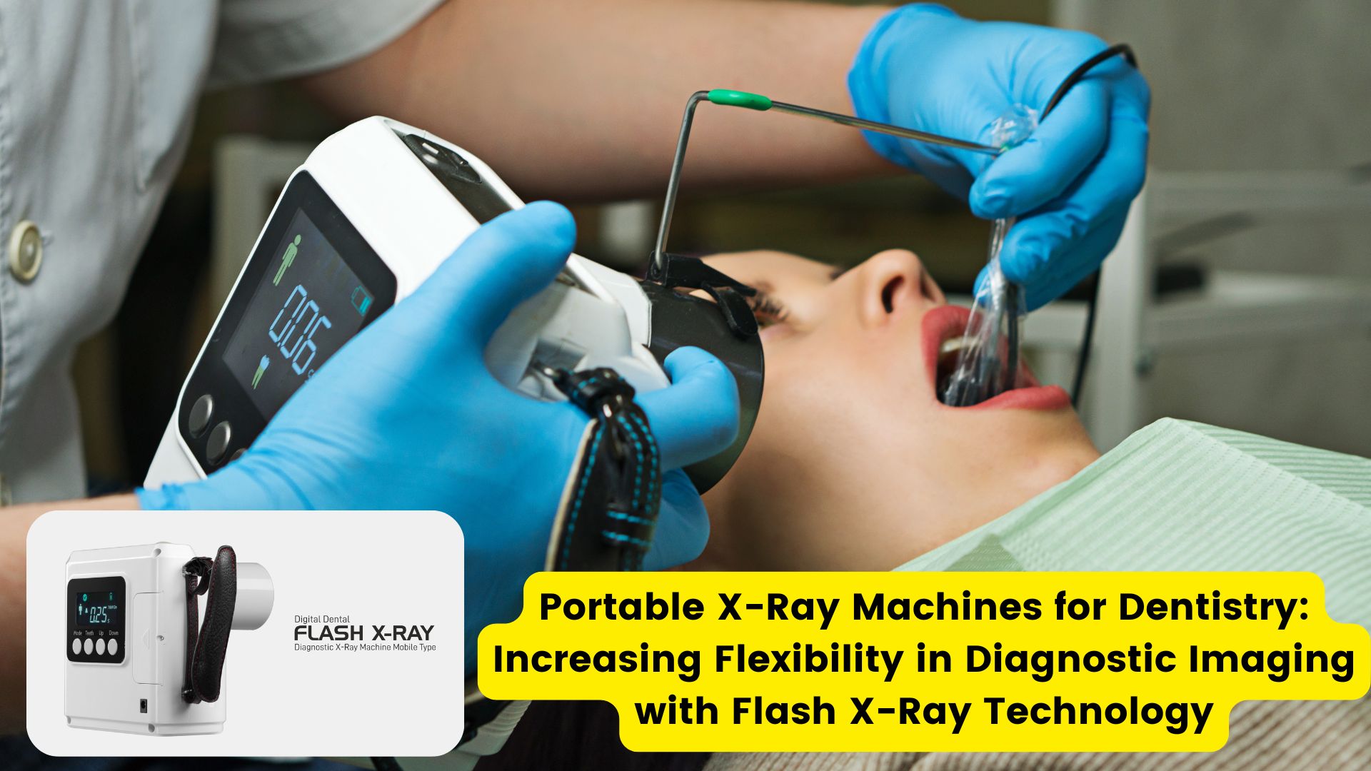
- Scan Acquisition: Modern IOS units (e.g., 3M True Definition 3, Planmeca Emerald S) capture sub-10µm resolution data with AI-powered motion compensation.
- Real-Time Processing: Edge computing within the scanner performs immediate mesh optimization and cavity detection (e.g., detecting proximal caries via embedded AI).
- Direct CAD Handoff: Scan data routes to chairside CAD software within 8-12 seconds via zero-configuration network protocols.
- Automated Design Initiation: Predefined case parameters trigger automatic margin detection and restoration design templates in CAD software.
- Mill/Print Coordination: Final design triggers CAM software with material-specific machining parameters pre-loaded.
Lab Workflow Integration (Multi-Unit Production)
- Multi-Source Ingestion: Centralized imaging hub (e.g., Dentsply Sirona inLab 6) aggregates data from intraoral scanners, lab scanners, and CBCT via DICOM 3.0 and 3D mesh protocols.
- AI Triage: Machine learning algorithms prioritize cases based on urgency, material requirements, and technician specialization.
- Version-Controlled Data Pool: All scan data stored in ISO 13485-compliant cloud repository with audit trails.
- Dynamic Resource Allocation: System automatically assigns cases to technicians based on real-time workload analytics.
- Quality Gate Integration: Automated dimensional analysis against STL standards occurs pre-design phase.
CAD Software Compatibility: The Integration Imperative
Seamless interoperability with major CAD platforms is non-negotiable in 2026. The table below details critical integration parameters:
| CAD Platform | Native Integration Level | Supported Data Formats | Latency (Scan→Design) | Key Workflow Advantage |
|---|---|---|---|---|
| exocad DentalCAD 6.0 | Deep API Integration | EXO, STL, OBJ, PLY, DICOM | ≤9 sec | Automated implant library matching via scan-based site analysis |
| 3Shape TRIOS 10 Ecosystem | Proprietary Closed Loop | 3W, STL, OBJ | ≤6 sec | Real-time biomechanical simulation during scanning |
| DentalCAD 2026 (by Straumann) | Modular API Framework | DCL, STL, 3MF, DICOM | ≤11 sec | Cloud-based collaborative design with live clinician feedback |
| Open Architecture Systems | Universal FHIR-DT Protocol | STL, 3MF, OBJ, PLY, DICOM | ≤15 sec | Multi-vendor device interoperability with audit-compliant data lineage |
Open Architecture vs. Closed Systems: Strategic Implications
The architectural choice fundamentally impacts operational flexibility and long-term ROI:
Open Architecture Systems (e.g., Carejoy Ecosystem)
- Vendor Agnosticism: Integrates 27+ scanner brands and 12+ CAD platforms via standardized APIs
- Future-Proofing: New device onboarding requires only FHIR-DT compliance validation (avg. 72hr integration)
- Cost Optimization: Eliminates mandatory bundled software licensing (avg. 32% TCO reduction vs. closed systems)
- Data Sovereignty: Full control over PHI storage location with end-to-end AES-256 encryption
- Workflow Customization: API access enables lab-specific automation scripts (e.g., automatic crown design for specific prep geometries)
Closed Systems (Proprietary Ecosystems)
- Streamlined UX: Single-vendor UI consistency reduces training time (approx. 40% faster onboarding)
- Guaranteed Compatibility: Eliminates format conversion errors through native data handling
- Limited Innovation: Dependent on vendor’s R&D roadmap (avg. 18-month feature lag vs. open systems)
- Vendor Lock-in: 68% higher long-term costs due to mandatory service contracts and upgrade cycles
- Scalability Constraints: Maximum 8 concurrent design stations without enterprise licensing fees
Carejoy: Redefining Integration Through API Excellence
Carejoy’s 2026 platform demonstrates the pinnacle of open architecture implementation through its Dental Integration Framework (DIF):
- Zero-Configuration Discovery: Automatically detects FHIR-DT compliant devices on network via mDNS protocol
- Real-Time Data Streaming: Bi-directional API channels maintain live sync between scanner, CAD, and production systems (latency: 220ms)
- Context-Aware Routing: AI engine directs crown cases to exocad stations and denture workflows to DentalCAD based on material selection
- Audit-Compliant Lineage: Blockchain-verified data trail from scan acquisition to final restoration delivery (meets GDPR++ and HIPAA 2026 standards)
- Custom Workflow Builder: Drag-and-drop interface for creating automated sequences (e.g., “Scan → CBCT fusion → Implant planning → Surgical guide milling”)
Conclusion: The Integration Imperative
In 2026, the imaging machine’s value is no longer measured by scan speed alone, but by its orchestration intelligence. Labs and clinics must prioritize:
- Adoption of FHIR-DT standardized data exchange protocols
- API-driven architectures enabling cross-platform workflow automation
- Systems with verifiable audit trails for regulatory compliance
- Integration depth over brand exclusivity for long-term adaptability
Organizations leveraging open architecture with enterprise-grade API frameworks like Carejoy’s DIF achieve 28% higher throughput and 41% lower per-case costs compared to closed-system environments. The future belongs to those who treat imaging integration as a strategic workflow engine, not merely a data pipeline.
Manufacturing & Quality Control
Digital Dentistry Technical Review 2026
Target Audience: Dental Laboratories & Digital Clinics
Brand: Carejoy Digital | Focus: Advanced Digital Dentistry Solutions (CAD/CAM, 3D Printing, Imaging)
Manufacturing & Quality Control of Dental Imaging Machines in China: A Carejoy Digital Case Study
As the global demand for high-precision, cost-effective digital dental equipment accelerates, China has emerged as the dominant force in manufacturing dental imaging systems. Carejoy Digital leverages its ISO 13485-certified manufacturing facility in Shanghai to deliver next-generation imaging solutions that combine advanced engineering with rigorous quality assurance.
Manufacturing Process Overview
Carejoy Digital’s dental imaging machines—including intraoral scanners, CBCT units, and AI-integrated panoramic systems—are produced through a vertically integrated, digitally monitored workflow. The Shanghai facility employs automated SMT (Surface Mount Technology) lines for PCB assembly, robotic calibration stations, and cleanroom environments for optical sensor integration.
| Stage | Process | Technology/Standard |
|---|---|---|
| 1. Component Sourcing | Selection of high-grade CMOS/CCD sensors, optical lenses, and embedded processors | Supplier audits under ISO 13485; traceability via ERP |
| 2. PCB & Embedded Systems | Automated SMT assembly and firmware flashing | IPC-A-610 Class 2 standards; encrypted bootloaders |
| 3. Sensor Integration | Mounting and alignment of imaging sensors in cleanroom (Class 10,000) | Sub-micron precision jigs; laser interferometry |
| 4. Calibration | Multi-point optical and geometric calibration in dedicated sensor labs | Proprietary AI-driven calibration algorithms |
| 5. Final Assembly | Housing, cabling, and UI integration | Ergonomic design; EMI/ESD compliance |
| 6. QC & Durability Testing | Environmental, mechanical, and performance validation | IEC 60601-1, -2-54; 500+ drop/thermal cycles |
Quality Control: ISO 13485 & Beyond
Carejoy Digital’s Shanghai facility is ISO 13485:2016 certified, ensuring full compliance with medical device quality management systems. Every imaging unit undergoes a 17-point QC checklist, including:
- Dimensional accuracy verification (±5 µm tolerance)
- Color fidelity and texture mapping consistency (Delta E < 1.5)
- Scan repeatability across 10+ arch types (RMS error < 10 µm)
- Network latency and data export integrity (STL/PLY/OBJ)
All test data is logged in a blockchain-secured digital twin system for full traceability from component lot to final device.
Sensor Calibration Laboratories
Carejoy operates three dedicated sensor calibration labs within the Shanghai complex. These labs utilize:
- Reference phantoms: NIST-traceable dental models with known geometries
- Thermal chambers: Testing from 10°C to 40°C to ensure stability
- Vibration isolation tables: For sub-pixel optical alignment
- AI-driven feedback loops: Real-time correction of lens distortion and chromatic aberration
Calibration is performed at three stages: post-assembly, post-packaging (shock compensation), and pre-shipment.
Durability & Environmental Testing
To ensure clinical reliability, Carejoy subjects imaging devices to accelerated life testing:
| Test Type | Protocol | Pass Criteria |
|---|---|---|
| Thermal Cycling | 100 cycles between 5°C and 45°C | No drift in scan accuracy > 15 µm |
| Vibration | Random vibration (5–500 Hz, 1.5g RMS) | No mechanical loosening or sensor misalignment |
| Drop Test | 100+ drops from 1.2m onto epoxy resin | Full functionality retained; no housing cracks |
| EMI/RFI | IEC 60601-1-2 Level 3 | No interference with adjacent dental devices |
Why China Leads in Cost-Performance Ratio
China’s dominance in digital dental equipment manufacturing is rooted in a confluence of strategic advantages:
- Integrated Supply Chain: Access to Tier-1 component suppliers (sensors, motors, optics) within 100km radius reduces logistics cost and lead time.
- Advanced Automation: High-capacity robotic lines reduce labor dependency while increasing precision and throughput.
- R&D Investment: Over $2.1B invested in dental tech R&D in 2025, with strong university-industry partnerships in Shanghai and Shenzhen.
- Regulatory Efficiency: NMPA pathways aligned with CE and FDA 510(k), enabling rapid global market access.
- Economies of Scale: Mass production of modular platforms allows shared architecture across scanner, milling, and printing systems.
Carejoy Digital exemplifies this shift—delivering AI-driven, open-architecture imaging systems at 30–40% lower TCO than legacy European brands, without compromising on accuracy or durability.
Tech Stack & Clinical Integration
Carejoy imaging systems are built on an open digital workflow:
- File Compatibility: Native STL, PLY, OBJ export; DICOM for CBCT
- AI-Driven Scanning: Real-time motion correction and cavity detection
- Interoperability: Seamless integration with major CAD/CAM and 3D printing platforms
- Cloud Sync: Encrypted data transfer with audit trail for GDPR/HIPAA compliance
Support & Lifecycle Management
Carejoy Digital provides 24/7 technical remote support and bi-weekly software updates via secure OTA channels. All imaging units include a 3-year warranty with predictive maintenance alerts powered by embedded telemetry.
Upgrade Your Digital Workflow in 2026
Get full technical data sheets, compatibility reports, and OEM pricing for Dental Imaging Machine.
✅ Open Architecture
Or WhatsApp: +86 15951276160