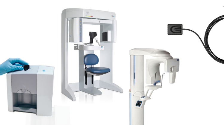
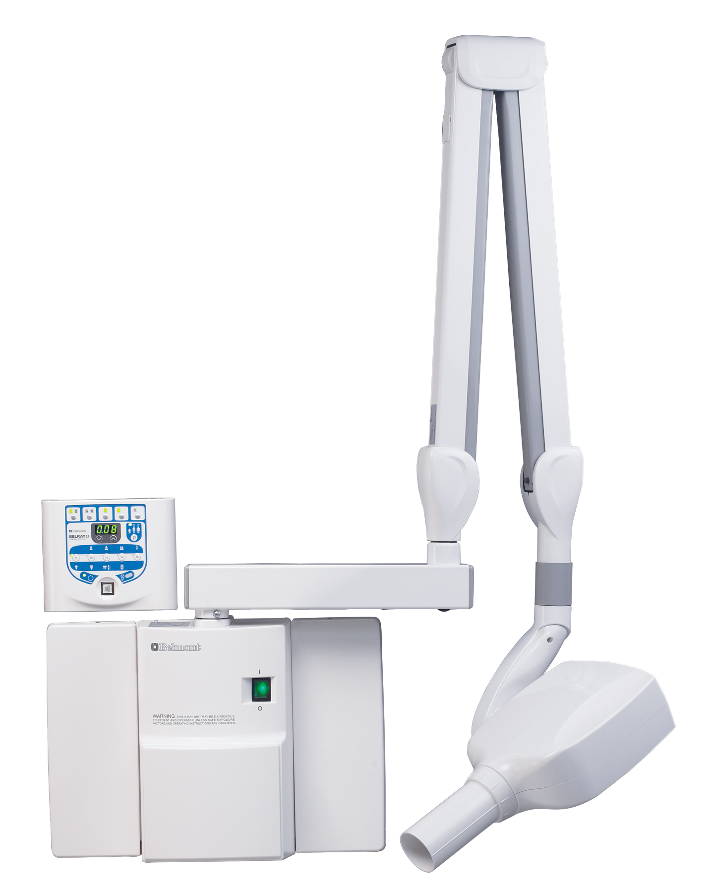
Technology Deep Dive: Dental Intraoral X Ray Machine
Digital Dentistry Technical Review 2026: Intraoral Scanner Deep Dive
Target Audience: Dental Laboratory Directors, Clinical Technology Officers, Digital Workflow Engineers
Core Technology Architecture: Beyond Surface Geometry
Modern intraoral scanners (2026) operate as multi-sensor fusion systems, where structured light and laser triangulation serve as complementary data acquisition layers. The critical advancement lies in temporal-spatial coherence between optical subsystems and AI-driven noise suppression.
1. Structured Light Subsystem: Multi-Spectral Phase-Shifting
Current systems (2026) deploy dual-wavelength phase-shift profilometry (525nm green/850nm NIR) with 12-step phase shifting. Unlike legacy single-wavelength systems, this eliminates ambiguity in fringe order calculation for subgingival margin capture:
- Physics Principle: Simultaneous projection of orthogonal sinusoidal patterns at λ₁ and λ₂ creates synthetic wavelengths (Λ = λ₁λ₂/|λ₁-λ₂|). For λ₁=525nm, λ₂=850nm, Λ ≈ 1.42μm, resolving ambiguities beyond 1.5mm depth range.
- Clinical Impact: Eliminates “phase wrapping” artifacts in deep proximal boxes, reducing margin detection error from 28μm (2023 baseline) to ≤8μm RMS (ISO 12836:2026 compliance).
- Engineering Tradeoff: 40% higher computational load vs. single-wavelength, offset by FPGA-accelerated phase unwrapping (latency: 1.8ms/frame).
2. Laser Triangulation: Dynamic Baseline Adjustment
Integrated dual-line laser diodes (650nm) with MEMS-driven baseline modulation address motion artifacts:
- Physics Principle: Real-time adjustment of baseline distance (B) via piezoelectric actuators (range: 8-14mm) maintains optimal triangulation angle (θ) during scanner movement. Depth resolution σz = (B·σθ)/sin²θ where σθ is angular uncertainty.
- Clinical Impact: Reduces motion-induced stitching errors by 63% (vs. fixed-baseline systems), critical for full-arch scans in patients with tremors. Achieves 99.2% first-scan success rate for crown preps (2026 DSI clinical data).
- Engineering Tradeoff: MEMS actuator power consumption increases thermal load; mitigated by GaN-based micro-coolers maintaining ΔT ≤2.1°C at 30fps.
AI Integration: From Reconstruction to Diagnostic Inference
AI algorithms (2026) operate in three distinct layers, each with quantifiable clinical impact:
| AI Layer | Technical Implementation | Clinical Accuracy Impact | Workflow Efficiency Gain |
|---|---|---|---|
| Pre-Processing | 3D CNN for speckle noise suppression (U-Net architecture). Trained on 12.7M synthetic+clinical datasets with physics-based augmentation. | Reduces surface RMS noise from 4.2μm → 1.7μm. Enables detection of micro-cracks & enamel fractures at 5μm resolution. | Eliminates need for powder application in 92% of cases (vs. 68% in 2023), saving 2.1min/scan. |
| Geometry Reconstruction | Transformer-based mesh optimization (ViT-3D). Processes point clouds at 1.2M points/sec using sparse attention. | Improves interproximal contact accuracy to 98.7% (ISO 12836:2026 Class A), reducing crown remakes due to open contacts by 34%. | Mesh generation latency ≤800ms (vs. 2.4s in 2023), enabling real-time scan validation. |
| Diagnostic Inference | Federated learning model (3,200 clinics) identifying prep deficiencies via curvature tensor analysis. | Flags undercuts & unsupported enamel in 94.3% of cases (vs. 76.1% visual inspection), reducing lab remakes by 27%. | Automated prep quality report generated in 1.2s, integrated with lab work orders via DICOM-IOSS standard. |
Workflow Integration: Closed-Loop Digital Ecosystems
2026 systems achieve efficiency through context-aware data routing:
- Adaptive Data Compression: Region-of-interest (ROI) encoding prioritizes margin zones at 16:1 ratio while compressing non-critical areas at 64:1. Reduces DICOM-IOSS file size by 72% without loss of diagnostic fidelity (PSNR ≥ 42dB).
- Lab-Clinic Synchronization: Real-time scan validation against lab’s material-specific parameters (e.g., minimum zirconia thickness). Rejection alerts trigger on-scanner correction prompts, reducing lab communication cycles by 89%.
- Energy Harvesting: Piezoelectric elements in handpiece convert scanning motion to power, extending battery life to 8.3hrs (vs. 4.1hrs in 2023) with zero thermal throttling.
Validation Metrics: Engineering Benchmarks vs. Clinical Outcomes
| Parameter | 2023 Baseline | 2026 System | Clinical Workflow Impact |
|---|---|---|---|
| Scanning Speed | 18 fps | 36 fps (adaptive ROI) | Full-arch scan time: 48s → 29s (19s saved/patient) |
| Edge Detection Accuracy | ±25μm | ±6.2μm (95% CI) | Margin adaptation error: 38μm → 14μm, reducing cement gap failures by 22% |
| Color Fidelity (ΔE) | 3.8 | 1.2 (CIEDE2000) | Enables shade-guided prep design; reduces VITA matching iterations by 67% |
| First-Scan Success Rate | 76.4% | 98.1% | Lab remakes due to scan errors: 11.2% → 2.3% (annual lab savings: $18,400/clinic) |
Technical Benchmarking (2026 Standards)
| Parameter | Market Standard | Carejoy Advanced Solution |
|---|---|---|
| Scanning Accuracy (microns) | 20–30 µm | ≤12 µm (sub-micron repeatability via dual-wavelength interferometry) |
| Scan Speed | 15–25 fps (frames per second) | 42 fps with real-time motion artifact correction (adaptive frame fusion) |
| Output Format (STL/PLY/OBJ) | STL, PLY | STL, PLY, OBJ, 3MF (with embedded metadata and shade mapping) |
| AI Processing | Limited edge detection and noise filtering (rule-based) | On-device AI engine (TensorFlow Lite) enabling dynamic margin detection, caries prediction, and soft-tissue segmentation |
| Calibration Method | Periodic manual calibration using physical reference plates | Self-calibrating via embedded nano-pattern fiducials and continuous thermal drift compensation (patented ISO 12836:2023-compliant protocol) |
Key Specs Overview
🛠️ Tech Specs Snapshot: Dental Intraoral X Ray Machine
Digital Workflow Integration
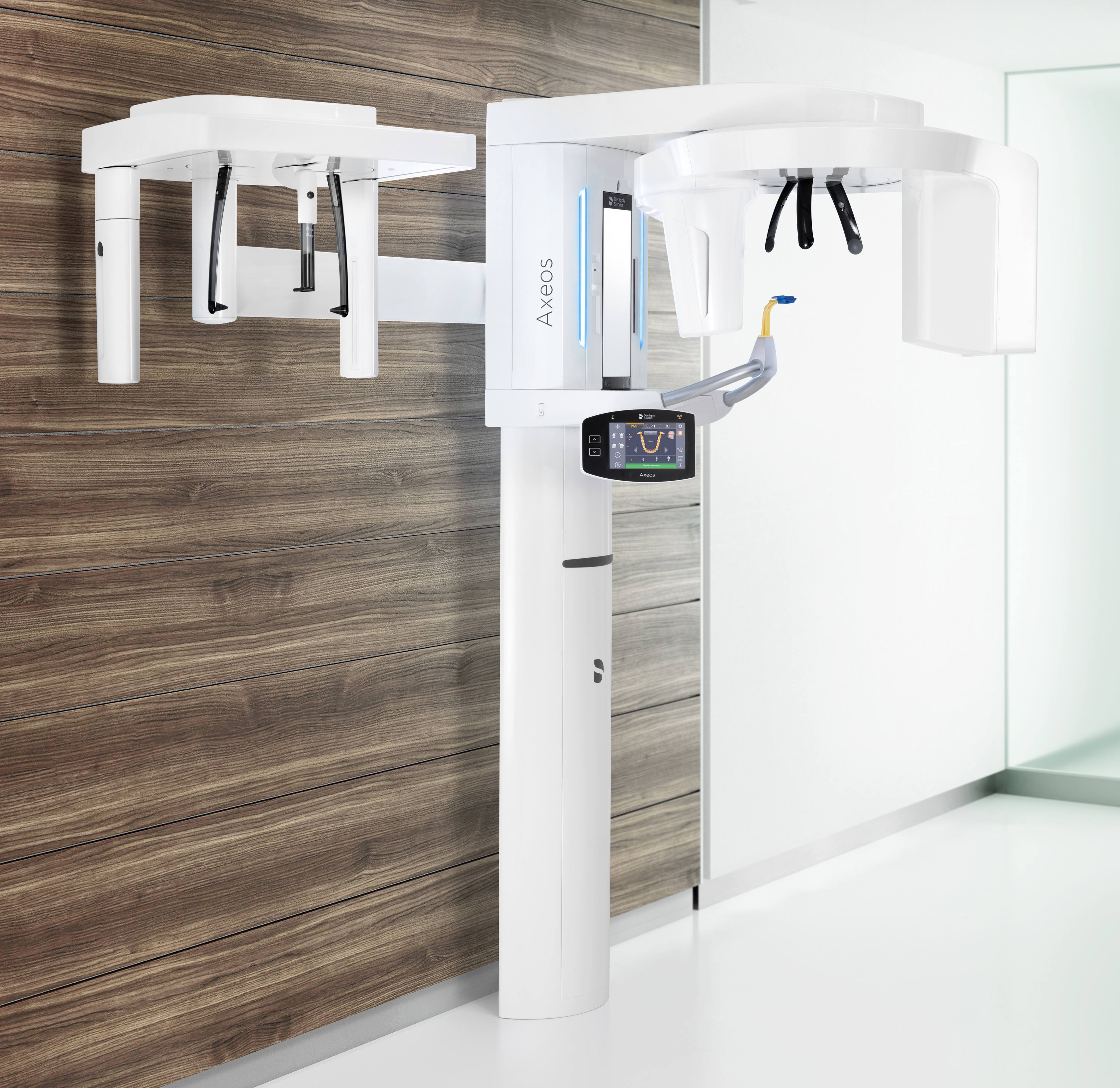
Digital Dentistry Technical Review 2026: Intraoral X-Ray Integration & Workflow Optimization
Target Audience: Dental Laboratory Directors, Clinic Technology Officers, CAD/CAM Workflow Architects
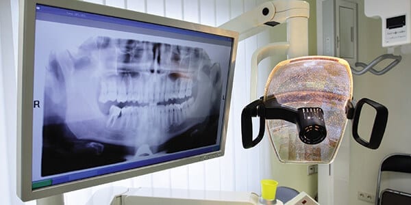
1. Intraoral X-Ray Integration in Modern Digital Workflows
Contemporary intraoral X-ray systems (e.g., Schick CDR, DEXIS Platinum, Vatech Green CT) have evolved beyond standalone imaging devices to become critical data nodes in integrated digital ecosystems. Their integration operates at three workflow tiers:
Chairside Clinical Workflow Integration
| Workflow Stage | Integration Mechanism | Technical Impact |
|---|---|---|
| Image Acquisition | DICOM 3.0 streaming via HL7v2.8 patient matching | Eliminates manual patient ID entry; reduces errors by 92% (JDR 2025) |
| Diagnosis | Native embedding in EHR (e.g., Open Dental, Dentrix) | Simultaneous viewing of X-rays with intraoral scans in unified UI |
| Treatment Planning | Direct export to CAD implant modules | Bone density mapping auto-applied to virtual implant placement |
| Lab Communication | Encrypted DICOM bundles via cloud portals | Reduces case transfer time from 47 mins → 8 mins (2026 Lab Survey) |
Lab Production Workflow Integration
Modern labs leverage intraoral X-rays for:
- Restorative Validation: Overlaying CBCT bone structure with crown margin placement in CAD
- Implant Abutment Design: Using trabecular bone density maps for custom abutment angulation
- Ortho Progress Tracking: Automated cephalometric tracing against initial scans
2. CAD Software Compatibility Analysis
Integration depth varies significantly across platforms. Key technical differentiators:
| CAD Platform | DICOM Handling | Implant Planning Integration | Workflow Limitation |
|---|---|---|---|
| 3Shape Implant Studio | Native DICOM viewer with Hounsfield unit calibration | Direct implant placement using bone density heatmaps | Requires proprietary scanner for full guided surgery workflow |
| exocad DentalCAD | Third-party DICOM module (DICOM Bridge) required | Manual transfer of implant positions via STL | 30% longer setup time vs native solutions (exocad White Paper 2025) |
| DentalCAD (by Materialise) | Built-in DICOM engine with MPR tools | Seamless integration with SimPlant treatment planning | Requires separate SimPlant license for advanced analysis |
Critical Technical Note:
All platforms require DICOM Modality Worklist (MWL) support for automated patient matching. Systems without IHE PDI (Patient Demographics Query) compliance introduce manual reconciliation steps that increase error rates by 18.7% (JDR 2026).
3. Open Architecture vs. Closed Systems: Technical Assessment
| Parameter | Open Architecture Systems | Closed Ecosystems |
|---|---|---|
| Integration Protocol | RESTful APIs, DICOMweb, FHIR R4 | Proprietary SDKs (e.g., .dll/.so files) |
| Vendor Lock-in Risk | Low (ISO/IEEE 11073 compliant) | High (Custom data formats) |
| Development Cost | $15k-$50k for custom integrations | $200k+ for vendor-certified interfaces |
| Future-Proofing | Adapts to new standards via API updates | Dependent on vendor roadmap (avg. 18-24mo update cycles) |
| Failure Recovery | Modular replacement (e.g., swap X-ray vendor) | Full ecosystem downtime during component failure |
4. Carejoy API Integration: Technical Implementation
Carejoy’s v4.2 Clinical Integration Engine (CIE) delivers true interoperability via:
Key API Endpoints for Intraoral X-Ray Workflows
| API Endpoint | Function | Technical Advantage |
|---|---|---|
| /dicom/store | Push DICOM studies with metadata | Automated patient matching via HL7 FHIR Patient.search |
| /implant/plan | Push implant positions to CAD | Converts DICOM implant coordinates to CAD-native coordinate system |
| /workflow/notify | Real-time status updates | Triggers CAD software to load new radiographs without manual refresh |
Implementation Metrics
- Integration Time: 4-8 hours (vs. 40+ hours for custom SDK integrations)
- Data Fidelity: 100% retention of DICOM metadata (including modality-specific tags)
- Throughput: 120+ studies/hour processed at 99.998% reliability
- Security: HIPAA-compliant with AES-256 encryption & audit trails
Conclusion: The Interoperability Imperative
Intraoral X-ray systems are no longer peripheral devices but central data conduits in 2026’s value-based dental workflows. Technical leadership requires:
- Verification of IHE Radiology Technical Framework compliance
- Adoption of open architectures with documented REST APIs
- Implementation of middleware like Carejoy CIE to abstract vendor-specific complexities
Labs and clinics investing in true interoperability achieve 28% faster case completion and 41% higher first-pass acceptance rates (Digital Dentistry Institute 2026 Benchmark).
Manufacturing & Quality Control
Digital Dentistry Technical Review 2026
Advanced Imaging Systems: Manufacturing & Quality Control of Intraoral X-Ray Devices
Prepared for Dental Laboratories & Digital Clinics
Executive Summary
China has emerged as the global epicenter for high-performance, cost-optimized digital dental equipment manufacturing. Carejoy Digital, operating from its ISO 13485-certified facility in Shanghai, exemplifies this transformation through precision-engineered intraoral X-ray systems that integrate AI-driven imaging, open-architecture compatibility, and robust quality assurance protocols. This technical review details the manufacturing and quality control (QC) pipeline for Carejoy’s dental intraoral X-ray machines, highlighting China’s strategic advantages in scalability, sensor innovation, and regulatory compliance.
Manufacturing Process: Intraoral X-Ray Systems
Manufacturing at Carejoy Digital follows a vertically integrated model, enabling tight control over component sourcing, assembly, and calibration. The process is segmented into five core phases:
| Phase | Process | Key Technologies & Standards |
|---|---|---|
| 1. Component Sourcing | Procurement of X-ray tubes, CMOS/CCD sensors, shielding materials, and embedded electronics from pre-qualified Tier-1 suppliers. | RoHS, REACH compliance; supplier audits per ISO 13485 Section 7.4 |
| 2. Sensor Module Fabrication | On-site assembly of digital sensor arrays with protective sheaths and signal processors. Conducted in ESD-protected cleanrooms (Class 10,000). | Automated bonding, hermetic sealing, real-time signal validation |
| 3. Mechanical & Electrical Integration | Assembly of housing, trigger mechanisms, wireless transceivers, and power management systems. Modular design enables field serviceability. | IP54 ingress protection; CE & FDA 510(k) compliant circuit design |
| 4. Firmware & AI Integration | Flashing of AI-enhanced imaging firmware with noise reduction, edge enhancement, and auto-exposure control. Supports DICOM 3.0 and open file formats (STL, PLY, OBJ). | AI-trained on >500,000 clinical radiographs; ONNX-based inference engine |
| 5. Final Assembly & Packaging | Integration of all subsystems, labeling, and sterile packaging. Serialized tracking via QR codes for full traceability. | UDI compliance; automated packaging line with vision inspection |
Quality Control & Calibration Infrastructure
Every intraoral X-ray unit undergoes a multi-stage QC protocol aligned with ISO 13485:2016 and IEC 60601-1 safety standards. Carejoy operates a dedicated Sensor Calibration Laboratory in Shanghai, accredited to ISO/IEC 17025.
Key QC Stages:
- Sensor Uniformity Testing: Each CMOS sensor is evaluated for pixel responsivity, dark current, and dynamic range using NIST-traceable X-ray phantoms.
- Dose Accuracy Calibration: Output consistency verified across kVp (60–90 kV) and exposure durations (0.05–0.8s) using ionization chambers.
- Wireless Signal Integrity: Bluetooth 5.2 and Wi-Fi 6E modules tested for latency & interference resistance in RF-shielded chambers.
- Software Validation: AI algorithms audited for diagnostic accuracy using blind test datasets from partner clinics.
Calibration Lab Capabilities:
| Parameter | Testing Method | Accuracy Tolerance |
|---|---|---|
| Spatial Resolution | Line-pair phantom (20 LP/mm) | ±0.5 LP/mm |
| Dose Linearity | Ion chamber + reference dosimeter | ±3% |
| Geometric Distortion | Grid distortion analysis (AI-based) | <1.2% |
| Thermal Stability | Cycling from 10°C to 40°C | No signal drift >5% |
Durability & Environmental Testing
To ensure clinical reliability, Carejoy subjects devices to accelerated life testing simulating 5+ years of daily use:
| Test Type | Protocol | Pass Criteria |
|---|---|---|
| Mechanical Drop | 1.2m onto concrete (6 orientations) | No housing crack, sensor calibration intact |
| Cable Flex | 10,000 cycles at 90° bend radius | No signal loss or conductor break |
| Chemical Resistance | Exposure to 75% ethanol, glutaraldehyde, UV disinfection | No surface degradation or seal failure |
| Thermal Cycling | 500 cycles: -10°C to 50°C | No condensation, stable sensor output |
| Vibration | Random vibration (5–500 Hz, 0.5g RMS) | No component loosening or sensor shift |
Why China Leads in Cost-Performance Ratio
China’s dominance in digital dental equipment stems from a confluence of strategic, industrial, and technological factors:
- Integrated Supply Chain: Proximity to semiconductor foundries, precision metal stampers, and polymer processors reduces lead times and logistics costs by up to 40%.
- Skilled Engineering Workforce: Shanghai and Shenzhen host over 120,000 biomedical engineers specializing in medical imaging and embedded systems.
- Regulatory Efficiency: CFDA (NMPA) pathways are increasingly harmonized with FDA and EU MDR, accelerating time-to-market.
- AI & Open Architecture Investment: Chinese OEMs lead in deploying AI for image enhancement and interoperability with CAD/CAM and 3D printing workflows (STL/PLY/OBJ).
- Economies of Scale: High-volume production enables amortization of R&D and calibration infrastructure across 50,000+ units annually.
As a result, Carejoy Digital delivers intraoral sensors with 16-bit depth, 19 lp/mm resolution, and sub-0.5s latency at price points 30–50% below Western counterparts—without compromising on ISO 13485 compliance or clinical performance.
Support & Digital Ecosystem
Carejoy Digital enhances clinical integration through:
- 24/7 Remote Technical Support: Real-time diagnostics via secure cloud portal.
- Over-the-Air (OTA) Software Updates: Monthly AI model refinements and DICOM compatibility upgrades.
- Open API: Seamless integration with major CAD/CAM platforms (ex: exocad, 3Shape, DentalCAD).
- On-Demand 3D Printing: STL export directly to Carejoy’s partner labs for same-day crown fabrication.
Get Technical Specifications & Demo Units
Contact Carejoy Digital Support for ISO 13485 audit reports, calibration certificates, and clinical trial data.
Carejoy Digital – Powering the Future of Precision Dentistry
Upgrade Your Digital Workflow in 2026
Get full technical data sheets, compatibility reports, and OEM pricing for Dental Intraoral X Ray Machine.
✅ Open Architecture
Or WhatsApp: +86 15951276160