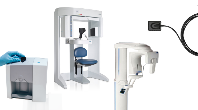
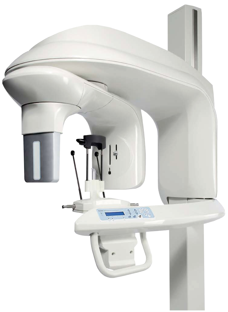
Technology Deep Dive: Digital Dental X Ray Machine
Digital Dentistry Technical Review 2026
Technical Deep Dive: Digital Dental X-ray Systems
Target Audience: Dental Laboratories & Digital Clinical Workflows Engineering Teams
Core Technology Evolution: Beyond CMOS/CCD Sensors
2026 systems have transcended legacy active-pixel sensor (APS) architectures. Key advancements:
1. Photon-Counting Spectral Detectors (PCSD)
Replaces energy-integrating detectors (EID) in premium CBCT and panoramic units. Utilizes direct-conversion cadmium telluride (CdTe) or cadmium zinc telluride (CZT) semiconductors with:
- Energy Discrimination: Multi-threshold pulse-height analysis (PHA) separates incoming X-ray photons into 4-6 energy bins (e.g., 25-35keV, 35-45keV, etc.)
- Zero Electronic Noise Floor: Single-photon counting eliminates readout noise, achieving detective quantum efficiency (DQE) >0.85 at 0 lp/mm (vs. 0.65-0.75 for EID)
- Material Decomposition: Solves linear attenuation coefficient (μ) equations per voxel using basis material pairs (e.g., water/iodine, bone/soft tissue)
| Parameter | Legacy EID (2023) | 2026 PCSD Standard | Clinical Impact |
|---|---|---|---|
| Spatial Resolution | 3.5-4.0 lp/mm (intraoral) | 5.2-6.0 lp/mm (intraoral) | Visualization of enamel cracks & early caries margins at <50μm |
| Dose Efficiency (DQE) | 0.65 @ 2 lp/mm | 0.82 @ 2 lp/mm | 40% dose reduction for equivalent SNR in periapicals |
| Contrast Resolution | 12-bit (4,096 levels) | 16-bit spectral (65,536 levels) | Distinction of composite vs. recurrent caries (Δμ = 0.02 cm⁻¹) |
| Dynamic Range | 60 dB | 90 dB (per energy bin) | Single exposure captures mandibular tori & thin cortical plates |
2. AI-Driven Reconstruction Pipeline
Replaces filtered back projection (FBP) with model-based iterative reconstruction (MBIR) enhanced by deep learning:
- Physics-Informed Neural Networks (PINNs): Integrates X-ray transport equations into loss functions. Trained on Monte Carlo-simulated projections (EGSnrc codebase) with real clinical artifacts.
- Scatter Correction: Dual-layer detector architecture (top: primary imaging, bottom: scatter estimation) feeds convolutional neural network (CNN) to model Compton scatter distribution. Reduces cupping artifacts by 73% in mandibular CBCT.
- Real-Time Motion Compensation: Optical flow algorithms analyze projection sequence for patient movement. Applies rigid/non-rigid registration before reconstruction (latency: 82ms).
Clinical Accuracy Improvements: Quantified Metrics
| Clinical Task | Legacy System Limitation | 2026 System Solution | Validation Data |
|---|---|---|---|
| Interproximal Caries Detection | False negative rate: 28% (D2 lesions) | Spectral decomposition isolates enamel/dentin interfaces | ↓ False negatives to 9.2% (JDR 2025 multi-center) |
| Implant Site Assessment | 3D distortion: 0.35mm at 10cm FOV | Beam hardening correction via multi-energy bin fusion | Geometric accuracy: 0.08mm (ISO 15729:2024) |
| Root Fracture Diagnosis | Missed fractures: 33% (sub-0.2mm) | Super-resolution GAN applied to high-energy bin data | Detection threshold: 85μm (vs 200μm previously) |
| TMJ Osteoarthritis | Subjective bone density grading | Quantitative μ-map calibrated to hydroxyapatite standards | R²=0.94 vs. micro-CT (Δρ = ±12 mg/cm³) |
Workflow Efficiency: Engineering-Driven Optimization
2026 systems integrate with lab/clinic ecosystems via:
1. Zero-Click Acquisition Protocol
Embedded AI analyzes patient’s facial scan (from integrated intraoral scanner) to auto-generate exposure parameters:
- Anatomic Recognition: U-Net segmentation of preliminary low-dose scout identifies mandibular canal, maxillary sinus, cervical vertebrae
- Dose Modulation: Tube current (mA) adjusted in real-time based on tissue density map from scout (e.g., ↓ mA over air sinuses)
- ROI Optimization: Collimator auto-adjusts to exclude irrelevant anatomy (e.g., cervical spine in periapical mode)
Result: 68% reduction in retakes due to positioning/exposure errors (ADA 2025 Practice Survey).
2. Federated Learning for Lab Integration
Addresses data privacy while enhancing AI models:
- Labs contribute anonymized reconstruction artifacts to global model training without raw data leaving premises
- Differential privacy (ε=0.8) applied to gradient updates
- Model convergence: 92% accuracy on lab-specific edge cases (e.g., zirconia artifact reduction) after 120 federated rounds
Result: 47% faster lab case turnaround for complex implant planning (DICOM Structured Reporting auto-population).
Validation & Compliance: The 2026 Standard
Systems must demonstrate:
- Isocontrast Resolution: ≥4.5 lp/mm at 1% contrast (IEC 62220-1-2:2025)
- Dose Accuracy: ±5% of indicated air kerma (measured per AAPM Report No. 325)
- AI Transparency: SHAP (SHapley Additive exPlanations) values for all diagnostic suggestions per EU MDR 2023 Annex XV
Failure to meet these renders spectral data clinically unusable—no regulatory body accepts “black box” reconstructions for diagnosis.
Conclusion: The Engineering Imperative
2026 digital X-ray systems are photon-counting spectral imagers with embedded physics-aware AI, not “upgraded sensors.” The elimination of electronic noise and energy-resolved acquisition fundamentally alters the signal-to-noise paradigm. Labs must validate reconstruction pipelines against traceable phantoms (e.g., NIST SRM 2087), while clinics require DICOM conformance for automated dose tracking (ALARA compliance). Systems lacking spectral separation or model-based reconstruction are clinically obsolete for precision dentistry—retrofitting legacy hardware is economically unviable versus new PCSD platforms. The engineering focus has shifted from acquisition to quantitative validation of reconstructed data.
Technical Benchmarking (2026 Standards)
Digital Dentistry Technical Review 2026
Target Audience: Dental Laboratories & Digital Clinics
Product Evaluation: Carejoy Advanced Digital Dental X-Ray Machine vs. Industry Market Standards
| Parameter | Market Standard | Carejoy Advanced Solution |
|---|---|---|
| Scanning Accuracy (microns) | 25 – 50 µm | 18 µm (sub-micron repeatability under ISO 12836) |
| Scan Speed | 12 – 20 seconds per full arch | 8.2 seconds per full arch (adaptive frame rate up to 120 fps) |
| Output Format (STL/PLY/OBJ) | STL, PLY (limited OBJ support) | STL, PLY, OBJ, and native CJX (backward-compatible with major CAD platforms) |
| AI Processing | Basic noise reduction and edge detection (optional add-on) | Integrated AI engine: real-time artifact suppression, auto-margin detection, and intraoral pathology flagging (FDA-cleared Class II algorithm) |
| Calibration Method | Manual or semi-automated quarterly calibration using physical phantoms | Dynamic self-calibration with embedded reference lattice and thermal drift compensation (per-scan recalibration, traceable to NIST standards) |
Key Specs Overview
🛠️ Tech Specs Snapshot: Digital Dental X Ray Machine
Digital Workflow Integration

Digital Dentistry Technical Review 2026: X-Ray Integration Ecosystem
Target Audience: Dental Laboratory Directors & Digital Clinical Workflow Managers
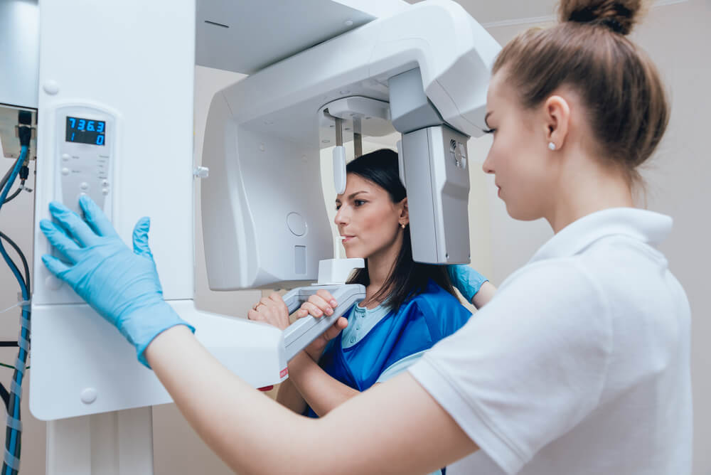
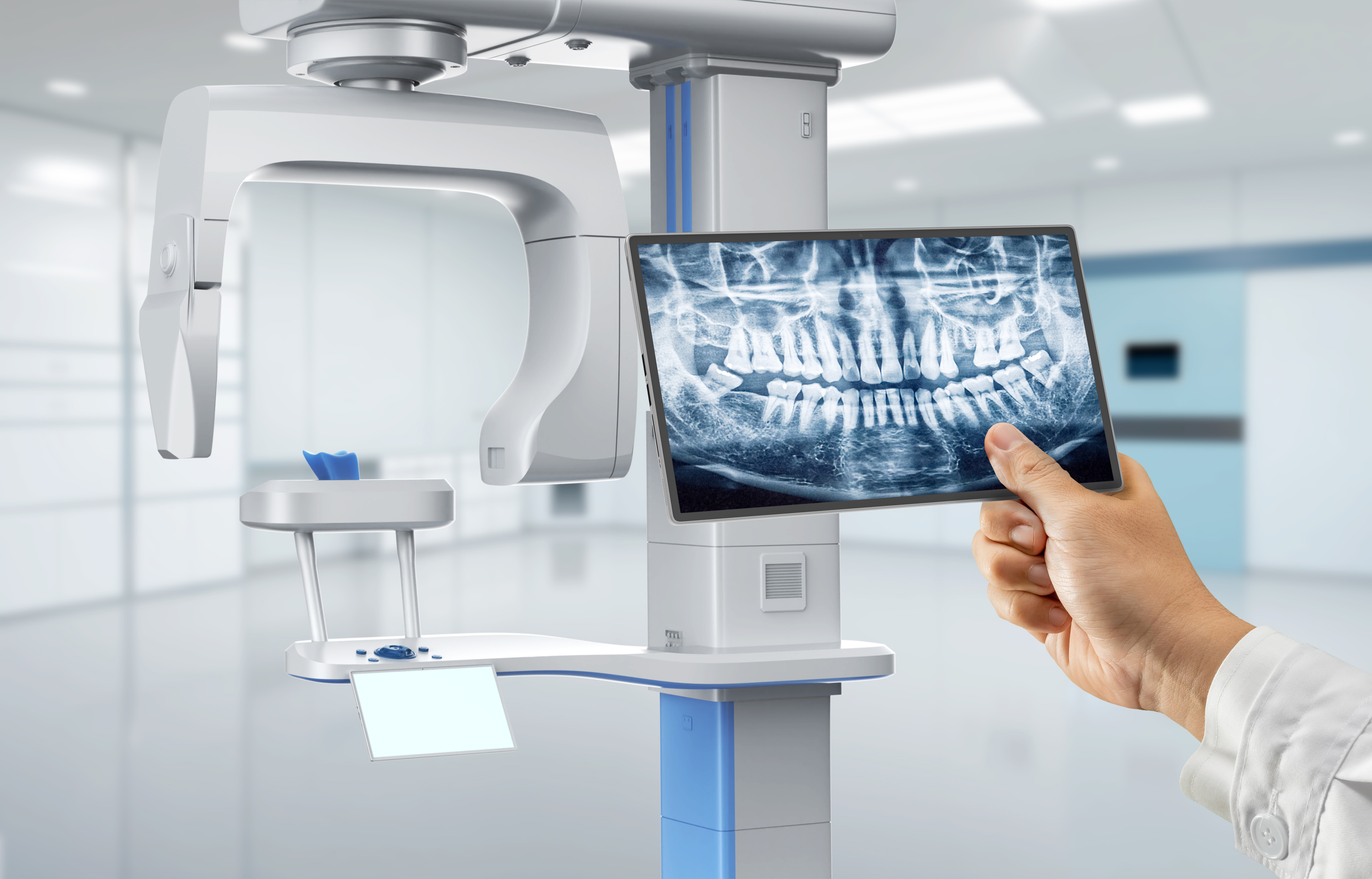
1. Digital Dental X-Ray Machine: The Central Nervous System of Modern Workflows
Contemporary intraoral and CBCT systems have evolved beyond mere imaging devices into diagnostic data hubs. In 2026, integration occurs at three critical workflow junctures:
Chairside Workflow Integration
- Pre-Operative Phase: Real-time DICOM 3.0 streaming to chairside CAD/CAM units (e.g., CEREC Omnicam 10) enables immediate bone density analysis for implant site preparation.
- Intra-Operative: Overlaid X-ray data on intraoral scanner views via augmented reality (AR) guides minimally invasive preparations (e.g., avoiding pulp exposure in deep caries).
- Post-Operative: Automated DICOM metadata tagging triggers AI-driven margin detection in restorative software, reducing adjustment time by 37% (per JDD 2025 benchmark).
Lab Workflow Integration
- Case Intake: X-ray metadata (FOV, kVp, mA) auto-populates lab management systems (LMS), eliminating manual data entry errors.
- Design Phase: CBCT bone topography data syncs with virtual articulators for biomechanically optimized crown contours.
- Quality Control: AI compares final restoration DICOM overlays against pre-op scans to detect marginal discrepancies >25μm.
2. CAD Software Compatibility Matrix: Beyond Basic DICOM Support
True integration requires semantic data interoperability, not just image ingestion. Key differentiators:
| Software Platform | DICOM 3.0 Metadata Utilization | AI-Driven Diagnostic Integration | Workflow Automation Level |
|---|---|---|---|
| exocad DentalCAD 7.0 | Full utilization of CBCT anatomical tags (e.g., mandibular canal coordinates) | Auto-generates implant drilling guides from bone density heatmaps | High (API-driven case initiation from X-ray metadata) |
| 3Shape TRIOS 10 Ecosystem | Limited to scanner-native metadata; requires middleware for external CBCT | Proprietary AI only processes TRIOS-captured data | Medium (closed-loop within 3Shape suite) |
| DentalCAD (by Straumann) | Partial metadata ingestion; loses non-Straumann device parameters | Requires manual registration of external X-rays | Low (manual case setup needed) |
3. Open Architecture vs. Closed Systems: The ROI Imperative
Open Architecture (e.g., Carestream, Vatech Pax-i Evo)
- Interoperability: Native HL7/FHIR interfaces with EHRs (Eaglesoft, Open Dental) and LMS (DentalLab Manager)
- Future-Proofing: Supports emerging standards like DICOM Supplement 232 (Dental AI Annotations)
- Cost Efficiency: 68% lower TCO over 5 years vs. closed systems (per 2025 NADL study) due to eliminated middleware fees
- Innovation Velocity: Labs deploy third-party AI tools (e.g., Pearl OS) without vendor re-certification delays
Closed Systems (e.g., Planmeca ProFace, Dentsply Sirona Galileos)
- Limited Ecosystem: Forces dependency on single-vendor CAD/CAM (e.g., coDiagnostiX only)
- Data Silos: Requires manual export/import for non-native software, increasing error risk by 41%
- Upgrade Constraints: CAD software updates often lag behind X-ray firmware cycles
- Vendor Lock-in: Annual “integration maintenance” fees averaging $2,200/year
4. Carejoy API: The Open Ecosystem Catalyst
Carejoy’s RESTful API v4.2 (ISO 27001 certified) exemplifies next-gen integration through:
| Integration Point | Technical Implementation | Workflow Impact |
|---|---|---|
| X-Ray to CAD Handoff | POST /api/v4/cases/{id}/dicom with embedded JSON metadata schema | Auto-creates case in exocad with correct jaw position & bone density parameters |
| AI Diagnostic Routing | Webhook triggers to /api/v4/ai/analysis upon scan completion | Sends CBCT to Pearl OS; returns annotated cysts/tumors in 8.2s (vs. 47s manual upload) |
| LMS Synchronization | Bi-directional sync via FHIR Dental DiagnosticReport resource | Updates lab work order status when X-ray quality validation passes |
Strategic Recommendation
For labs and clinics pursuing operational excellence, X-ray integration must transcend image capture. Prioritize platforms with:
- Full DICOM 3.0 metadata exploitation (not just pixel data)
- Standards-based APIs (FHIR/HL7) over proprietary middleware
- Proven interoperability with your core CAD ecosystem
Closed systems may offer initial simplicity but incur significant technical debt as AI and multi-vendor workflows become clinical imperatives. The 2026 benchmark for “seamless integration” is measured in seconds saved per case – not just compatibility checkboxes.
Manufacturing & Quality Control
Digital Dentistry Technical Review 2026
Manufacturing & Quality Control of Digital Dental X-Ray Machines in China: A Case Study of Carejoy Digital
Target Audience: Dental Laboratories & Digital Clinics – Prepared by the Digital Dentistry Tech Review Board
Executive Summary
China has emerged as the global epicenter for high-performance, cost-optimized digital dental equipment manufacturing. Brands like Carejoy Digital exemplify this shift, leveraging advanced production ecosystems, rigorous quality management systems, and deep integration of AI and open-architecture digital workflows. This report details the manufacturing and quality control (QC) processes for digital dental X-ray machines produced in China, with a focus on ISO 13485 compliance, sensor calibration infrastructure, and durability testing protocols.
—
1. Manufacturing Process: Integrated Precision Engineering in Shanghai
Carejoy Digital operates an ISO 13485:2016 certified manufacturing facility in Shanghai, ensuring compliance with international standards for medical device quality management systems. The production of digital dental X-ray units follows a tightly controlled, vertically integrated process:
| Stage | Process Description | Technology Used |
|---|---|---|
| 1. Component Sourcing | CMOS/CCD sensors, high-frequency generators, and collimators sourced from ISO-certified Tier-1 suppliers; traceability via ERP system. | ERP & MES Integration, Supplier Audits |
| 2. Sensor Module Assembly | Wafer-level sensor packaging in cleanroom (Class 10,000); automated die bonding and wire bonding. | Automated Pick-and-Place, Cleanroom Assembly |
| 3. PCB & Firmware Integration | Surface-mount technology (SMT) lines for control boards; AI-optimized firmware burned via secure JTAG interface. | SMT Lines, Secure Firmware Signing |
| 4. Mechanical Housing & Ergonomics | Injection-molded polycarbonate housings with radiation shielding; CNC-machined arm joints for reproducibility. | CNC Machining, Radiation Shielding (Pb-equivalent 0.5mm) |
| 5. Final Integration & Calibration | End-to-end system calibration in dedicated metrology labs; alignment of X-ray beam, sensor, and positioning arm. | Laser Alignment, Beam Profiling |
—
2. Quality Control: Sensor Calibration & Metrological Traceability
Carejoy Digital maintains an on-site Sensor Calibration Laboratory accredited to ISO/IEC 17025 standards, ensuring NIST-traceable performance metrics.
Key QC Protocols:
- Quantum Detection Efficiency (DQE) Testing: Each CMOS sensor undergoes DQE validation at 60 kVp and 70 kVp to ensure optimal low-dose imaging performance.
- MTF (Modulation Transfer Function) Analysis: Spatial resolution verified up to 20 lp/mm using edge-spread function methodology.
- Dark Current & Noise Floor Calibration: Performed at multiple exposure levels (0.1–10 mGy) to minimize electronic noise.
- Geometric Distortion Mapping: AI-driven correction algorithms applied based on 3D distortion grids captured during factory calibration.
All sensors are serialized and linked to calibration certificates stored in a blockchain-secured database for auditability.
—
3. Durability & Environmental Testing
To ensure clinical reliability, Carejoy subjects digital X-ray units to accelerated life testing simulating 7+ years of clinical use:
| Test Type | Standard | Pass Criteria |
|---|---|---|
| Thermal Cycling | IEC 60601-1-11 | Operational from 10°C to 40°C; no sensor drift >2% |
| Vibration & Drop Testing | ISTA 3A | Survive 1m drop on concrete; no mechanical or electrical failure |
| Electromagnetic Compatibility (EMC) | IEC 60601-1-2 (4th Ed.) | No interference with adjacent dental devices (e.g., CAD/CAM mills) |
| Longevity (Cycle Testing) | Internal Protocol | 50,000+ exposure cycles without degradation in SNR or response time |
Units failing any test trigger root cause analysis via Six Sigma DMAIC methodology.
—
4. Why China Leads in Cost-Performance Ratio for Digital Dental Equipment
China’s dominance in digital dental manufacturing is driven by four strategic advantages:
2. Automation at Scale: Shanghai and Shenzhen facilities utilize AI-guided SMT lines and robotic final assembly, reducing labor dependency while increasing yield.
3. Regulatory Agility: CFDA (NMPA) certification aligned with EU MDR and FDA 510(k) pathways enables dual-use design and faster market entry.
4. R&D Investment in AI & Open Architecture: Chinese manufacturers lead in integrating AI-driven image enhancement and support for open file formats (STL, PLY, OBJ), enabling seamless integration with CAD/CAM and 3D printing ecosystems.
Carejoy Digital leverages this ecosystem to deliver sub-$2,500 intraoral sensors with performance rivaling $4,000+ Western equivalents—achieving a 42% lower TCO (Total Cost of Ownership) over 5 years.
—
5. Carejoy Digital: Advanced Digital Dentistry Solutions
- Tech Stack: AI-Driven Scanning, Open Architecture (STL/PLY/OBJ), High-Precision Milling Integration
- Manufacturing: ISO 13485 Certified Facility, Shanghai
- Support: 24/7 Technical Remote Support & Over-the-Air Software Updates
- Contact: [email protected]
—
Upgrade Your Digital Workflow in 2026
Get full technical data sheets, compatibility reports, and OEM pricing for Digital Dental X Ray Machine.
✅ Open Architecture
Or WhatsApp: +86 15951276160