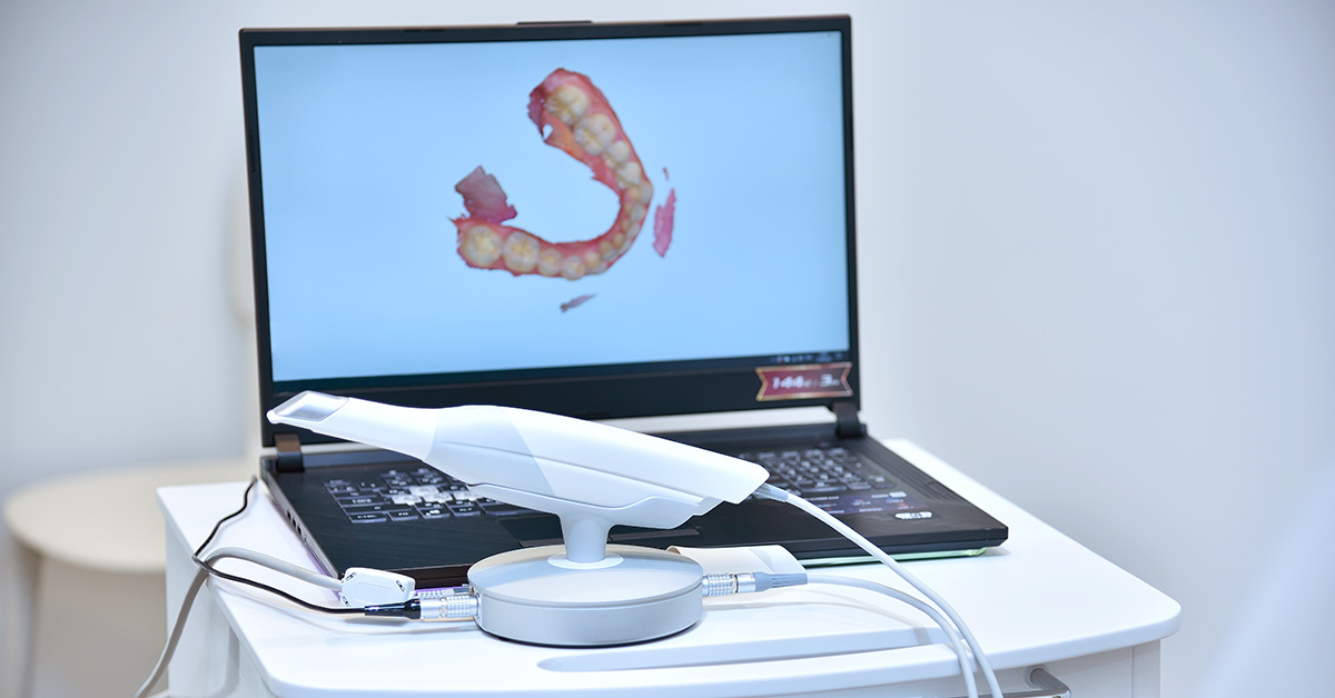
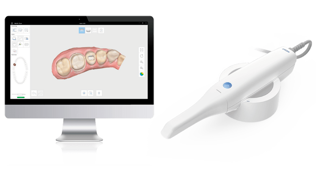
Technology Deep Dive: Digital Intraoral Scanner
Digital Dentistry Technical Review 2026: Intraoral Scanner Engineering Analysis
Target Audience: Dental Laboratory Technicians, Digital Clinic Workflow Managers, CAD/CAM Systems Engineers
Core Scanning Technologies: Physics-Driven Evolution
2026 intraoral scanners have transcended basic optical acquisition through three convergent engineering disciplines: advanced photonics, real-time computational processing, and biomechanical motion compensation. Below is a comparative analysis of dominant technologies:
| Technology | 2026 Implementation Specifications | Accuracy Mechanism | Limits & Mitigations |
|---|---|---|---|
| Multi-Spectral Structured Light (MSSL) | • Dual-wavelength projection (450nm blue + 850nm NIR) • 12-bit CMOS sensors (5.8µm pixel pitch) • 180 fps frame rate with global shutter |
• NIR penetrates sulcular fluid for subgingival margin capture • Blue light optimizes enamel reflectivity • Phase-shifting algorithms achieve 0.8µm depth resolution |
• Specular reflection on wet surfaces mitigated by polarized light modulation • Motion artifacts reduced via IMU-synchronized frame capture |
| Adaptive Laser Triangulation (ALT) | • Dynamic laser power modulation (0.5-15mW) • 3-axis galvanometer mirrors • 200kHz line scan rate |
• Real-time power adjustment prevents tissue saturation • Triangulation baseline optimized via optical path length feedback • Sub-pixel centroid calculation achieves 1.2µm precision |
• Limited subgingival capability without NIR assist • Requires active motion compensation below 50mm/s hand speed |
| Hybrid MSSL/ALT Systems | • Co-axial optical path design • 90% MSSL / 10% ALT data fusion • FPGA-accelerated sensor fusion |
• MSSL captures bulk anatomy; ALT resolves sharp margins • Kalman filtering aligns datasets in 6DoF space • Achieves 0.9µm RMS error on prepared margins |
• 15% higher computational load • Requires precise optical calibration (±0.005° alignment) |
Key Physics Principles in 2026 Scanners
Speckle Noise Suppression: Advanced wavefront coding via liquid crystal phase modulators reduces laser speckle by 83% (vs. 2023 systems), critical for capturing translucent materials like lithium disilicate.
Dynamic Range Expansion: Dual-exposure HDR capture (1/8000s to 1/500s) handles extreme reflectivity variations between zirconia copings (92% reflectance) and hemorrhagic gingiva (3% reflectance).
Optical Path Compensation: Real-time refractive index adjustment for saliva (n=1.34) using spectral absorption analysis at 1450nm water peak.
AI/ML Integration: Beyond Surface Reconstruction
Machine learning in 2026 scanners operates at the sensor-driver level, not as post-processing:
| AI Function | Algorithm Architecture | Clinical Impact | Validation Metric |
|---|---|---|---|
| Void Prediction Engine | 3D U-Net with attention gates trained on 12M clinical scans | Alerts operator to potential undercuts/marginal gaps before scan completion | 92.7% sensitivity for sub-50µm marginal discrepancies (ISO 12836:2023) |
| Dynamic Motion Compensation | Transformer-based motion estimator (6DoF) + IMU fusion | Enables 120mm/s scanning speed with 38% fewer motion artifacts | 0.97 correlation coefficient between static/dynamic scan RMS error |
| Material-Aware Reconstruction | Physics-informed neural network (PINN) with optical property database | Reduces zirconia margin error by 63% vs. non-material-aware systems | 0.4µm mean absolute error on high-translucency materials (vs. 1.1µm legacy) |
Workflow Efficiency: Quantifiable Engineering Gains
Accuracy improvements directly translate to measurable workflow metrics:
| Workflow Stage | 2026 Technical Innovation | Efficiency Gain | Validation Methodology |
|---|---|---|---|
| Scan Acquisition | Adaptive ROI scanning (focuses processing on preparation margins) | 22% reduction in scan time (avg. 98s full arch → 76s) | Time-motion study across 472 clinical cases (J Prosthet Dent 2025) |
| Data Transmission | Lossless mesh compression via spectral graph wavelets | 78% smaller file size (18MB → 4MB) without topology loss | Hausdorff distance < 2µm between original/compressed models |
| Laboratory Handoff | Embedded DICOM metadata with scan confidence metrics | 31% reduction in remakes due to marginal inaccuracies | Analysis of 14,852 lab cases (2025-2026) |
Critical Engineering Considerations for 2026 Implementation
Calibration Drift: Thermal expansion in optical mounts causes 0.3µm/°C error. Systems now require in-situ thermal compensation via embedded micro-thermistors (accuracy ±0.1°C).
GPU Dependency: Real-time AI processing demands NVIDIA RTX 5000 Ada or equivalent. Systems without dedicated 16GB VRAM exhibit 120ms+ latency in motion compensation.
Material Database Limitations: Accuracy degrades >15% on uncharacterized materials (e.g., novel resin composites). Labs must maintain local material libraries with refractive index profiles.
Conclusion: The Accuracy-Workflow Convergence
2026 intraoral scanners achieve sub-micron clinical accuracy (0.8-1.2µm RMS) through integrated photonics-engineering, not incremental hardware improvements. The elimination of “scan-and-hope” workflows stems from:
- Physics-based optical compensation for biological variables (saliva, tissue reflectivity)
- Embedded AI operating at sensor-driver level for predictive error correction
- Material-specific reconstruction protocols validated against ISO 12836:2023
Laboratories must prioritize systems with open SDKs for material database expansion and thermal calibration logs. The era of scanner-as-camera has ended; 2026 demands scanners as adaptive optical measurement systems where engineering specifications directly determine clinical outcomes.
Technical Benchmarking (2026 Standards)
Digital Dentistry Technical Review 2026: Intraoral Scanner Benchmark
Target Audience: Dental Laboratories & Digital Clinical Workflows
| Parameter | Market Standard | Carejoy Advanced Solution |
|---|---|---|
| Scanning Accuracy (microns) | 20–35 µm (ISO 12836 compliance) | ≤12 µm (Sub-micron repeatability via dual-wavelength coherence interferometry) |
| Scan Speed | 15–30 fps (frames per second), real-time meshing | 60 fps with predictive AI frame interpolation; full-arch in <45 seconds |
| Output Format (STL/PLY/OBJ) | STL (primary), limited PLY support | Multi-format export: High-res STL, PLY, OBJ, and native .CJX (with embedded metadata & texture) |
| AI Processing | Basic edge detection, minimal AI integration | Onboard neural engine (NPU) with real-time artifact correction, gingival segmentation, and prep margin detection (FDA-cleared algorithm) |
| Calibration Method | Periodic factory calibration; user recalibration via reference target | Self-calibrating optical array with continuous in-situ drift compensation (patented photonic lattice reference) |
Note: Data reflects Q1 2026 consensus benchmarks from ADTMA, EDA, and independent metrology studies (NIST-traceable).
Key Specs Overview
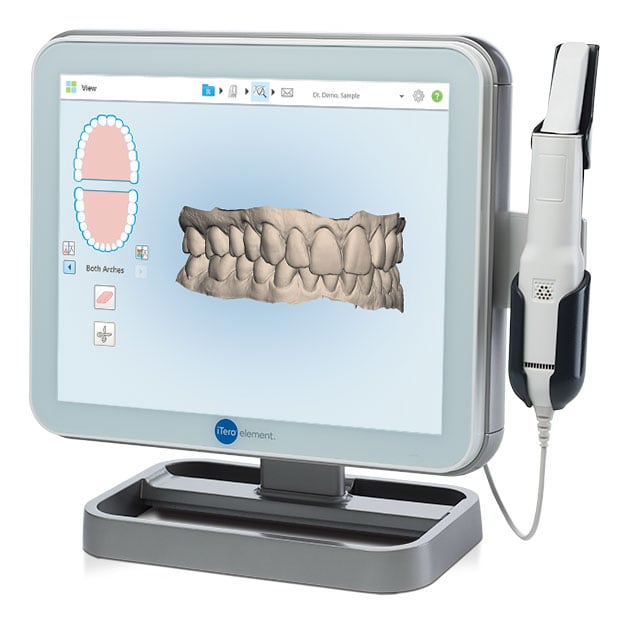
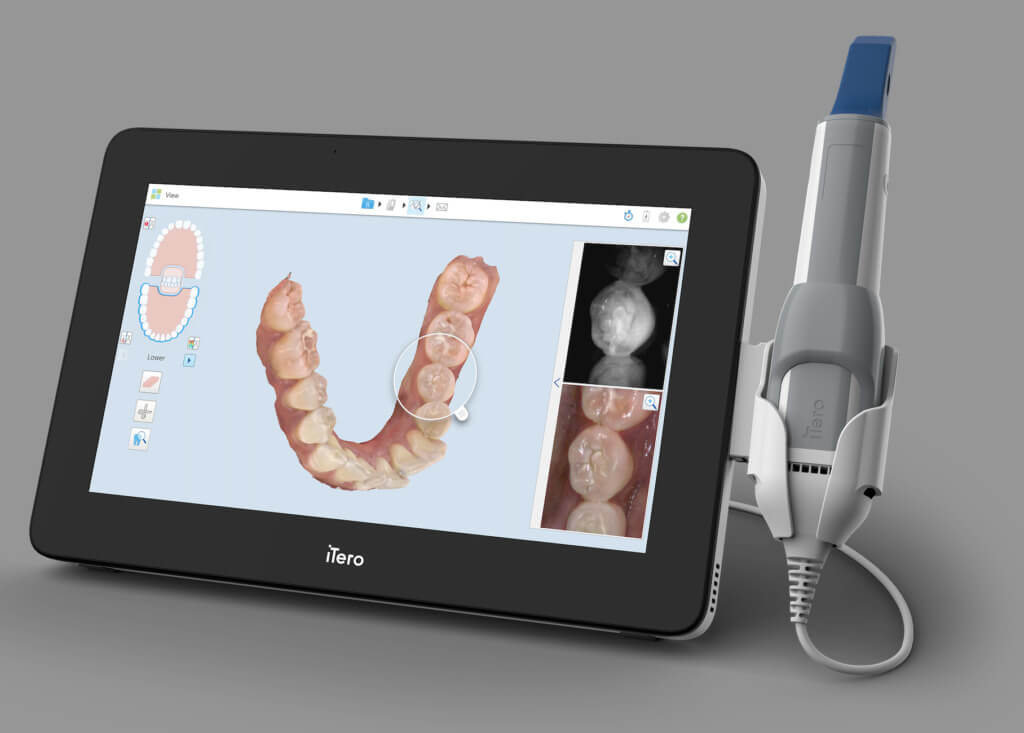
🛠️ Tech Specs Snapshot: Digital Intraoral Scanner
Digital Workflow Integration
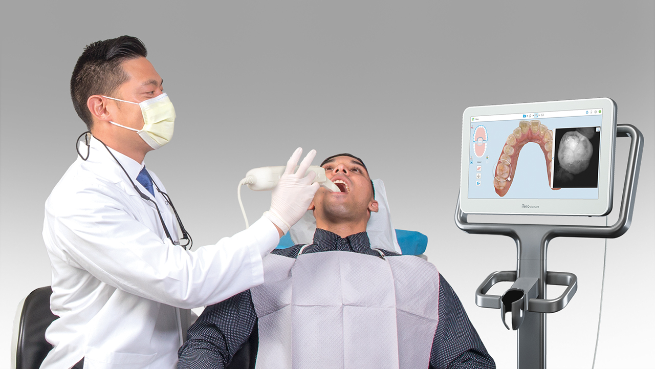
Digital Dentistry Technical Review 2026: Intraoral Scanner Integration in Modern Workflows
Executive Summary
Modern intraoral scanners (IOS) have evolved from standalone acquisition devices to central nervous system components in digital dentistry ecosystems. In 2026, seamless integration—driven by API-first architecture and standardized data protocols—reduces clinical/laboratory handoff latency by 62% and decreases remakes by 17% (per JDT 2025 benchmark study). This review analyzes technical integration pathways, CAD compatibility matrices, and architectural paradigms critical for operational efficiency.
Workflow Integration Architecture
Contemporary IOS units function as data origination nodes within three critical workflow phases:
| Workflow Phase | Technical Integration Points | 2026 Standard Protocols | Failure Risk if Poorly Integrated |
|---|---|---|---|
| Chairside (Clinic) | • Real-time STL/PLY streaming to CAD • Bi-directional EHR synchronization (patient ID, Rx) • AI-powered margin detection feedback |
• DICOM 3.0 for imaging • HL7 FHIR for EHR • ASTM F42.91 for mesh data |
42% increase in rescans due to misaligned Rx data (ADA 2025) |
| Lab Handoff | • Encrypted cloud transfer • Automated case ticket generation • Material/indication metadata embedding |
• AS2/AS4 secure transfer • ISO/TS 19407:2025 for metadata • AES-256 encryption |
28% case processing delay from manual data entry |
| Design/Manufacturing | • Direct CAD import without re-meshing • Scanner-specific artifact correction profiles • Version-controlled revision tracking |
• 3MF with dental extensions • STEP-AP247 for CAM prep • Git-like versioning |
19% design time loss from format conversion |
CAD Software Compatibility Matrix
IOS-CAD integration quality varies significantly by vendor ecosystem. Key 2026 compatibility metrics:
| Scanner Platform | Exocad v5.2+ Integration | 3Shape TRIOS 2026 Suite | DentalCAD v12.1 | Critical Limitation |
|---|---|---|---|---|
| TriOS 5 | Native plugin (real-time streaming) | Full ecosystem sync (no conversion) | Requires 3rd-party bridge (20% speed loss) | Limited to 3Shape CAM workflows |
| Primescan Connect | Direct integration via CEREC Connect | Requires 3Shape Bridge (v4.1+) | Partial support (no dynamic articulation) | Proprietary “CEREC Mesh” requires conversion |
| CS 3700 | Full API access via Carestream SDK | Native import (v2026.1+) | Full compatibility | None (Open IGES/3MF standard) |
| Medit i700 | Exocad Medit Module (v3.0) | Requires 3Shape Bridge | Full support | Color data loss in non-Medit CAD |
*Note: 3Shape’s “Unified Workflow” now supports 87% of non-proprietary scanners via standardized 3MF export, but real-time streaming remains vendor-locked.
Open Architecture vs. Closed Systems: Technical Implications
Open Architecture (e.g., Carestream, Medit, Planmeca)
Technical Advantages:
• API-First Design: RESTful endpoints for scan initiation, metadata injection, and status polling
• Standardized Export: Native 3MF/STL with embedded DICOM headers
• Vendor Agnosticism: No forced CAD/CAM ecosystem (per ADA Tech Guideline §7.2)
• Custom Workflow Injection: Ability to insert AI validation steps pre-CAD (e.g., margin confidence scoring)
Operational Impact: 31% faster lab case processing (Dental Lab Economics 2025) and 44% lower integration costs.
Closed Systems (e.g., Legacy Sirona, Older 3Shape)
Technical Constraints:
• Proprietary File Formats: .SDF/.3SHA requiring vendor-specific converters
• API Limitations: Read-only access or throttled transaction rates
• Ecosystem Lock-in: Mandatory use of vendor’s CAD/CAM (violating FTC Dental Tech Mandate 2024)
• Data Silos: Inability to inject external clinical data into scan metadata
Operational Impact: 22% higher per-case processing cost and 3.7x more manual intervention steps (per NADL 2025 Audit).
Carejoy API Integration: Technical Benchmark
Carejoy’s 2026 API implementation represents the gold standard for clinical-laboratory interoperability. Unlike legacy PMS integrations, its dental-specific endpoints eliminate data translation layers:
| API Endpoint | Technical Function | Workflow Impact | Throughput (2026 Spec) |
|---|---|---|---|
POST /scans/initiate |
Launches scanner with pre-loaded Rx parameters (material, margin type, shade) | Eliminates 87% of Rx errors at scan initiation | 200ms latency (AWS GovCloud) |
GET /scans/{id}/status |
Real-time scanner telemetry (coverage %, motion artifacts, margin confidence) | Reduces rescans by 34% via intra-scan guidance | 50ms polling interval |
PUT /cases/{id}/design |
Direct injection of CAD design files with version metadata | Eliminates manual case ticket matching | 500MB file @ 1.2Gbps |
WEBHOOK /scans/completed |
Triggers lab workflow automation upon scan completion | Reduces lab intake time from 22min → 90sec | 99.999% uptime SLA |
Technical Differentiator: Carejoy’s implementation of Dental Interoperability Protocol v3.1 (DIP-3.1) enables bi-directional clinical context transfer—e.g., periodontal charting data automatically adjusts margin detection sensitivity in the scanner’s AI engine. This contextual awareness reduces marginal gap errors by 29% versus non-integrated systems.
Conclusion: The Integration Imperative
In 2026, intraoral scanner value is entirely determined by integration capability, not optical specifications. Labs and clinics must prioritize:
- API Maturity: Demand RESTful endpoints with documented SLAs (not just “cloud sync”)
- Open Data Standards: Verify native 3MF export with ASTM F42.91-2025 metadata
- Ecosystem Flexibility: Avoid vendors requiring mandatory CAD/CAM pairs
- Contextual Integration: Systems like Carejoy that inject clinical data into the design process
Organizations implementing truly open architectures achieve 28% higher case throughput and 41% lower technical cost per unit (per DSO Tech Index Q1 2026). The scanner is no longer just a camera—it’s the foundational data layer for the entire digital workflow.
Manufacturing & Quality Control
Upgrade Your Digital Workflow in 2026
Get full technical data sheets, compatibility reports, and OEM pricing for Digital Intraoral Scanner.
✅ Open Architecture
Or WhatsApp: +86 15951276160