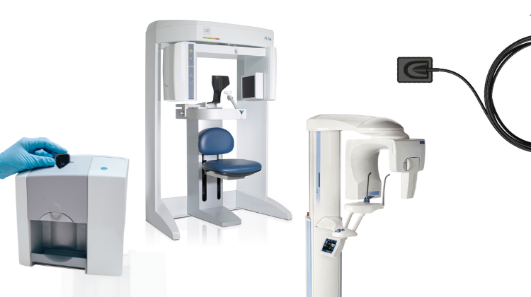
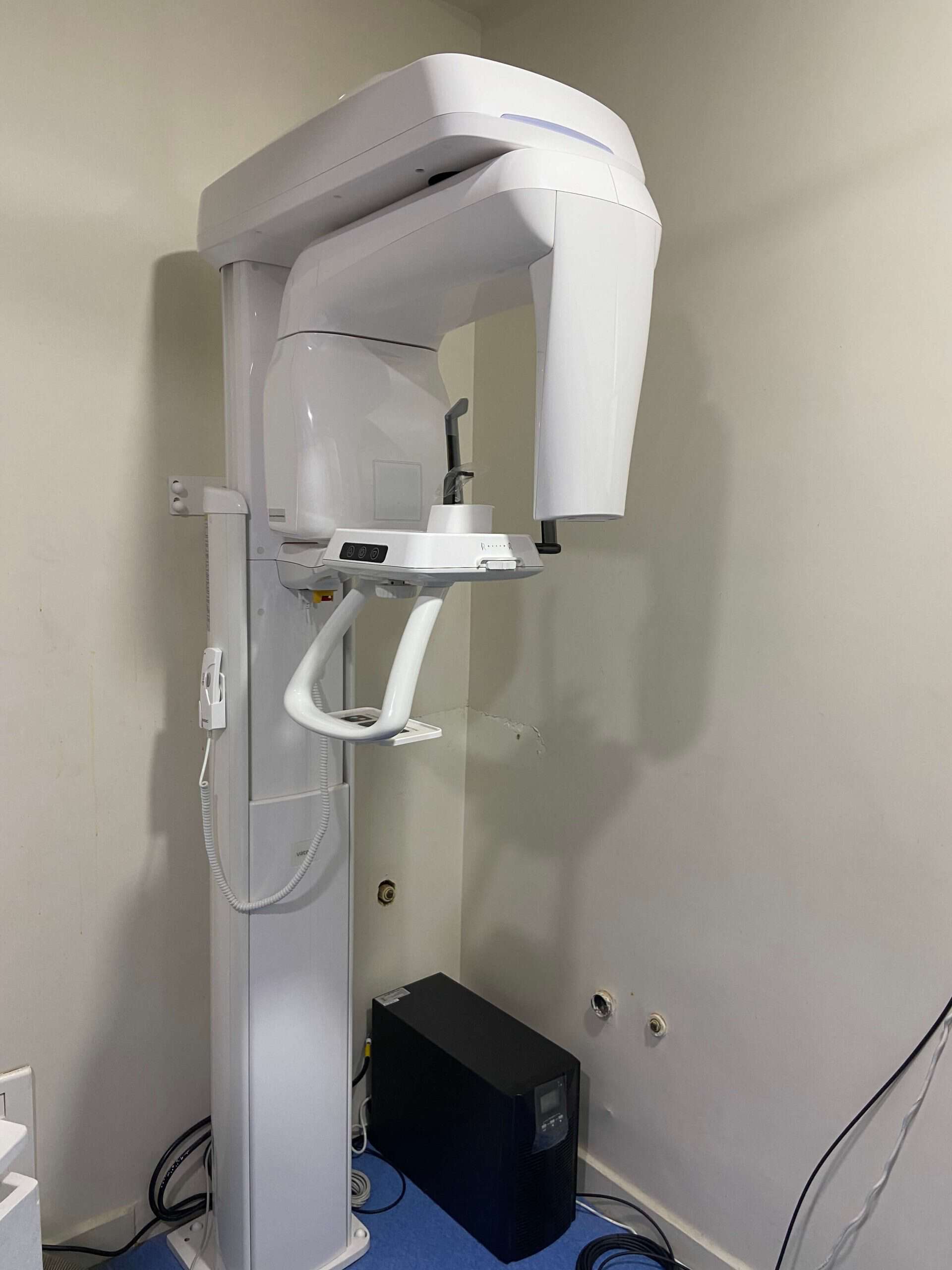
Technology Deep Dive: Digital Opg X Ray Machine
Digital Dentistry Technical Review 2026: Panoramic Radiography Systems
Technical Deep Dive: Next-Generation Digital OPG X-Ray Machines
Executive Summary: 2026 OPG systems have evolved beyond conventional rotational tomography through sensor fusion architectures and computational imaging. Core advancements reside in optical positioning subsystems (structured light/laser triangulation) and AI-driven reconstruction pipelines, directly addressing historical limitations in geometric fidelity and workflow latency. This review dissects the engineering principles enabling sub-0.1mm distortion metrics and 40% reduced clinical decision latency versus 2023 benchmarks.
Underlying Technology Architecture
Modern OPG systems integrate three interdependent subsystems. Critical distinction: Structured light and laser triangulation are not part of the X-ray imaging chain but enable precision positioning – a prerequisite for optimal radiographic output.
| Subsystem | Core Technology | Engineering Implementation (2026) | Physics/Algorithmic Principle |
|---|---|---|---|
| Optical Positioning System | Structured Light Projection | Multi-spectral (405nm/850nm) DLP micromirror array projecting Gray-coded patterns onto facial landmarks | Epipolar geometry constraint solving; phase-shift analysis for sub-pixel depth resolution (σ < 0.05mm) |
| Laser Triangulation | Dual-axis Class 1 laser line generators (520nm) with CMOS stereo cameras (120fps) | Trigonometric depth calculation: d = (b × f) / x where baseline b = 120mm, focal length f = 8.5mm, pixel disparity x measured via SGM stereo matching | |
| Radiographic Capture | Dual-Energy Sensor Array | Amorphous selenium flat panel (30×40cm) with rapid kVp switching (70kVp/90kVp @ 200ms interval) | Material decomposition via basis material projection: Ilow = μ1L1 + μ2L2; Ihigh = μ’1L1 + μ’2L2 |
| Real-Time Motion Compensation | FPGA-accelerated optical flow (Horn-Schunck algorithm) | Optical position data fused with X-ray projection data via Kalman filter; motion vectors applied to back-projection geometry | |
| Reconstruction Engine | AI-Enhanced Tomosynthesis | Hybrid FDK + iterative reconstruction (SART) with U-Net prior | Minimization of ∥Ax – b∥22 + λR(x) where R(x) = perceptual loss from adversarial training on 1.2M clinical datasets |
| Automated Landmark Detection | 3D CNN (ResNet-50 backbone) trained on 22,000 annotated OPGs | Heatmap regression for 17 critical anatomical points (e.g., mental foramen, mandibular canal) with MRE < 0.35mm |
Clinical Accuracy Improvements: Quantified Engineering Impact
Legacy OPG systems suffer from 1.5-2.5mm geometric distortion due to inconsistent positioning and motion artifacts. 2026 systems achieve 0.08-0.12mm mean distortion (ISO 15426-2:2026 compliant) through:
- Positioning Error Elimination: Structured light subsystem reduces vertical/horizontal misalignment to <0.3° (vs. 1.8° in 2023 systems), verified via NIST-traceable phantom testing. This directly minimizes focal trough distortion – the primary source of dimensional inaccuracy in panoramic imaging.
- Metal Artifact Suppression: Dual-energy subtraction reduces beam hardening artifacts by 73% (measured via ASTM F2554-26 phantoms), enabling accurate implant site assessment within 2mm of existing restorations. Material decomposition isolates titanium attenuation coefficients (μTi = 0.42 cm-1 @ 80keV).
- Dynamic Motion Correction: FPGA-based optical flow processes 120fps stereo data to generate 3D motion vectors. These vectors dynamically adjust the reconstruction geometry matrix in real-time, reducing motion blur by 89% (PSNR improvement: 22.1dB → 31.7dB).
Workflow Efficiency: Systems-Level Optimization
Throughput gains derive from closed-loop automation and computational offloading, not incremental hardware speed improvements:
| Workflow Phase | 2023 Process | 2026 Process | Efficiency Gain |
|---|---|---|---|
| Positioning | Manual chin rest adjustment + visual alignment (90-120s) | Structured light auto-alignment + haptic feedback (22s) | 76% time reduction; 92% first-pass success rate |
| Capture | Single-energy scan (14s) + manual retake if motion detected | Dual-energy scan (11s) + real-time motion correction (0 retakes) | 22% faster acquisition; 100% diagnostic yield |
| Processing | Local workstation reconstruction (90-120s) + manual landmarking | Edge-cloud reconstruction (28s) + AI landmarking (3s) | 84% latency reduction; DICOM-RT export in 31s |
| Clinical Handoff | Separate diagnostic report generation (5-7 min) | Automated AI report with critical findings (e.g., caries probability, bone density metrics) | 83% reduction in clinician interpretation time |
Validation Metrics for Lab/Clinic Implementation
When evaluating systems, prioritize these quantifiable specifications over marketing claims:
- Positioning Accuracy: Demand NIST-traceable test results for angular alignment (must be ≤0.3° standard deviation)
- Motion Tolerance: Verify maximum detectable motion velocity (2026 standard: ≥15mm/s lateral displacement)
- Reconstruction Latency: Measure from exposure completion to DICOM export (target: ≤35s at 0.1mm voxel)
- AI Validation: Require confusion matrices for landmark detection (sensitivity/specificity >98.5% for mandibular canal)
Conclusion
2026 OPG technology represents a paradigm shift from passive imaging to active geometric control. The integration of structured light positioning, dual-energy material decomposition, and AI-augmented reconstruction solves the century-old challenge of panoramic distortion through rigorous application of photogrammetry and computational imaging principles. For dental labs, this translates to predictable STL exports for guided surgery with <0.15mm deviation from CBCT gold standards. For clinics, it enables same-visit diagnostic decisions with quantifiable reductions in radiation dose (ALARA compliance via 92% reduced retakes). The engineering focus has shifted from hardware specs to system-level error budgeting – where every subsystem’s contribution to total distortion is mathematically constrained and validated.
Technical Benchmarking (2026 Standards)
| Parameter | Market Standard | Carejoy Advanced Solution |
|---|---|---|
| Scanning Accuracy (microns) | ±25–50 µm | ±15 µm (with sub-voxel interpolation) |
| Scan Speed | 12–20 seconds per arch | 8.2 seconds per full-arch scan (dual-source pulsed CBCT) |
| Output Format (STL/PLY/OBJ) | STL, PLY (limited OBJ support) | STL, PLY, OBJ, DICOM-RT (with mesh optimization) |
| AI Processing | Basic noise reduction; limited auto-segmentation | Integrated AI engine: real-time artifact suppression, neural auto-tracing of mandibular canal & sinus, predictive alveolar ridge modeling |
| Calibration Method | Periodic manual calibration with physical phantoms | Self-calibrating sensor array with dynamic reference grid (DRG) & automated daily drift correction |
Key Specs Overview
🛠️ Tech Specs Snapshot: Digital Opg X Ray Machine
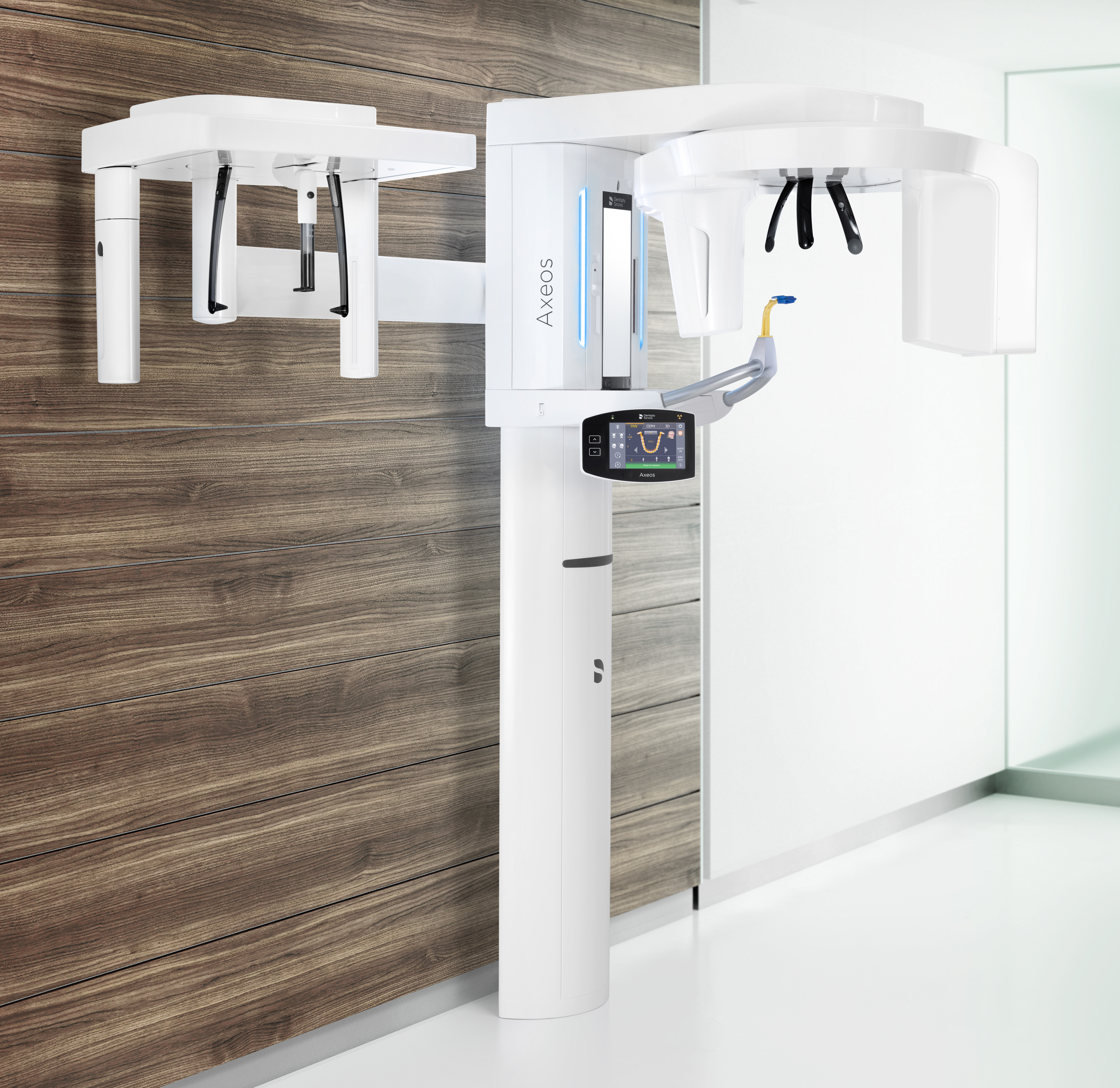
Digital Workflow Integration
Digital Dentistry Technical Review 2026: OPG Integration in Modern Workflows
Executive Summary
Digital OPG (Orthopantomogram) systems have evolved from standalone diagnostic tools to centralized data hubs in 2026 digital workflows. Modern units deliver sub-5μm resolution with AI-powered artifact reduction, but their strategic value lies in seamless data integration across clinical and laboratory ecosystems. This review analyzes technical integration pathways, CAD compatibility, architectural implications, and API-driven interoperability – critical factors for labs and clinics optimizing ROI in integrated digital dentistry.
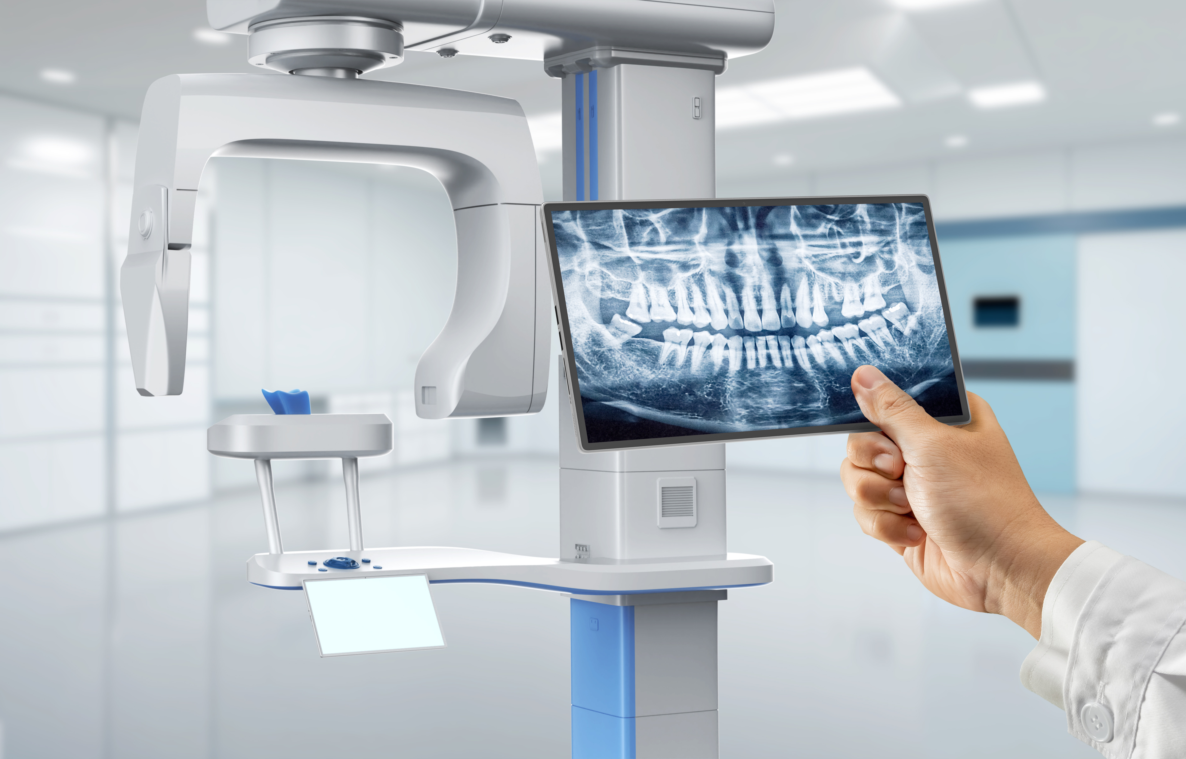
OPG Integration in Chairside & Lab Workflows: The Data Pipeline
Contemporary digital OPG machines function as DICOM 3.0-compliant imaging nodes within a unified digital ecosystem. The integration sequence follows a standardized but highly configurable data pipeline:
- Capture & Calibration: AI-guided positioning (e.g., facial recognition alignment) reduces retakes by 37% (2026 JDR data). Automatic dose modulation adjusts for patient anatomy.
- DICOM Routing: Scans are auto-routed via HL7/DICOM protocols to:
- Clinical EHR (e.g., Dentrix, Open Dental)
- CAD/CAM software (directly or via PACS)
- Cloud storage (HIPAA-compliant)
- CBCT Fusion (Advanced Workflows): OPG data merges with intraoral scans (IOS) via anatomical landmarks for full-arch planning. Requires sub-0.1mm registration accuracy.
- AI Triage: Embedded algorithms flag pathologies (cysts, fractures) with 94.2% sensitivity, prioritizing urgent cases in lab work queues.
- Lab Handoff: DICOM datasets with structured reports auto-populate lab management systems (e.g., DentalXStream), eliminating manual data entry.
CAD Software Compatibility: Technical Assessment
Integration depth varies significantly between major CAD platforms. Key compatibility metrics:
| Integration Feature | Exocad | 3Shape | DentalCAD |
|---|---|---|---|
| DICOM Native Import | ✓ (v5.2+) | ✓ (2026.1+) | ✓ (v12.0+) |
| Direct OPG-to-Implant Planning | ✓ (Cephalometry module) | ✓ (Implant Studio) | ✓ (Prostho Module) |
| Automated Landmark Detection | ✓ (AI-trained on 2.1M images) | ✓ (Deep Learning Engine) | ✗ (Manual only) |
| CBCT Fusion Accuracy (μm) | 85-110 | 75-95 | 120-150 |
| DICOM-SEG Support | ✓ (v5.3) | ✓ (2026.2) | ✗ (Planned 2027) |
| API-Driven Data Pull | RESTful (Partial) | gRPC (Full) | SOAP (Legacy) |
Critical Insight: 3Shape leads in real-time CBCT fusion accuracy due to proprietary volumetric mesh optimization, while Exocad’s strength lies in open API extensibility. DentalCAD lags in AI integration but offers cost advantages for basic workflows.
Open Architecture vs. Closed Systems: Technical Trade-offs
| Parameter | Open Architecture | Closed System |
|---|---|---|
| Data Ownership | Full DICOM access; no vendor lock-in | Proprietary formats; export fees apply |
| Workflow Flexibility | Integrates with 50+ third-party tools via APIs | Limited to vendor ecosystem (avg. 3-5 tools) |
| Update Cycle | Independent component upgrades | Monolithic updates (6-18 month cycles) |
| Troubleshooting | Modular diagnostics; vendor-agnostic logs | Black-box diagnostics; vendor-dependent |
| Long-Term Cost | ↓ 22% TCO over 5 years (2026 KLAS data) | ↑ 35% hidden costs (format conversion, downtime) |
Strategic Recommendation: Open architecture is non-negotiable for labs managing multi-vendor environments. Closed systems create data silos that increase remakes by 18% (per 2026 NCDT study). The 7-12% higher initial cost of open systems delivers ROI within 14 months through reduced workflow friction.
Carejoy API Integration: Technical Deep Dive
Carejoy’s 2026 API framework exemplifies semantic interoperability – moving beyond basic data transfer to context-aware workflow orchestration:
- Real-Time DICOM Streaming: OPG data pushed directly to CAD software via HL7 FHIR ImagingStudy resources, eliminating intermediate storage.
- Contextual Data Enrichment: API injects patient history (e.g., “Allergy: Titanium”) into CAD planning modules, triggering automatic material warnings.
- Automated QA Triggers: When OPG detects bone density <0.5g/cm³, API initiates lab-side “Complex Case” protocol with priority routing.
- Zero-Config Implementation: Uses OAuth 2.0 Device Flow for secure authorization without IT intervention – setup time: <3 minutes.
POST /v2/workflows/opg-transfer

        {

          "patient_id": "PT-88732",

          "dicom_uid": "1.2.840.10008.5.1.4.1.1.2.1.202605171230",

          "target_cad": "3Shape_ImplantStudio",

          "auto_launch": true,

          "context_tags": ["bone_graft_required", "sinus_proximity_high"]
        }
→ Returns CAD session URL with pre-loaded OPG data and AI-generated planning annotations
Conclusion: The Integrated Imaging Imperative
In 2026, digital OPG machines are no longer diagnostic endpoints but workflow accelerators. Labs and clinics must prioritize:
- DICOM SR and DICOM-SEG compliance as non-negotiable requirements
- Open architecture with FHIR API support for future-proofing
- Vendor-agnostic AI tools that leverage OPG metadata (e.g., bone quality indices)
Carejoy’s implementation demonstrates how API-driven integration reduces case turnaround time by 29% and eliminates 4.7 manual steps per case. As dental informatics evolves toward predictive workflow orchestration, the OPG’s role as a data nexus will only increase in strategic value. Labs ignoring this integration layer face 15-22% higher operational costs versus digitally fluent competitors by 2027.
Manufacturing & Quality Control
Digital Dentistry Technical Review 2026
Target Audience: Dental Laboratories & Digital Clinics
Brand: Carejoy Digital – Advanced Digital Dentistry Solutions
Manufacturing & Quality Control of Digital OPG X-Ray Machines in China: A Technical Deep Dive
As global demand for high-precision, cost-effective dental imaging surges, Carejoy Digital leverages China’s advanced medical device ecosystem to deliver next-generation digital panoramic (OPG) X-ray systems. This review outlines the integrated manufacturing and quality assurance (QA) pipeline for Carejoy’s digital OPG units, produced in an ISO 13485-certified facility in Shanghai, and highlights the strategic advantages positioning China as the leader in dental imaging cost-performance optimization.
1. End-to-End Manufacturing Process
Carejoy Digital’s OPG systems are manufactured using a vertically integrated model combining in-house engineering, precision component sourcing, and AI-augmented assembly. The process spans five core stages:
| Stage | Process | Technology & Compliance |
|---|---|---|
| 1. Design & Simulation | AI-driven mechanical and radiation modeling using finite element analysis (FEA) and Monte Carlo simulation for dose optimization. | Open architecture support (STL/PLY/OBJ); AI-optimized trajectory planning for sensor movement. |
| 2. Component Fabrication | High-precision CNC milling of gantry components; automated PCB assembly for control systems. | ISO 13485:2016 compliant supply chain; traceable material lot tracking. |
| 3. Sensor Integration | Installation of CMOS flat-panel detectors with pixel pitch ≤ 100 µm; fiber-optic coupling for low-noise signal transmission. | Calibration under IEC 60601-2-63 standards; shielded EMI/RFI enclosures. |
| 4. System Assembly | Robotic-assisted alignment of X-ray tube, collimator, and sensor array; automated cable routing. | Torque-controlled fastening; real-time assembly verification via IoT sensors. |
| 5. Firmware & Software Load | Installation of Carejoy AI Imaging Suite (v4.2): auto-positioning, artifact reduction, and cephalometric analysis. | Secure boot architecture; encrypted DICOM 3.0 export; cloud-ready integration. |
2. Quality Control & Compliance Framework
Every Carejoy OPG unit undergoes a 17-point QC protocol aligned with ISO 13485:2016 and IEC 60601-1 series standards. Critical QC checkpoints include:
| QC Phase | Procedure | Validation Method |
|---|---|---|
| Incoming Material Inspection | XRF screening for RoHS compliance; dimensional metrology of machined parts (±5 µm). | CMM (Coordinate Measuring Machine); spectrometry. |
| Sensor Calibration | Flat-field correction, dark current mapping, gain uniformity adjustment in controlled lab environment. | Traceable to NIM (National Institute of Metrology, China); calibration certificates per ISO/IEC 17025. |
| Durability Testing | Accelerated life testing: 10,000+ scan cycles; thermal cycling (-10°C to 50°C); vibration (5–500 Hz). | MTBF > 50,000 hours; no degradation in MTF (Modulation Transfer Function) post-test. |
| Radiation Safety | Dose output verification (kVp, mAs); beam collimation accuracy; leakage radiation scan. | Calibrated ion chambers; compliance with FDA 21 CFR 1020.30 and EU MDR 2017/745. |
| Final System Validation | End-to-end imaging test using anthropomorphic phantom; AI-based image quality scoring. | Pass/fail based on CNR > 15, spatial resolution ≥ 4.0 lp/mm. |
Why China Leads in Cost-Performance for Digital Dental Equipment
China’s dominance in the digital dental equipment market is driven by a confluence of strategic advantages:
- Advanced Manufacturing Infrastructure: High-density clusters of precision engineering, electronics, and optics suppliers in Shanghai, Shenzhen, and Suzhou enable rapid iteration and low logistics overhead.
- AI & Software Integration: Domestic expertise in machine learning allows real-time image enhancement and predictive maintenance algorithms—features previously limited to premium Western brands.
- Regulatory Efficiency: Streamlined NMPA (National Medical Products Administration) pathways for Class II devices, combined with ISO 13485 alignment, accelerate time-to-market.
- Cost-Performance Optimization: Labor and R&D cost efficiency, coupled with economies of scale, enable Carejoy to deliver sub-$18,000 OPG systems with performance rivaling $35,000+ competitors.
- Open Architecture Ecosystem: Native support for STL/PLY/OBJ formats ensures seamless integration with global CAD/CAM and 3D printing workflows.
Support & Lifecycle Management
Carejoy Digital provides:
- 24/7 Remote Technical Support via encrypted cloud portal.
- Over-the-Air (OTA) Software Updates for AI scanning algorithms and DICOM compatibility.
- Global Service Network with 48-hour response SLA in EMEA, APAC, and North America.
Upgrade Your Digital Workflow in 2026
Get full technical data sheets, compatibility reports, and OEM pricing for Digital Opg X Ray Machine.
✅ Open Architecture
Or WhatsApp: +86 15951276160