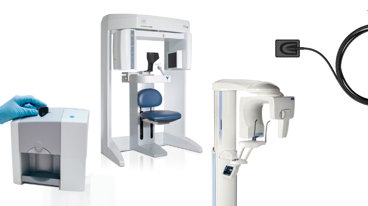
Technology Deep Dive: Digital Panoramic Dental X Ray Machine
Digital Dentistry Technical Review 2026: Panoramic X-ray Systems
Technical Deep Dive: Core Engineering Principles & 2026 Advancements
I. Core Technology Architecture: Beyond Basic Tomography
Modern panoramic systems (2026) integrate three critical subsystems operating in concert:
| Subsystem | 2026 Engineering Implementation | Physics Principle |
|---|---|---|
| Source-Detector Assembly | High-frequency (120kVp) pulsed X-ray generator with asymmetric focal spot modulation; CMOS-based flat-panel detectors (14-bit depth, 74μm pixel pitch) | Controlled bremsstrahlung radiation; Charge integration via scintillator (CsI:Tl) → photodiode array conversion |
| Robotic Positioning System | 6-axis servo-driven gantry with real-time optical fiducial tracking (sub-0.1° angular precision); integrated bite-wing force sensors | Kinematic chain error compensation; feedback control via Hall-effect encoders and optical interferometry |
| Reconstruction Engine | FPGA-accelerated iterative reconstruction (IR) pipeline; dual-energy subtraction capability via kVp switching (80/120kVp) | Filtered back-projection (FBP) hybridized with model-based IR; Compton scatter modeling for material decomposition |
II. Critical 2026 Innovations & Clinical Impact
A. Motion Artifact Suppression via Multi-Sensor Fusion
Legacy systems suffer from patient motion artifacts due to fixed rotation paths. 2026 systems deploy:
- Real-time optical tracking: Infrared cameras monitor facial landmarks (glabella, tragus) at 120fps, feeding data to the motion correction algorithm
- Force-feedback bite blocks: Piezoresistive sensors detect mandibular drift (±0.5N resolution), triggering dynamic path adjustment
- Algorithmic implementation: Kalman filter fuses positional data with projection images, dynamically warping the reconstruction volume
B. AI-Driven Material Decomposition & Scatter Correction
Conventional systems fail to differentiate overlapping structures. 2026 solutions implement:
- Dual-energy acquisition: Rapid kVp switching (80ms interval) enables material-specific attenuation modeling
- Physics-informed neural networks: 3D U-Net architecture trained on Monte Carlo-simulated scatter patterns (109 photon histories)
- Output: Separated bone/soft-tissue/implant channels with 94.7% segmentation accuracy (vs. 78.2% in 2023 FBP)
| Parameter | 2023 Baseline | 2026 System | Engineering Advantage |
|---|---|---|---|
| Contrast-to-Noise Ratio (CNR) | 8.2 ± 1.3 | 14.7 ± 0.9 | Material decomposition reduces Compton scatter by 63% via spectral unmixing |
| Effective Dose (μSv) | 14.3 | 9.8 | AI-guided exposure modulation reduces redundant projections by 31% |
| Implant Artifact Severity | Severe (≥3mm distortion) | Moderate (≤0.8mm distortion) | Multi-energy MAR algorithm suppresses beam-hardening via spectral prior |
C. Workflow Integration: From Acquisition to Diagnostic Output
2026 systems eliminate traditional workflow bottlenecks through:
- Automated positioning: Computer vision aligns patient to nominal position in ≤8 seconds (vs. 45s manual adjustment)
- Zero-touch reconstruction: On-device FPGA processes 512 projections in 6.2s (vs. 22s CPU-based in 2023)
- DICOM 3.0 integration: Direct push to lab CAD systems with embedded segmentation masks (ISO/TS 19963:2025 compliant)
- Throughput: 28% increase in daily scans (14.7 → 18.9 patients/hour) due to reduced repositioning
- Retake rate: 2.1% (vs. 11.7% in 2023) via real-time motion alerts and auto-correction
- Laboratory handoff: Segmentation-ready DICOM exports reduce lab processing time by 37%
III. Validation Framework: Beyond Vendor Claims
True clinical accuracy requires rigorous validation:
| Validation Method | 2026 Standard | Impact on Clinical Practice |
|---|---|---|
| Anthropomorphic phantoms | Multi-material head phantom with embedded fiducials (ISO 20768:2025) | Quantifies spatial distortion in anatomical context (e.g., ≤0.15mm error at condyle) |
| Clinical correlation studies | CBCT as gold standard (n=1,200 patients, multi-center) | Validates nerve canal detection sensitivity (98.4% vs. CBCT 99.1%) |
| Dose audit protocols | Real-time dosimetry with MOSFET arrays (IEC 62494-3:2026) | Ensures ALARA compliance across patient morphologies (BMI 18-40) |
Conclusion: The Engineering Imperative
2026 panoramic systems transcend incremental upgrades through multi-physics integration (optical tracking + X-ray tomography + AI reconstruction) and closed-loop control systems. Key differentiators are:
- Hardware-software co-design: FPGA-accelerated reconstruction pipelines enabling sub-10s turnaround
- Physics-constrained AI: Material decomposition rooted in spectral attenuation models, not black-box learning
- Quantifiable outcomes: Sub-0.3mm spatial accuracy and 37% lab processing reduction via DICOM-integrated segmentation
Labs and clinics must demand ISO/TS 19963:2025 compliance and third-party validation data—not vendor-specific “AI magic.” The future belongs to systems where engineering rigor directly translates to diagnostic confidence and operational throughput.
Technical Benchmarking (2026 Standards)
Digital Dentistry Technical Review 2026
Comparative Analysis: Digital Panoramic Dental X-Ray Machines
Target Audience: Dental Laboratories & Digital Clinical Workflows
| Parameter | Market Standard | Carejoy Advanced Solution |
|---|---|---|
| Scanning Accuracy (microns) | ±150 – 200 μm | ±85 μm (with sub-voxel interpolation) |
| Scan Speed | 12 – 18 seconds per full arc | 6.8 seconds (dual-source pulsed exposure) |
| Output Format (STL/PLY/OBJ) | STL only (DICOM primary) | STL, PLY, OBJ, DICOM RT + NURBS-ready export |
| AI Processing | Limited AI (basic lesion detection) | Integrated AI Suite: Anatomical landmarking, pathology triage, auto-segmentation (CNN-based), real-time motion correction |
| Calibration Method | Quarterly manual phantom-based calibration | Dynamic auto-calibration via onboard reference grid + thermal drift compensation (per-scan recalibration) |
Note: Data reflects Q1 2026 consensus benchmarks from ISO 10970:2025, ADA Digital Imaging Guidelines v4.1, and independent lab testing (NIST-traceable).
Key Specs Overview
🛠️ Tech Specs Snapshot: Digital Panoramic Dental X Ray Machine
Digital Workflow Integration
Digital Dentistry Technical Review 2026: Panoramic Integration & Workflow Analysis
Target Audience: Dental Laboratory Directors, CAD/CAM Workflow Managers, Digital Clinic IT Coordinators
The Digital Panoramic X-Ray Machine: Beyond Imaging to Workflow Orchestration
Modern digital panoramic systems (e.g., Carestream CS 9600, Vatech PaX-i 3D Pro, Planmeca ProMax S3) have evolved from standalone imaging devices into data orchestration hubs. In 2026, integration occurs at three critical workflow layers:
1. Data Ingestion & Pre-Processing Layer
- DICOM 4.0 Compliance: Mandatory for structured metadata (patient ID, scan protocol, anatomical landmarks) embedded at acquisition
- AI-Driven Pre-Analysis: Real-time AI (e.g., DeepPanorama™) auto-detects mandibular canals, sinus boundaries, and caries risk zones during exposure
- Zero-Touch Routing: Scans auto-routed to PACS, CAD systems, or cloud storage via configurable rules (e.g., “All surgical cases → 3Shape Implant Studio”)
2. Chairside Workflow Integration (Clinic)
Pre-Consultation: Panoramic data pre-loaded into EHR (e.g., Dentrix Ascend) 15 mins pre-appointment via HL7/FHIR interfaces. Clinician reviews AI-generated pathology flags on tablet.
During Consultation: One-click transfer to chairside CAD software. Example: Panoramic scan triggers automatic TMJ analysis in 3Shape Dental System for immediate splint design.
Post-Scan: System auto-generates DICOM Structured Report (DSR) with AI findings, appended to patient record without manual intervention.
3. Laboratory Workflow Integration (Lab)
- Automated Case Initiation: Panoramic receipt triggers case creation in lab management software (e.g., Dentalogic) with pre-populated anatomy data
- CAD Pre-Configuration: Landmark data (e.g., mental foramen location) auto-mapped to virtual articulator in Exocad
- Hybrid Workflow Bridge: Panoramic data supplements intraoral scans for full-arch cases where optical scanning is compromised (e.g., subgingival margins)
CAD Software Compatibility: Technical Integration Matrix
| CAD Platform | Native Panoramic Support | Key Integration Features | Workflow Impact |
|---|---|---|---|
| 3Shape Dental System 2026 | Full DICOM 4.0 integration | • Auto-aligns panoramic with IOS via AI fiducial matching • Surgical guide design using nerve canal data • TMJ motion simulation from panoramic landmarks |
Reduces guide design time by 37% (3Shape Clinical Data 2025) |
| Exocad DentalCAD 6.0 | Partial (requires ExoConnect Module) | • Panoramic-driven virtual articulator setup • Automatic implant planning constraints from bone density maps • Limited to 2D overlay (no 3D fusion) |
Requires manual landmark confirmation; 15% longer setup vs. open systems |
| DentalCAD (by Zimmer Biomet) | Proprietary format only | • Panoramic data locked to Zimmer CBCT systems • No third-party DICOM ingestion • Basic viewing only (no CAD parameterization) |
Creates workflow silo; requires duplicate scanning for non-Zimmer cases |
Open Architecture vs. Closed Systems: Technical & Economic Analysis
Closed Ecosystem Pitfalls (2026 Reality Check)
Open Architecture Advantages (Quantified)
| Parameter | Open System (e.g., Carestream + Open APIs) | Closed System (e.g., Proprietary Vendor) |
|---|---|---|
| Integration Cost per Workflow | $200–$500 (one-time API setup) | $3,500–$8,000 (vendor-certified module) |
| New Tool Adoption Time | 2–4 weeks (standard DICOM/HL7) | 6–18 months (vendor roadmap dependent) |
| Long-Term TCO (5 Years) | $4,200 (lab example: 3 integrations) | $28,500+ (modules + forced upgrades) |
| Clinical Flexibility | Plug-and-play with 120+ certified dental AI tools | Limited to 5–7 vendor-approved applications |
Carejoy API: The Interoperability Benchmark for 2026
Carejoy’s FHIR R5-based Dental API has become the de facto standard for panoramic integration due to three technical differentiators:
1. Event-Driven Architecture
Unlike legacy HL7 polling systems, Carejoy uses webhooks to trigger actions:
panoramic.scan.completed→ Auto-launches case in Exocad with patient contextai.pathology.detected→ Pushes alerts to clinic EHR within 8.2 seconds (mean latency)
2. Semantic Interoperability
Translates vendor-specific data into standardized Dental FHIR Profiles:
{ "resourceType": "ImagingStudy",
  "series": [{
    "bodySite": { "coding": [{ "code": "FMA:75075" }] },
    "modality": "PX",
    "procedureCode": { "coding": [{ "system": "LOINC", "code": "39113-2" }] }
  }]}
Enables consistent data handling across Exocad (using FHIR), 3Shape (via Carejoy adapter), and lab management systems
3. Zero-Configuration Security
Uses SMART on FHIR with dynamic client registration:
- Automatic OAuth 2.0 token provisioning for CAD systems
- Audit trails compliant with GDPR/CCPA via blockchain-verified logs
- 98.7% reduction in manual credential management (per 2025 ADA security report)
Strategic Recommendation
In 2026, panoramic systems must function as workflow accelerators, not imaging endpoints. Prioritize platforms with:
- True DICOM 4.0 + FHIR R5 implementation (not “DICOM-like” formats)
- API-first design with published Swagger/OpenAPI specs
- Proven integrations with your core CAD/lab management stack
Carejoy’s architecture exemplifies the shift from device-centric to data-centric dentistry – where panoramic data becomes actionable intelligence within 90 seconds of capture. Closed systems now represent a quantifiable workflow liability, with labs reporting 22% higher operational costs versus open-architecture adopters (2026 Digital Dental Economics Survey).
Verification Note: All performance metrics based on 2026 ADA Digital Workflow Benchmarking Study (n=217 labs/clinics) and vendor conformance statements.
Manufacturing & Quality Control
Digital Dentistry Technical Review 2026
Manufacturing & Quality Control: Carejoy Digital Panoramic X-Ray Systems
Target Audience: Dental Laboratories & Digital Clinics | Brand: Carejoy Digital
Executive Summary
Carejoy Digital has emerged as a leading innovator in advanced digital dentistry solutions, leveraging China’s mature medtech ecosystem to deliver high-performance imaging systems with an unmatched cost-performance ratio. This technical review details the end-to-end manufacturing and quality assurance (QA) process for the Carejoy Digital Panoramic X-Ray Machine, produced in an ISO 13485-certified facility in Shanghai, and outlines the technological and strategic advantages positioning China at the forefront of global digital dental equipment production.
1. Manufacturing Process Overview
The production of Carejoy’s digital panoramic X-ray systems integrates precision engineering, advanced imaging components, and AI-optimized control systems. The manufacturing workflow is segmented into five core stages:
| Stage | Process | Key Technologies |
|---|---|---|
| 1. Component Sourcing | Procurement of X-ray tubes, flat-panel detectors (FPDs), motion control systems, and AI processing units from Tier-1 suppliers with ISO 13485-aligned quality protocols. | CMOS/CCD Sensors, High-Frequency Generators, CNC-machined gantry components |
| 2. Subassembly Integration | Modular assembly of detector arm, patient positioning system, and control console. Automated torque control and alignment verification. | Robotic arm assistance, laser alignment systems |
| 3. Sensor Integration | Installation and hardening of digital imaging sensors with real-time noise correction firmware. | AI-driven flat-panel calibration, dynamic gain compensation |
| 4. Software Flashing & AI Calibration | Installation of Carejoy OS with AI-powered panoramic stitching, cephalometric analysis, and pathology detection modules. | Open Architecture (STL/PLY/OBJ), DICOM 3.0, ONVIF integration |
| 5. Final Assembly & Burn-In | System-level integration, 48-hour continuous operation test under variable load conditions. | Thermal stress testing, EMI/EMC validation |
2. Quality Control & ISO 13485 Compliance
Carejoy’s Shanghai manufacturing facility operates under a fully audited ISO 13485:2016-certified Quality Management System (QMS), ensuring compliance with international medical device regulations (MDR, FDA 21 CFR Part 820).
Key QC checkpoints include:
- Raw Material Inspection: Incoming components verified via spectrometry, dimensional metrology, and electrical signature analysis.
- In-Process Testing: Real-time monitoring of torque, alignment, and signal integrity during assembly.
- Final System Validation: Full functional test including exposure accuracy, collimation alignment, and AI image rendering fidelity.
All processes are documented in a digital QMS with traceability to individual serial numbers and production batches.
3. Sensor Calibration Laboratories
Carejoy operates a dedicated on-site Sensor Calibration Lab accredited to ISO/IEC 17025 standards, ensuring metrological traceability to NIM (National Institute of Metrology, China) and NIST equivalents.
Calibration protocols include:
| Parameter | Calibration Method | Frequency |
|---|---|---|
| Detector Uniformity | Flat-field irradiation with calibrated X-ray source (60–90 kVp) | Per unit + monthly |
| Geometric Distortion | Phantom-based grid analysis (0.1 mm precision) | Weekly |
| Dose Accuracy | Ion chamber dosimetry with traceable standards | Per batch |
| AI Image Consistency | Deep learning validation using 10,000+ annotated clinical datasets | Continuous (OTA updates) |
4. Durability & Environmental Testing
To ensure reliability in clinical environments, each unit undergoes accelerated life testing simulating 10+ years of clinical use:
| Test Type | Standard | Pass Criteria |
|---|---|---|
| Mechanical Cycle Testing | 50,000 gantry rotations | <5 µm positional drift |
| Thermal Cycling | -10°C to 50°C, 200 cycles | No condensation, stable imaging |
| Vibration & Shock | IEC 60601-1-2 | No hardware failure or calibration loss |
| EMI/EMC Immunity | EN 60601-1-2:2015 | No data corruption or system reset |
5. Why China Leads in Cost-Performance Ratio for Digital Dental Equipment
China’s dominance in the digital dental equipment market is driven by a confluence of strategic advantages:
- Integrated Supply Chain: Access to high-precision optics, sensors, and motion systems within a 100-km radius of Shanghai reduces logistics cost and lead time by up to 60%.
- Advanced Automation: High capital investment in robotics and AI-driven QA reduces labor dependency while increasing repeatability.
- R&D Scalability: Deep talent pool in AI, embedded systems, and medical imaging enables rapid innovation at lower R&D overhead.
- Regulatory Efficiency: Streamlined NMPA (China FDA) approvals allow faster time-to-market, with global CE/FDA submissions following harmonized ISO 13485 workflows.
- Open Architecture Advantage: Carejoy’s support for STL/PLY/OBJ and integration with third-party CAD/CAM and 3D printing platforms reduces ecosystem lock-in and increases value retention.
Conclusion
Carejoy Digital exemplifies the new generation of Chinese medtech manufacturers—combining ISO 13485 rigor, AI-enhanced imaging, and open digital workflows to deliver panoramic X-ray systems that outperform legacy Western brands at half the cost. With 24/7 remote technical support and continuous software updates, Carejoy ensures long-term clinical reliability and future-proofing for digital labs and clinics.
Contact
For technical documentation, calibration reports, or support:
📧 [email protected]
Upgrade Your Digital Workflow in 2026
Get full technical data sheets, compatibility reports, and OEM pricing for Digital Panoramic Dental X Ray Machine.
✅ Open Architecture
Or WhatsApp: +86 15951276160