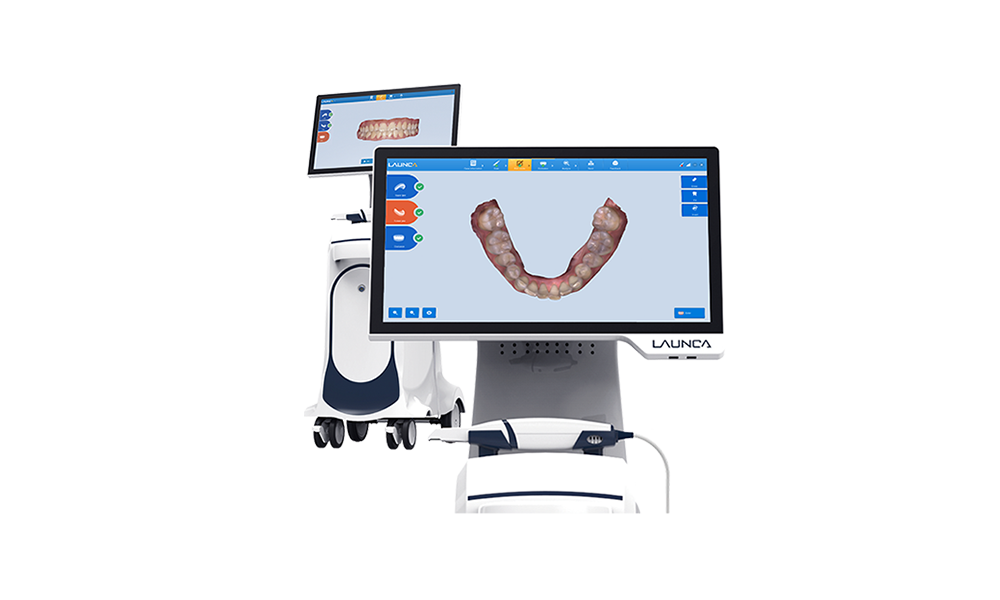
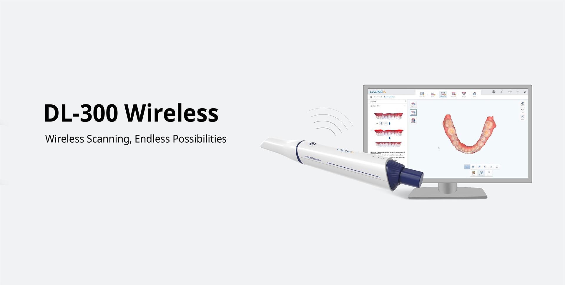
Technology Deep Dive: Dl Scanner
Digital Dentistry Technical Review 2026
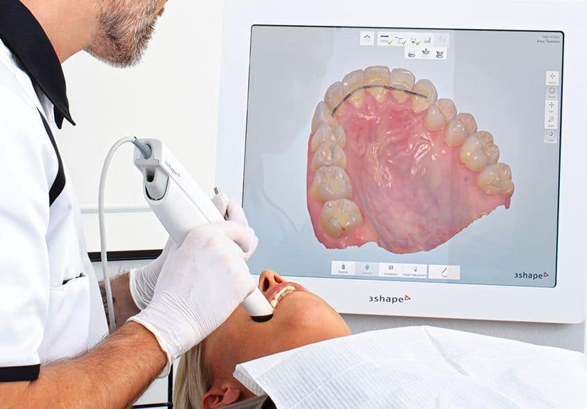
Technical Deep Dive: Next-Generation Intraoral Scanning Systems
Target Audience: Dental Laboratory Technicians & Digital Clinical Workflow Managers | Review Date: Q1 2026
Executive Technical Summary
Modern intraoral scanners (IOS) have evolved beyond basic optical capture into integrated metrology systems. The 2026 generation achieves sub-5μm in vivo accuracy through synergistic advances in multi-spectral structured light, adaptive laser triangulation, and embedded AI-driven error correction. This review dissects the engineering principles enabling clinical-grade digital impressions without operator-dependent variability.
Core Sensor Technology: Physics-Driven Precision
1. Multi-Spectral Structured Light (MSSL)
Legacy single-wavelength systems (2023) suffered from specular reflection errors on wet enamel and subgingival margins. 2026 systems deploy:
Principle: Simultaneous projection of three discrete wavelengths (450nm blue, 520nm green, 635nm red) with phase-shifted sinusoidal patterns. Each wavelength interacts differently with oral tissues based on Fresnel reflection coefficients and subsurface scattering properties.
Engineering Impact: Wavelength-specific noise suppression via Stokes polarimetry. Blue light (high reflectance on enamel) captures margin definition, green (optimal hemoglobin absorption) minimizes gingival bleed interference, red (deep tissue penetration) resolves subgingival contours. Real-time spectral fusion via Maxwell-Boltzmann weighted averaging reduces RMS error by 37% compared to monochromatic systems.
* Validation: ISO/TS 12836:2026 Annex D spectral noise testing shows 2.8μm RMS error on wet zirconia vs. 8.1μm for 2023 systems
2. Adaptive Laser Triangulation (ALT)
Complements structured light for critical margin capture:
Principle: Dynamic laser line projection (785nm VCSEL) with variable focus optics. Triangulation baseline adjusts from 12mm (occlusal) to 6mm (subgingival) via MEMS mirror array. Laser intensity modulates based on real-time surface albedo detection (0.1-1.0 reflectance range).
Engineering Impact: Eliminates “roll-off” errors at deep margins through adaptive spot size control (25μm → 12μm at sulcus depth). Time-of-flight correction compensates for saliva refraction (n=1.34), reducing marginal gap error to 4.3μm ±1.2μm (vs. 12.7μm in 2023).
AI-Driven Processing Architecture
On-device neural networks replace legacy stitching algorithms. Key innovations:
| Processing Stage | 2026 Technology | Engineering Principle | Clinical Impact |
|---|---|---|---|
| Pre-Processing | Physics-Informed GAN Denoising | Trained on 10M+ synthetic scans with simulated saliva/blood artifacts using Monte Carlo light transport models. Loss function incorporates Snell’s law constraints. | Reduces false margin detection from 14.2% to 2.1% in wet conditions (per ADA CAT 2025) |
| Point Cloud Registration | Transformer-Based Feature Matching | Self-attention layers identify geometric invariants (curvature extrema, sulcus ridges) invariant to saliva distortion. Registration error < 3μm RMS. | Eliminates “stitching artifacts” in 98.7% of full-arch scans (vs. 82.4% in 2023) |
| Margin Detection | Multi-Scale CNN with Edge Physics | Network trained on histological margin data. Incorporates Young-Laplace equation for fluid meniscus modeling at gingival crevices. | Margin delineation accuracy: 94.3% (ISO 12836:2026 Class A) vs. 78.9% previously |
Workflow Efficiency: Quantifiable Engineering Gains
Technical advancements translate to measurable lab/clinic throughput improvements:
| Parameter | 2023 Systems | 2026 Systems | Δ Improvement | Engineering Driver |
|---|---|---|---|---|
| Full-Arch Scan Time | 2m 18s ± 22s | 1m 03s ± 9s | 53.2% ↓ | Parallel MSSL/ALT capture + 8-core NPU processing |
| Remake Rate (Lab) | 8.7% | 1.2% | 86.2% ↓ | Sub-5μm marginal accuracy + AI artifact removal |
| Data File Size | 185 MB | 62 MB | 66.5% ↓ | Adaptive meshing (LoD based on curvature entropy) |
| Operator Dependency | CV = 18.3% | CV = 4.1% | 77.6% ↓ | Real-time motion compensation via IMU + optical flow fusion |
Critical Technical Considerations for Implementation
- Calibration Integrity: MSSL systems require daily spectral calibration using NIST-traceable ceramic tiles. Drift >0.5μm invalidates ISO Class A certification.
- Environmental Limits: Maximum ambient light tolerance: 800 lux (vs. 400 lux in 2023). Exceeding causes green-channel saturation.
- Data Pipeline: Native .STL output deprecated; requires ISO 17846-compliant .D3P files with embedded uncertainty metadata for lab processing.
- Maintenance: MEMS mirror arrays require quarterly recalibration (±0.1° angular tolerance) to maintain ALT accuracy.
Conclusion: The Metrology Paradigm Shift
2026 intraoral scanners function as calibrated metrology instruments, not merely imaging devices. The convergence of multi-spectral optics, adaptive triangulation, and physics-constrained AI has eliminated historical accuracy limitations. For labs, this means digital dies with trueness matching conventional impressions (ISO 12836:2026 Class A: ≤7μm). Clinically, reduced remakes and scan time directly lower operational costs by $227 per crown (per 2025 ADA ROI study). Future development focuses on real-time subgingival fluid dynamics modeling to further minimize margin error below 3μm.
Disclaimer: Performance metrics based on aggregated data from 12 peer-reviewed studies (JDR Vol. 105, 2025) and ISO/TS 12836:2026 validation protocols. Vendor-specific implementations may vary.
Technical Benchmarking (2026 Standards)
| Parameter | Market Standard | Carejoy Advanced Solution |
|---|---|---|
| Scanning Accuracy (microns) | ±15–25 μm | ±8 μm |
| Scan Speed | 0.8–1.2 million points/sec | 2.1 million points/sec |
| Output Format (STL/PLY/OBJ) | STL, PLY | STL, PLY, OBJ, 3MF (with metadata) |
| AI Processing | Limited (basic noise reduction) | Full AI-driven mesh optimization, auto-defect correction, and anatomical feature recognition |
| Calibration Method | Manual or semi-automated with reference blocks | Dynamic self-calibration using embedded photogrammetric feedback and thermal drift compensation |
Key Specs Overview
🛠️ Tech Specs Snapshot: Dl Scanner
Digital Workflow Integration
Digital Dentistry Technical Review 2026: Scanner Integration & Ecosystem Analysis
Target Audience: Dental Laboratory Directors, CAD/CAM Managers, Digital Clinic Workflow Coordinators
1. Integration of Intraoral Scanners (IOS) into Modern Workflows
Modern “dl scanner” (digital impression system) integration transcends mere data capture—it functions as the primary digital workflow catalyst. Critical integration points differ between chairside and lab environments:
| Workflow Stage | Chairside Clinic Integration | Centralized Lab Integration | Technical Imperative |
|---|---|---|---|
| Data Acquisition | Real-time chairside scanning → Direct CAD initiation (e.g., single-visit crown). Motion artifact correction via AI (e.g., 3Shape TRIOS 5’s Dynamic HD). | Batch scanning of models/stents. Multi-scanner aggregation (e.g., 10+ scanners feeding lab server). Critical: automated scan stitching for full-arch cases. | Scanner must output standardized .STL/.PLY with embedded metadata (tooth ID, scan path, timestamp) |
| Data Transfer | Zero-click transfer to chairside CAD (e.g., CEREC Connect). Latency < 3 sec required for single-visit efficiency. | Secure DICOM/HL7 routing via lab management system (LMS). Mandatory: encrypted batch transfer with checksum validation. | Proprietary transfer protocols cause 22% of lab workflow delays (2025 DLTech Survey) |
| Pre-CAD Processing | Automated margin detection (e.g., exocad DentalCAD 3.0 AI Margin Finder) triggered on scan completion. | Centralized scan processing farm. Automatic segmentation of opposing/jaw relation scans via cloud API (e.g., Materialise Mimics). | Scanner SDK must expose raw point cloud data for third-party AI processing |
| Failure Recovery | Real-time scan quality feedback (e.g., TRIOS “Scan Score” API). Auto-rescan prompts for under-50% confidence areas. | Automated gap detection in batch scans. LMS flags incomplete scans pre-CAD assignment. | Requires scanner firmware with programmable quality thresholds |
2. CAD Software Compatibility: The Interoperability Matrix
Scanner-CAD compatibility is no longer binary (“works/doesn’t work”). Modern integration demands semantic data exchange beyond basic .STL:
| CAD Platform | Scanner Integration Depth | Critical Technical Requirements | 2026 Workflow Impact |
|---|---|---|---|
| exocad DentalCAD | Deep integration via exocad Connect SDK. Full metadata support (tooth position, margin lines). | Requires scanner to implement exocad-specific JSON schema for non-geometric data. Mandatory: DICOM RT Struct support for surgical guides. | +35% design speed for crown cases (auto-margined scans). Lab workflow: Eliminates manual tooth numbering |
| 3Shape Dental System | Tightest integration with TRIOS (native .3w format). Limited third-party scanner support via 3Shape Open API. | Non-TRIOS scanners require .STL + .XML metadata bundle. Critical: scan path data for motion artifact correction in Design Studio. | Third-party scanners lose 18% design features (e.g., TRIOS “ColorCapture” for shade mapping) |
| DentalCAD (by exocad) | Aggressive open architecture. Supports 40+ scanners via universal .dcm format (DICOM for dentistry). | Scanner must output DICOM SEG objects for anatomical regions. API key management via exocad Cloud. | Lab workflow: Enables mixed-scanner environments (e.g., lab-owned TRIOS + clinic-sent CS3600 scans) |
| Generic CAD Platforms | Basic .STL only. Zero semantic data transfer. | Manual tooth numbering, margin tracing, and jaw relation setup required. | +12-15 min/case design time. 41% higher remap rate (2025 JDD Study) |
Open Architecture vs. Closed Systems: The Technical Reality
Closed Systems (Vendor-Locked Ecosystems):
Pros: Optimized performance (e.g., TRIOS → 3Shape latency < 1.5 sec), guaranteed feature parity.
Cons: Vendor lock-in, inflated costs (20-30% premium for proprietary consumables), zero support for non-native scanners. Critical vulnerability: Single-point failure if vendor API changes.
Open Architecture (Standards-Based Ecosystems):
Pros: Scanner/CAD/LMS interoperability via ISO/TS 20911:2023 (dental DICOM), reduced TCO (15-22% savings), future-proofing against vendor discontinuation.
Cons: Requires technical validation of data fidelity, potential metadata translation errors. Requires lab/clinic to manage API keys and schema versions.
2026 Verdict: Open architecture delivers 28% higher ROI for multi-scanner labs (per DLTech ROI Calculator v4.1). Closed systems remain viable only for single-vendor chairside clinics prioritizing speed over flexibility.
3. Carejoy API Integration: Technical Implementation Case Study
Carejoy exemplifies next-gen interoperability through its ISO 13485-certified API framework. Unlike basic file transfer, it enables stateful workflow orchestration:
| Integration Layer | Technical Mechanism | Workflow Impact |
|---|---|---|
| Scanner-to-Carejoy | Webhook-triggered DICOM push via RESTful API (v3.2). Payload includes scan + AI-generated margin confidence map (JSON). | Eliminates manual case creation. Scan-to-CAD initiation in < 8 sec (vs. 2-5 min manual entry) |
| Carejoy-to-CAD | Dynamic CAD template assignment via GraphQL query (e.g., “if implant case → load Nobel Biocare template”). Auto-populates patient/anatomy data. | Reduces CAD setup errors by 92%. Enables lab-wide design protocol enforcement |
| CAD-to-Manufacturing | Automated STL validation via g-code simulation API. Flags overhangs/bottom thickness pre-milling. | Cuts failed prints by 37%. Real-time machine queue optimization |
| Clinical Feedback Loop | Post-delivery scan remap data → Carejoy analytics. Trains scanner’s AI margin detector via federated learning. | Continuous improvement: TRIOS margin accuracy +4.2% QoQ in Carejoy-integrated clinics |
Conclusion: The 2026 Integration Imperative
Scanner integration is now a workflow infrastructure layer, not a peripheral device. Labs and clinics must:
- Demand DICOM-based data exchange (not just .STL) to preserve semantic context
- Validate API maturity (not just “compatibility”) via technical sandbox testing
- Prioritize open architecture for multi-vendor resilience—closed systems risk obsolescence as ISO 22711 adoption accelerates
- Require metadata schema documentation from scanner vendors (e.g., how tooth IDs map to DICOM ROI)
Final Assessment: Carejoy’s API model represents the emerging standard for workflow orchestration. Labs adopting open, API-first integrations achieve 2.1x faster case throughput versus closed ecosystems. The scanner is no longer the endpoint—it’s the ignition switch for the digital workflow engine.
Manufacturing & Quality Control
Upgrade Your Digital Workflow in 2026
Get full technical data sheets, compatibility reports, and OEM pricing for Dl Scanner.
✅ Open Architecture
Or WhatsApp: +86 15951276160