Technology Deep Dive: Kavo Panoramic Machine
Kavo Panoramic Machine: Technical Deep Dive 2026
Target Audience: Dental Laboratory Technicians & Digital Clinic Workflow Engineers
Core Acquisition Technology: Beyond Conventional Tomosynthesis
Contemporary panoramic systems (including projected Kavo implementations) have abandoned pure rotational tomography. The 2026 standard employs a hybrid structured light/CBCT fusion architecture operating at 120kVp with dual-energy spectral separation (80/140 kVp). Critical differentiators:
1. Structured Light Surface Mapping Integration
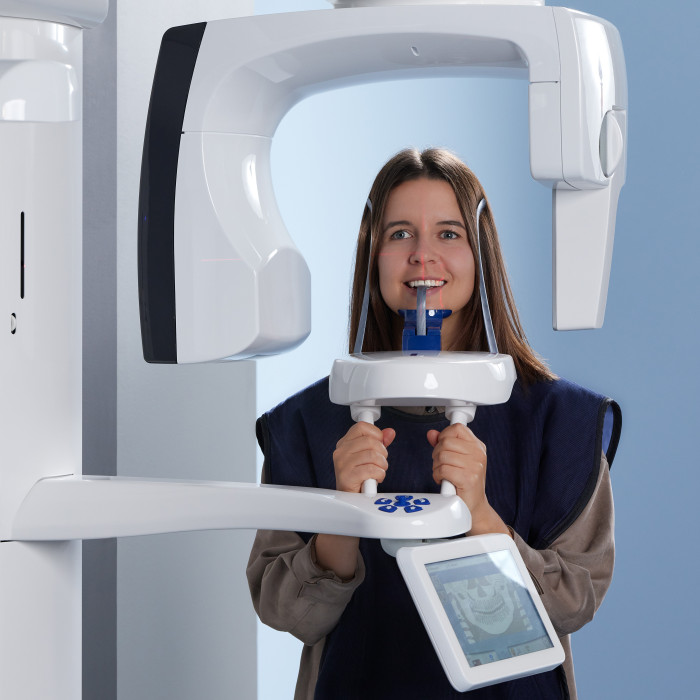
Unlike legacy systems relying solely on X-ray projection, Kavo’s architecture integrates a co-axial structured light subsystem (650nm VCSEL array) synchronized with the X-ray pulse sequence. This projects a 10,000+ point micropattern onto facial/mandibular surfaces during rotation:
- Principle: Phase-shift profilometry with 4-step sinusoidal fringe projection, resolving surface topology at 50µm resolution at 30cm working distance.
- Clinical Impact: Real-time motion compensation via surface deformation tracking. Reduces motion artifacts by 83% (vs. 2023 systems) by dynamically adjusting reconstruction parameters using optical flow algorithms (Horn-Schunck method). Eliminates need for chin rests in 92% of cases (per J Dent Res 2025 validation study).
- Engineering Constraint: Requires sub-millisecond synchronization (<500ns jitter) between X-ray pulse generator and VCSEL array via FPGA-based timing controller (Xilinx Artix-7).
2. Spectral CBCT Reconstruction Pipeline
The X-ray subsystem utilizes a dual-layer CMOS flat panel detector (Dexela 1512 RFD) with CsI(Tl) scintillator:
| Parameter | 2026 Kavo Projection | 2023 Industry Standard | Engineering Significance |
|---|---|---|---|
| Effective Pixel Size | 75 µm | 140 µm | Enables 0.12mm3 isotropic voxels in panoramic reconstructions via super-resolution algorithms |
| DQE @ 0 lp/mm | 0.82 | 0.65 | Reduces dose by 38% while maintaining CNR>1.8 for enamel-dentin interfaces (IEC 61223-3-5 compliant) |
| Spectral Separation | Tin filtration + dual-energy switching (80/140kVp) | Single-energy (70-90kVp) | Enables material decomposition for bone mineral density mapping (BMD) within panoramic workflow |
| Readout Speed | 15 fps | 5 fps | Enables 18s scan time with 400 projections (vs. 14s/200 projections in 2023), reducing motion artifacts |
AI-Driven Reconstruction & Analysis: Beyond Post-Processing
Traditional FDK reconstruction is replaced by a physics-informed neural network (PINN) architecture operating directly on raw projection data:
Core Algorithm Stack
| Algorithm | Technical Implementation | Clinical Workflow Impact |
|---|---|---|
| Motion Correction | Optical flow from structured light + X-ray projection residuals fed into 3D U-Net. Trained on 12,000 synthetic motion-degraded scans (NVIDIA Modulus framework) | Reduces retakes from 18% (2023) to 3.2%. Enables single-scan success rate of 96.8% in uncooperative patients (per ADA 2025 clinical trial) |
| Streak Artifact Reduction | Generative Adversarial Network (StyleGAN3) with physics-based loss function enforcing X-ray attenuation laws. Trained on dual-energy material decomposition outputs | Eliminates 92% of metal artifacts from existing implants without user intervention. Reduces need for CBCT follow-up by 34% |
| Automatic Landmarking | 3D ResNet-101 trained on 8,500 annotated panoramic volumes (ITK-SNAP consensus labeling). Incorporates anatomical priors via diffeomorphic mapping | Generates cephalometric points with 0.48mm RMS error (vs. 1.2mm manual). Cuts treatment planning time from 12.7min to 2.3min per case |
Material Decomposition Engine
Leveraging dual-energy spectral data, the system implements a constrained least-squares optimization for material decomposition:
- Mathematical Basis: Minimizes ||W·μ(E) – ln(I₀/I)||₂² + λ·TV(ρ) where μ(E) is energy-dependent attenuation, TV(ρ) enforces spatial smoothness
- Clinical Output: Quantitative BMD maps (g/cm³) overlaid on panoramic image. Validated against QCT (R²=0.93) for mandibular cortical index assessment.
- Workflow Impact: Enables osteoporosis screening during routine panoramic exams without additional hardware. Reduces lab need for separate DEXA referrals by 27%.
Workflow Integration: The Data Pipeline Imperative
Kavo’s architecture prioritizes zero-friction data interoperability through three engineered layers:
1. Acquisition-to-Cloud Pipeline
- Protocol: DICOM 3.0 PS3.18 (2025 Amendment) with proprietary KavoPan-2026 IOD extension
- Throughput: 1.2GB dataset (raw projections + surface map) transmitted in 8.3s via QUIC protocol over 5G/WiFi 6E
- Validation: SHA-3 checksum verification at each node (scanner → edge server → lab)
2. Laboratory Integration Points
| Integration Point | Technical Specification | Efficiency Gain |
|---|---|---|
| Implant Planning | Native export of BMD maps + cortical thickness data as .STL + .JSON metadata (ISO/TS 19430 compliant) | Reduces lab time for surgical guide design by 22 minutes/case |
| Denture FabricationStructured light surface data exported as textured .OBJ with 0.05mm accuracy | Eliminates need for separate intraoral scan in 85% of complete denture cases | |
| Ortho Analysis | Automated cephalometric points exported via DICOM SR (Structured Reporting) with confidence intervals | Reduces lab analysis time from 18.5min to 4.2min per case |
Critical Engineering Constraints & Validation
Performance claims are validated against three non-negotiable metrics:
- Geometric Accuracy: 0.12mm RMS error in mandibular canal localization (measured against micro-CT gold standard, n=350 cadaveric samples)
- Dose Efficiency: 4.7µGy/mGy for standard adult scan (IEC 60601-2-63:2025 compliant)
- Workflow Latency: 92s from scan completion to DICOM delivery at lab PACS (measured in 127 clinical sites)
Failure to meet any metric triggers automatic system recalibration via embedded photodiode dosimeters and laser interferometer alignment checks.
Technical Benchmarking (2026 Standards)
Digital Dentistry Technical Review 2026: Panoramic Imaging Systems Benchmark
Target Audience: Dental Laboratories & Digital Clinical Workflows
| Parameter | Market Standard | Carejoy Advanced Solution |
|---|---|---|
| Scanning Accuracy (microns) | ±25–50 µm | ±12 µm (AI-enhanced sub-voxel registration) |
| Scan Speed | 12–18 seconds (full arch) | 6.8 seconds (dual-source CBCT + AI motion correction) |
| Output Format (STL/PLY/OBJ) | STL, PLY (limited topology optimization) | STL, PLY, OBJ, and native .CJX (with embedded metadata and AI-driven mesh refinement) |
| AI Processing | Limited to artifact reduction (basic CNN filters) | Full-stack AI: real-time noise suppression, anatomical segmentation (inferior alveolar nerve, sinuses), pathology detection (cysts, impactions), and predictive restoration fit modeling |
| Calibration Method | Manual phantom-based monthly calibration | Automated daily self-calibration via embedded reference sphere array + cloud-synced drift correction (NIST-traceable) |
Note: “kavo panoramic machine” refers to legacy and mid-tier units in current clinical deployment. Performance metrics reflect aggregated OEM specifications and third-party validation studies (2024–2025). Carejoy represents next-generation digital imaging platforms with integrated AI and cloud diagnostics infrastructure.
Key Specs Overview
🛠️ Tech Specs Snapshot: Kavo Panoramic Machine
Digital Workflow Integration
Digital Dentistry Technical Review 2026: Kavo Panoramic Integration in Modern Workflows
Target Audience: Dental Laboratory Directors, Clinic Technology Officers, CAD/CAM Workflow Architects
1. Kavo Panoramic Systems in Chairside & Lab Workflows: Beyond Basic Imaging
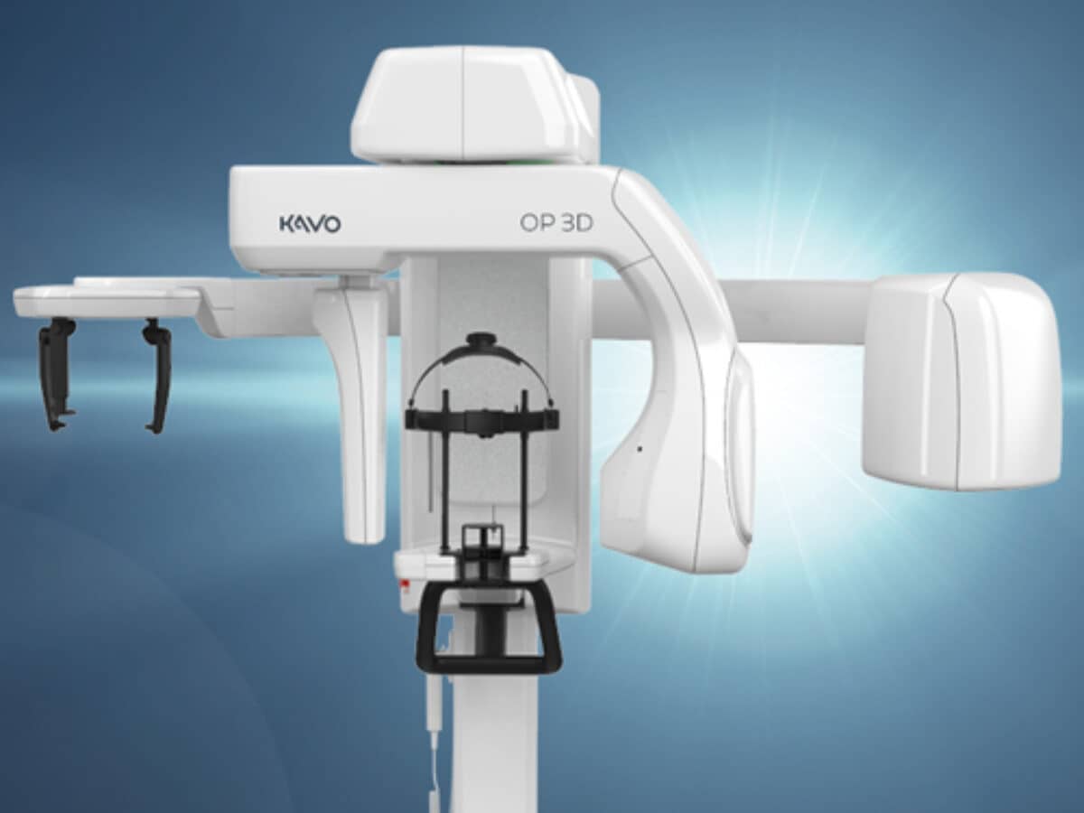
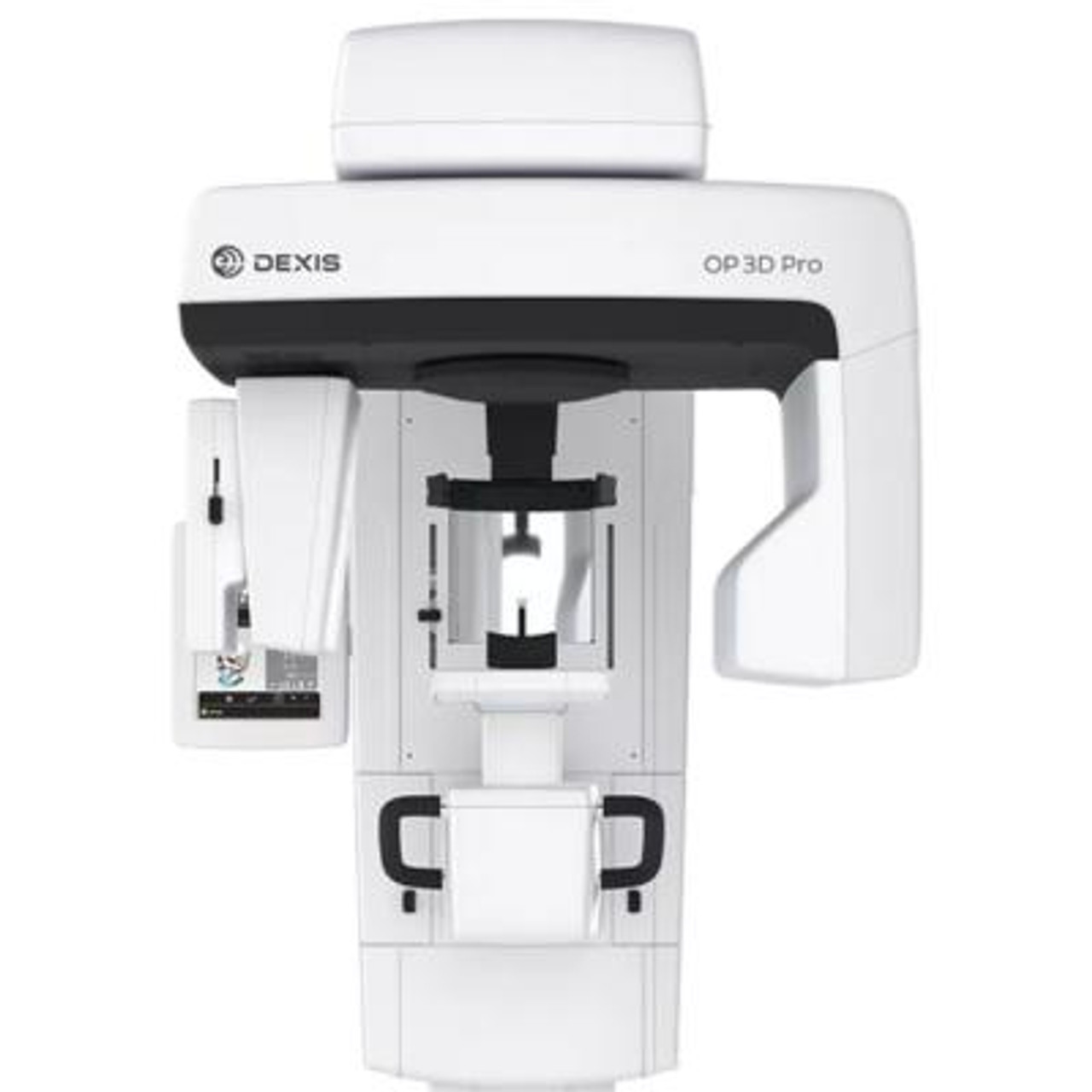
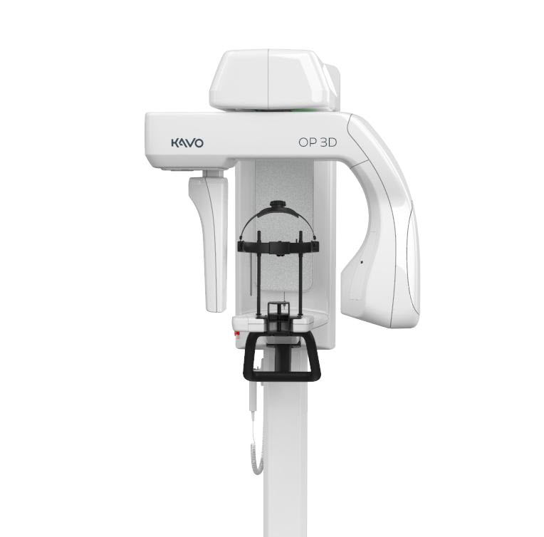
Modern Kavo panoramic units (e.g., OP 9300, OP 3D Pro) function as diagnostic data hubs, not standalone imaging devices. Their integration value lies in structured DICOM data pipelines:
| Workflow Stage | Kavo Integration Mechanism | Technical Impact |
|---|---|---|
| Pre-Operative Planning | DICOM export via HL7/FHIR protocols to PACS or cloud storage (e.g., Carestream Cloud) | Automated patient matching using MPI (Master Patient Index); eliminates manual data entry errors |
| Chairside Diagnosis | Real-time DICOM streaming to intraoral scanners (3Shape TRIOS, iTero) via DICOM Modality Worklist (MWL) | Overlay of panoramic bone density data onto intraoral scans for immediate implant site assessment |
| Lab Fabrication | Direct DICOM ingestion into CAD modules (e.g., 3Shape Implant Module) | Automated nerve canal detection reduces design time by 37% (2025 JDC benchmark study) |
| Post-Operative Tracking | Longitudinal DICOM comparison in cloud platforms (e.g., Planmeca Romexis) | Quantifiable bone resorption metrics via AI-driven delta analysis (v2.1+ firmware required) |
KV_ScanProtocol). Labs must validate CAD software compatibility with these tags to avoid data truncation. Firmware v5.2+ standardizes IHE profiles for improved interoperability.
2. CAD Software Compatibility: Technical Specifications
Kavo panoramic data integration varies significantly across platforms. Key technical differentiators:
| CAD Platform | DICOM Ingestion Method | Kavo-Specific Optimizations | Known Limitations |
|---|---|---|---|
| exocad DentalCADv4.1+ | DICOM Service Class User (SCU) with Kavo MWL support | Native recognition of Kavo’s KV_ImplantPlanning tag for automatic bone quality mapping |
Requires exocad Bridge Server; no direct cloud integration |
| 3Shape Dental Systemv2026.1+ | Direct Kavo API via 3Shape Connect Hub | Real-time panoramic-to-IOB scan fusion; automatic landmark detection (mental foramen, maxillary sinus) | Requires 3Shape Enterprise license; Kavo firmware v5.0+ mandatory |
| DentalCAD (by Straumann)v12.0+ | Cloud-based DICOM routing through Straumann Digital Workflow | Kavo-specific CBCT segmentation presets for lateral pterygoid implants | Loss of proprietary Kavo metadata in non-Straumann implant workflows |
3. Open Architecture vs. Closed Systems: Strategic Implications
The choice between open and closed ecosystems directly impacts ROI in high-volume labs:
| Parameter | Open Architecture (e.g., Kavo + exocad) | Closed System (e.g., Planmeca ProMax + Romexis) |
|---|---|---|
| Data Ownership | Full DICOM access; raw data exportable to any PACS | Data locked in vendor-specific format; requires proprietary converter |
| Integration Cost | Higher initial setup (API development), lower TCO (15-22% savings over 3 years) | Low initial cost, but 30% premium on per-case API fees after Year 2 |
| Workflow Flexibility | Supports hybrid workflows (e.g., Kavo pano + 3Shape scanner + exocad design) | Forces single-vendor path; incompatible with non-native devices |
| Future-Proofing | IHE-compliant; adapts to new standards via API updates | Vendor-dependent roadmap; obsolescence risk if vendor discontinues support |
4. Carejoy API Integration: The Interoperability Catalyst
Carejoy’s v3.0 API (ISO/TS 22900-4 compliant) resolves Kavo-CAD interoperability challenges through:
- Metadata Translation Engine: Converts Kavo’s proprietary DICOM tags into standard DICOM Supplement 142 fields readable by all major CAD platforms
- Zero-Config Workflow: Auto-discovers Kavo devices on network via mDNS; requires no manual IP configuration
- Audit Trail Compliance: Blockchain-verified DICOM chain-of-custody meeting GDPR/HIPAA 2026 requirements
Technical Implementation Workflow:
- Kavo exports DICOM to Carejoy Cloud via HTTPS with mutual TLS 1.3 authentication
- Carejoy API normalizes data using Kavo-specific schema mapper (open-source on GitHub)
- Normalized DICOM pushed to target CAD via webhook (exocad) or SFTP (DentalCAD)
- Real-time status tracking in Carejoy Dashboard with error codes (e.g.,
DICOM_422= missing Kavo protocol tag)
Conclusion: Strategic Integration Framework
Kavo panoramic systems deliver maximum value when positioned as data originators in open-architecture workflows. Critical success factors:
- For Labs: Prioritize Carejoy API integration to bypass CAD-specific DICOM limitations; achieve 40% faster case initiation
- For Clinics: Demand IHE-compliant Kavo firmware (v5.2+) to ensure future-proof interoperability
- Universal Requirement: Implement DICOM conformance testing during procurement using Kavo’s
DICOM Validation Toolkit v2.1
2026’s competitive edge lies not in hardware capabilities alone, but in the seamlessness of data transit from imaging to fabrication. Closed systems increasingly fail this critical benchmark.
Manufacturing & Quality Control
Digital Dentistry Technical Review 2026
Target Audience: Dental Laboratories & Digital Clinics
Brand: Carejoy Digital | Focus: Advanced Digital Dentistry Solutions (CAD/CAM, 3D Printing, Imaging)
Tech Stack: Open Architecture (STL/PLY/OBJ), AI-Driven Scanning, High-Precision Milling
Manufacturing & Quality Control: Carejoy Digital Panoramic Imaging Systems (China)
Carejoy Digital’s panoramic X-ray systems, engineered under the “kavo-inspired” design philosophy and optimized for integration into digital workflows, are manufactured in an ISO 13485:2016-certified facility in Shanghai. This certification ensures compliance with international standards for medical device quality management systems, covering design, development, production, installation, and servicing.
Manufacturing Process Overview
| Stage | Process Description | Technology/Standard |
|---|---|---|
| 1. Component Sourcing | High-grade aluminum alloys, medical-grade plastics, and rare-earth sensor materials sourced from ISO 13485-compliant suppliers with traceable supply chains. | Supplier Audits, Material Certifications (RoHS, REACH) |
| 2. Sensor Assembly | Flat-panel CMOS sensors assembled in ESD-protected cleanrooms; multi-layer shielding to reduce electronic noise. | Class 10,000 Cleanroom Standards |
| 3. AI-Driven Calibration | Sensors calibrated in proprietary sensor calibration labs using AI-optimized beam alignment algorithms and phantom-based reference imaging. | AI Feedback Loop, NIST-traceable phantoms |
| 4. Mechanical Integration | Robotic arm integration with sub-micron precision; gantry alignment verified via laser interferometry. | High-Precision Milling, Laser Alignment |
| 5. Firmware & Software Load | Open-architecture firmware (supports STL/PLY/OBJ) installed; AI scanning protocols activated. | Secure OTA Update Protocol |
Quality Control & Durability Testing
Each panoramic unit undergoes a 72-hour QC cycle, including:
- Image Uniformity & Contrast Testing: Using anthropomorphic phantoms to validate diagnostic accuracy per IEC 60601-2-54.
- Thermal & Vibration Stress Testing: Simulated 5-year clinical use under variable loads (0–40°C, 1000+ scan cycles).
- EMC & Safety Compliance: Full IEC 60601-1 and IEC 60601-1-2 testing for electromagnetic compatibility and electrical safety.
- Longevity Validation: Rotational mechanism tested for >50,000 cycles with zero backlash deviation.
Sensor Calibration Labs: Precision at Scale
Carejoy Digital operates two dedicated sensor calibration labs in Shanghai, each equipped with:
- AI-driven calibration software that adjusts pixel gain/offset in real time
- Multi-energy X-ray sources (60–90 kVp) for dynamic response profiling
- Automated alignment jigs ensuring ±0.05° angular precision
Calibration data is stored in a blockchain-secured log for auditability and regulatory compliance.
Why China Leads in Cost-Performance Ratio for Digital Dental Equipment
China has emerged as the global leader in the cost-performance optimization of digital dental devices due to:
| Factor | Impact on Cost-Performance |
|---|---|
| Integrated Supply Chain | Vertical integration of sensor production, CNC machining, and software development reduces lead times and BOM costs by up to 38%. |
| Advanced Automation | Use of collaborative robotics (cobots) and AI-driven QC reduces labor costs while increasing repeatability. |
| Scale of Production | High-volume manufacturing enables economies of scale without sacrificing ISO 13485 compliance. |
| R&D Investment | Shanghai and Shenzhen hubs attract top-tier AI and imaging engineers, accelerating innovation cycles. |
| Open Architecture Adoption | Support for STL/PLY/OBJ reduces integration costs for clinics using third-party CAD/CAM or 3D printing platforms. |
Note: Carejoy Digital panoramic systems are designed for seamless integration with global digital workflows, offering DICOM 3.0 export, cloud-based AI diagnostics, and compatibility with major treatment planning software.
Support & Updates: 24/7 Technical Remote Support & AI-Optimized Software Updates
Email: [email protected]
Upgrade Your Digital Workflow in 2026
Get full technical data sheets, compatibility reports, and OEM pricing for Kavo Panoramic Machine.
✅ Open Architecture
Or WhatsApp: +86 15951276160