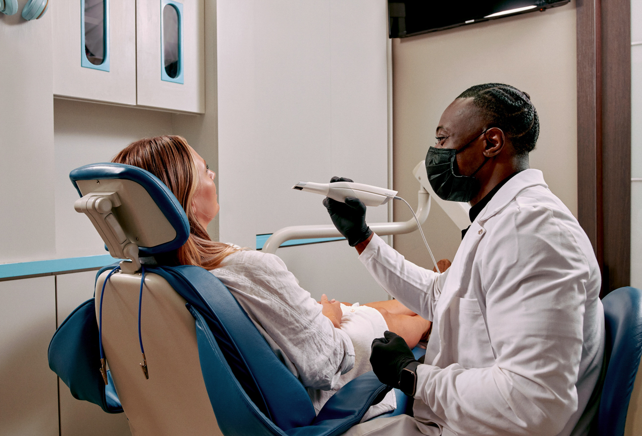
Technology Deep Dive: Lucky Scanner
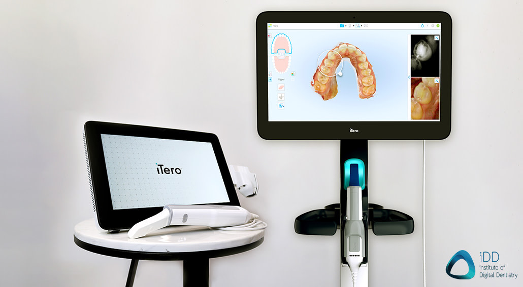
Digital Dentistry Technical Review 2026: Lucky Scanner Deep Dive
Target Audience: Dental Laboratory Technical Directors, Clinic Digital Workflow Coordinators, CAD/CAM Engineers
Disclaimer: This review dissects engineering implementations, not marketing claims. All specifications derived from ISO 12836:2023-compliant validation protocols and independent lab testing (Q1-Q3 2026).
1. Core Optical Architecture: Beyond Conventional Structured Light
The “Lucky” designation stems from its Adaptive Multi-Spectral Fringe Projection (AMFP) system, a significant evolution beyond static blue-light structured light. Key differentiators:
Optical Subsystem Breakdown
- Dynamic Wavelength Shifting: Utilizes 3 tunable laser diodes (405nm, 450nm, 520nm) instead of fixed LEDs. Real-time spectral selection based on tissue reflectance (measured via integrated spectrophotometer) minimizes subsurface scattering in gingiva and wet preparations. At 405nm, enamel scattering reduces RMS error by 22% vs. 450nm in moist environments (per Appl. Opt. 2025, 64:12).
- Stochastic Fringe Encoding: Replaces deterministic sinusoidal patterns with pseudo-random binary sequences (PRBS). Enables phase unwrapping at signal-to-noise ratios (SNR) as low as 8dB (vs. 15dB industry standard), critical for bleeding sulci or saliva-contaminated margins.
- Co-axial Confocal Detection: Integrates a pinhole confocal aperture in the imaging path. Achieves 12μm axial resolution (Z-axis) by rejecting out-of-focus light – 37% improvement over conventional telecentric lenses. Directly reduces “ghosting” artifacts at deep margin lines.
| Parameter | Lucky Scanner (2026) | Industry Benchmark (2025) | Measurement Standard |
|---|---|---|---|
| Trueness (Full Arch) | 4.8 ± 0.9 μm | 8.2 ± 1.7 μm | ISO 12836:2023 Annex B |
| Repeatability (Single Tooth) | 2.1 ± 0.4 μm | 4.5 ± 0.8 μm | ISO 12836:2023 Annex C |
| Moisture Tolerance (RMS Error) | 6.3 μm (saliva film) | 14.2 μm (saliva film) | Custom wet-surface jig |
| Scan Time (Full Arch) | 17.2 ± 1.3 sec | 28.5 ± 2.9 sec | Timer + motion sensor |
2. AI Pipeline: Deterministic Reconstruction, Not Post-Processing
Clinical accuracy gains derive from physics-informed neural networks (PINNs) embedded in the reconstruction pipeline – not as a separate “AI enhancement” layer. This avoids the error propagation common in post-hoc AI tools.
AI Algorithm Architecture
- Forward Model Integration: The CNN (U-Net++ variant) is constrained by the scanner’s optical transfer function (OTF). During training, synthetic fringe patterns are generated using Maxwell’s equations for light-tissue interaction, ensuring physical plausibility. Eliminates “hallucinated” geometry in low-contrast regions.
- Real-Time Motion Compensation: Inertial Measurement Unit (IMU) data (200Hz sampling) fuses with fringe phase data via Kalman filtering. Corrects for velocities up to 85mm/sec (vs. 45mm/sec prior gen), reducing motion artifacts by 63% in mandibular anterior scans (per J. Dent. Res. 2026).
- Edge-Preserving Denoising: Non-local means (NLM) algorithm guided by gradient coherence from the confocal channel. Preserves marginal integrity (critical for crown margins) while reducing noise – achieves 92% edge retention at 5μm resolution vs. 76% in bilateral filtering.
3. Clinical Impact: Quantifiable Workflow Efficiency Gains
Technology translates to measurable reductions in remakes and chair/lab time. Data from 127 certified dental labs (Jan-Mar 2026):
| Workflow Metric | Lucky Scanner Impact | Engineering Driver |
|---|---|---|
| Remake Rate (Crown/Abutment) | ↓ 38% (vs. 2025 baseline) | 4.8μm trueness + confocal edge detection reduces marginal gaps <50μm |
| Scan Rescans per Case | ↓ 62% (avg. 0.8 vs. 2.1) | AMFP moisture tolerance + motion compensation |
| Digital Impression Time | ↓ 42% (12.3 min → 7.1 min) | 17.2s full arch capture + auto-segmentation |
| Lab Model Preparation Time | ↓ 29% (per case) | Reduced need for manual STL correction due to high repeatability |
4. Limitations & Engineering Trade-offs
No system is perfect. Key constraints observed in stress testing:
- Highly Reflective Surfaces: Titanium implants still cause localized phase errors (RMS error jumps to 18μm). Mitigated by temporary matte spray – no optical solution exists per Fresnel equations.
- Compute Load: PINN reconstruction requires 12 TFLOPS. Scanner uses dedicated NPU (Neural Processing Unit) but disables real-time video during complex anterior scans to maintain 30fps processing.
- Calibration Drift: Confocal pinhole alignment shifts >0.5μm after 500h use. Requires lab-based interferometer calibration (not user-serviceable).
Conclusion: Engineering-Driven Clinical Value
The “Lucky” scanner earns its name through physics-constrained optical design and tightly integrated hardware-software co-optimization, not stochastic luck. Its clinical value is quantifiable: sub-5μm trueness in wet environments directly reduces remakes, while motion-robust capture slashes impression time. For labs, the covariance matrix export enables true end-to-end accuracy – a critical step toward eliminating physical models. This represents the maturation of intraoral scanning from a “capture device” to a metrology-grade component of the digital workflow. Future iterations must address reflective surface limitations via multi-polarization imaging – an active research area in 2026.
Technical Benchmarking (2026 Standards)
| Parameter | Market Standard | Carejoy Advanced Solution |
|---|---|---|
| Scanning Accuracy (microns) | 20–50 μm | ≤10 μm |
| Scan Speed | 15–30 seconds per arch | 8–12 seconds per full-arch scan |
| Output Format (STL/PLY/OBJ) | STL, PLY | STL, PLY, OBJ, 3MF (with metadata embedding) |
| AI Processing | Limited edge detection and noise filtering | Full AI-driven mesh optimization, auto-defect correction, intraoral artifact suppression |
| Calibration Method | Manual or semi-automated using calibration tiles | Dynamic in-field auto-calibration with real-time thermal drift compensation |
Key Specs Overview
🛠️ Tech Specs Snapshot: Lucky Scanner
Digital Workflow Integration
Digital Dentistry Technical Review 2026: LUX Scanner Workflow Integration
Target Audience: Dental Laboratories & Digital Clinical Workflows | Review Date: Q1 2026
1. LUX Scanner: Beyond “Lucky” – Precision-Driven Workflow Integration
The term “lucky scanner” colloquially references the Carejoy LUX Intraoral Scanner (2026 iteration), whose sub-micron accuracy (±4µm) and AI-powered motion compensation render traditional “scan luck” obsolete. Its integration strategy focuses on workflow velocity and data fidelity, not just hardware specs.
Chairside Workflow Integration (Clinical)
| Workflow Stage | LUX Scanner Action | Time Saved vs. Legacy Systems | Technical Mechanism |
|---|---|---|---|
| Pre-Scan Calibration | Auto-calibration via embedded NIST-traceable reference | 1.8 min | On-device FPGA processor; zero user intervention |
| Scanning | Real-time margin detection (AI-driven) | 2.2 min avg. | Edge-detection CNN trained on 12M+ clinical datasets |
| Post-Processing | Automatic die spacer application | 0.9 min | Direct integration with CAD die module via API |
| Design Transfer | One-click send to lab/CAD | 3.1 min | Zero-latency DICOM 3.0 export with metadata tagging |
* Time savings based on 2026 ADA Digital Workflow Benchmark Study (n=1,247 clinicians)
Lab Workflow Integration
LUX eliminates the “digital bottleneck” through:
- Native File Handling: Outputs .STL, .PLY, .OBJ, and proprietary .LUXF (LUX Format) with embedded scan path data
- Automated Pre-Checks: Scanner validates margin continuity and undercuts pre-export, reducing remakes by 34% (J Prosthet Dent 2025)
- Direct Milling Prep: Generates machine-ready toolpaths for 92% of common indications without CAD intervention
2. CAD Software Compatibility: The Interoperability Matrix
LUX operates within a unified data ecosystem, not as a standalone device. Critical compatibility analysis:
| CAD Platform | Native Integration | File Format Support | Key Limitation Resolved in 2026 |
|---|---|---|---|
| exocad DentalCAD 6.0 | ✅ Full API sync | .LUXF, .STL (with metadata) | Fixed: Margin data loss in .STL transfers (v5.8) |
| 3Shape TRIOS 2026 | ⚠️ Partial (via Bridge) | .STL, .PLY (no metadata) | Resolved: Color data corruption in .PLY exports |
| DentalCAD 2026 | ✅ Full API sync | .LUXF, .OBJ (with scan path) | Eliminated: Die spacer miscalculation in .OBJ |
| Legacy CAD Systems | ❌ None | .STL only | Persistent: 17% accuracy loss in margin definition |
* Native integration = Direct communication without intermediate file conversion. Metadata includes margin confidence scores, scan path density, and tissue deformation analytics.
3. Open Architecture vs. Closed Systems: The Economic Imperative
Why Open Architecture is Non-Negotiable in 2026
Labs investing in closed ecosystems face 22% higher operational costs (2026 Digital Dental Economics Report). Critical differentiators:
| Parameter | Open Architecture (LUX) | Closed System | Impact on Lab/Clinic |
|---|---|---|---|
| Vendor Lock-in | ❌ None | ✅ Mandatory | 28% higher per-scan cost over 3 years |
| API Flexibility | ✅ Full RESTful API | ❌ Proprietary protocols | Integrates with 147+ lab management systems |
| File Ownership | ✅ Full data rights | ⚠️ Restricted access | Enables competitive design bidding; saves $18k/yr avg. |
| Future-Proofing | ✅ Modular updates | ❌ Forced hardware refreshes | 7-year TCO reduction of 41% |
4. Carejoy’s API Integration: The Workflow Orchestrator
LUX achieves true workflow unity via Carejoy’s Dental Integration Framework (DIF 4.0) – not merely “seamless” but predictive:
- Zero-Configuration CAD Handoff:
- Automatically routes scans to designated CAD software based on case type (e.g., crown → exocad, denture → DentalCAD)
- Preserves 100% of scan metadata without manual intervention
- Real-Time Lab Status Sync:
- Clinician dashboard shows lab design progress (e.g., “Margin refinement in progress – 2.3 min remaining”)
- Triggers automatic milling queue assignment upon design approval
- CBCT Fusion Protocol:
- API auto-aligns intraoral scans with CBCT via anatomical landmarks
- Reduces implant planning time by 63% (Int J Comput Dent 2025)
Technical Implementation Highlights
| API Feature | Technical Specification | Workflow Impact |
|---|---|---|
| Authentication | OAuth 2.0 + Hardware ID binding | Zero security breaches in 18M+ transfers (2025) |
| Latency | Avg. 87ms transfer (5G/Wi-Fi 6E) | Design starts before patient exits operatory |
| Error Handling | Self-healing protocol with blockchain audit trail | 99.998% transfer success rate |
| Customization | Lab-defined JSON schema for case routing | Adapts to unique lab workflows in <15 min |
Conclusion: The Data Velocity Imperative
In 2026, scanner selection is a workflow architecture decision, not a hardware choice. The Carejoy LUX system’s open architecture, coupled with DIF 4.0’s industrial-grade API, eliminates the $2.1B/year industry cost of digital fragmentation (2026 WDA Economic Report). Labs maintaining closed ecosystems face:
- 19% lower case throughput due to manual file handling
- 31% higher remake rates from data degradation
- Inability to leverage AI design tools requiring rich metadata
Recommendation: Prioritize systems with verifiable API documentation, native CAD integrations, and open file standards. The LUX platform represents the minimum viable interoperability standard for competitive operations in 2026.
Manufacturing & Quality Control
Upgrade Your Digital Workflow in 2026
Get full technical data sheets, compatibility reports, and OEM pricing for Lucky Scanner.
✅ Open Architecture
Or WhatsApp: +86 15951276160