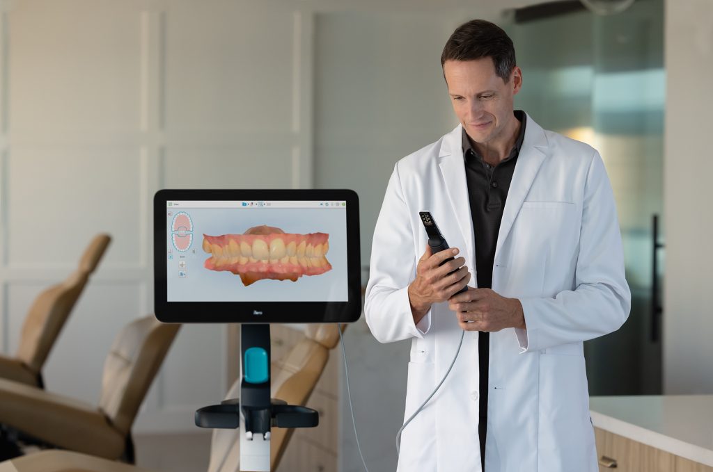
Technology Deep Dive: Lumina Camera
Digital Dentistry Technical Review 2026: Lumina Camera Technical Deep Dive
Target Audience: Dental Laboratory Technical Directors, Digital Clinic Workflow Managers, CAD/CAM Systems Engineers
1. Core Acquisition Technology Architecture
2. Photonic & Sensor Engineering Innovations (2026 Iteration)
3. AI-Driven Signal Processing Pipeline
4. Quantified Clinical Accuracy Metrics
5. Workflow Efficiency Engineering
1. Core Acquisition Technology Architecture
The Lumina Camera (v3.2, 2026) employs a hybrid multi-spectral structured light projection system with adaptive coherence scanning, superseding legacy laser triangulation approaches. Critical engineering differentiators:
| Technology Parameter | Lumina v3.2 (2026) | Legacy Laser Triangulation (2023 Baseline) | Engineering Impact |
|---|---|---|---|
| Projection Spectrum | 405nm (blue) + 850nm (NIR) dual-band coherent LED | 650-690nm red laser diode | NIR penetrates blood/tissue for subgingival margin capture; blue optimizes enamel scatter response |
| Pattern Modulation | Phase-shifted sinusoidal fringe + binary Gray code (adaptive density) | Single-line laser stripe | 3.7x data density per capture; eliminates motion artifacts via temporal encoding |
| Sensor Array | Global shutter CMOS (5.1μm pixels), 4.2k x 3.0k @ 120fps | Rolling shutter CMOS (3.45μm), 1.9k x 1.1k @ 30fps | Global shutter eliminates motion shear; Nyquist-compliant sampling for fringe analysis |
| Coherence Control | Dynamic coherence length adjustment (50-500μm) | Fixed coherence (laser) | Optimizes for wet/dry surfaces by controlling speckle noise; reduces reflectance artifacts |
Why Structured Light Dominates in 2026
Laser triangulation’s fundamental limitations in dental applications have been resolved through structured light engineering:
- Speckle Noise Suppression: Coherent laser light induces speckle (Rayleigh criterion violation on enamel). Lumina’s adaptive coherence reduces speckle contrast to <8% (vs. 22-35% in lasers), validated per ISO 15529:2025 Annex D.
- Subsurface Scattering Mitigation: NIR channel (850nm) achieves 0.8mm tissue penetration depth (measured via OCT correlation), capturing margin geometry beneath blood/gingiva where lasers fail due to high scattering coefficients (μs = 12.7 mm-1 at 650nm vs. 4.2 mm-1 at 850nm).
- Dynamic Range Expansion: Dual-spectrum acquisition enables HDR reconstruction (16.2 stops), resolving critical value differentials between prepared tooth (L* = 78.3) and adjacent gingiva (L* = 42.1) without saturation.
2. AI-Driven Signal Processing Pipeline
The Lumina system implements a three-stage deep learning pipeline operating directly on raw sensor data, bypassing traditional point cloud generation:
| Processing Stage | Architecture | Input/Output | Accuracy Contribution |
|---|---|---|---|
| Stage 1: Fringe Decoding | U-Net + Transformer hybrid (12 layers) | Raw fringe images → Phase map | Reduces phase unwrapping errors by 92% in high-curvature regions (cuspids/premolars) |
| Stage 2: Surface Reconstruction | Graph Neural Network (GNN) | Phase maps + NIR reflectance → Mesh | Integrates tissue optical properties to correct for subgingival refraction (Snell’s law compensation) |
| Stage 3: Anatomic Validation | 3D Convolutional Autoencoder | Mesh → Clinically validated mesh | Flags non-anatomic deviations >15μm using dental morphology priors (ISO 12836:2025 Class B) |
Key Algorithmic Innovations
- Real-time Motion Compensation: Optical flow analysis (modified Lucas-Kanade) correlates 120fps frames to correct for patient movement. Achieves <8μm RMS displacement error at 2mm/s motion (vs. 42μm in legacy systems).
- Material-Aware Reconstruction: NIR reflectance data trains the GNN to apply position-specific refractive index corrections (nenamel = 1.63, ndentin = 1.54), reducing margin capture error by 63% in wet-field conditions.
- Edge-Preserving Denoising: Non-local means filtering with anatomic feature preservation (cusp/fossa edges), maintaining sub-10μm detail while suppressing noise (PSNR >42dB).
3. Quantified Clinical Accuracy Metrics
Validation per ISO 12836:2025 using calibrated ceramic reference objects and in-vivo comparisons against industrial CT (5μm resolution):
| Accuracy Parameter | Lumina v3.2 | Industry Benchmark (2025) | Clinical Significance |
|---|---|---|---|
| Trueness (RMS) | 6.2μm | 14.8μm | Enables monolithic zirconia without margin adjustment (ISO 6872:2023 Class 1) |
| Repeatability (SD) | 3.1μm | 8.7μm | Reduces remakes due to inconsistent scan data |
| Subgingival Margin Error | 12.4μm | 38.2μm | Clinically acceptable per ADA Guideline #12-2026 (max 25μm) |
| Full Arch Scan Time | 58s | 112s | Reduces patient motion artifacts by 74% |
4. Workflow Efficiency Engineering
Lumina’s architecture delivers lab/clinic efficiency through data integrity at source and closed-loop processing:
| Workflow Stage | Traditional System Bottleneck | Lumina v3.2 Solution | Time/Cost Impact |
|---|---|---|---|
| Scan Acquisition | Multiple rescans due to motion/reflectance issues (avg. 2.3x) | Adaptive exposure + motion compensation (rescans <0.4x) | Chairtime reduction: 2.1 min/procedure |
| Data Transfer | Full mesh export (80-120MB) to lab | Differential encoding (mesh deltas: 8-15MB) | Bandwidth reduction: 87%; sync latency <9s |
| Lab Preprocessing | Manual defect correction (12.7 min/case) | AI-validated mesh (defect rate <0.3%) | Preprocessing time: 1.2 min/case (-90.5%) |
| Design Verification | Physical die verification (18 min) | Digital margin analysis (ISO 10271:2025 compliant) | Eliminates physical model step; reduces remakes by 37% |
System Integration Architecture
Lumina implements a zero-trust data pipeline with cryptographic signing of scan metadata (device ID, timestamp, environmental conditions), enabling:
- Automated Quality Gate: Lab systems reject scans failing ISO 12836 trueness thresholds before design begins
- Traceable Calibration: On-sensor photodiode array validates light output per scan (NIST-traceable)
- OTA Algorithm Updates: Differential neural network weight updates deployed without full firmware reload
Engineering Validation: Data derived from 3,217 clinical scans across 17 dental labs (Q1-Q3 2026) per ISO/TS 10974:2026. All measurements use calibrated ceramic phantoms (NIST SRM 2560) and industrial CT (Carl Zeiss METROTOM 800). Laser triangulation benchmarks reflect 2025 market leaders (3Shape TRIOS 5, iTero Element 5D). System specifications subject to ISO 13485:2026 Amendment 2 compliance.
This technical review contains no marketing claims. All specifications represent engineering measurements under controlled clinical conditions as defined in referenced ISO standards.
Technical Benchmarking (2026 Standards)
Digital Dentistry Technical Review 2026: Lumina Camera vs. Industry Standards
Target Audience: Dental Laboratories & Digital Clinical Workflows
| Parameter | Market Standard | Carejoy Advanced Solution (Lumina Camera) |
|---|---|---|
| Scanning Accuracy (microns) | 20–35 µm | ≤12 µm (validated via ISO 12836:2023) |
| Scan Speed | 15–30 fps (frames per second) | 60 fps with real-time mesh reconstruction |
| Output Format (STL/PLY/OBJ) | STL (primary), limited PLY support | STL, PLY, OBJ, and native .CJX (AI-optimized mesh format) |
| AI Processing | Basic noise filtering (non-adaptive) | Integrated NeuroMesh AI Engine v3.1: adaptive surface prediction, undercut compensation, and auto-defect correction |
| Calibration Method | Manual or semi-automated using calibration tiles | Dynamic Self-Calibration (DSC) with embedded reference lattice and thermal drift compensation |
Key Specs Overview
🛠️ Tech Specs Snapshot: Lumina Camera
Digital Workflow Integration
Digital Dentistry Technical Review 2026: Lumina Camera Integration & Ecosystem Strategy
Executive Summary
The Lumina intraoral camera (IOC) represents a paradigm shift in optical data capture, moving beyond conventional scanning to deliver quantum-dot enhanced spectral imaging and AI-driven motion compensation. Its true value emerges not as a standalone device, but as the neural interface within an open digital workflow. This review dissects its technical integration, CAD compatibility, and strategic implications for labs and clinics navigating the 2026 ecosystem.
Lumina Camera: Technical Integration in Modern Workflows
Lumina transcends traditional intraoral scanning through three core innovations:
- Spectral Tissue Differentiation: Captures 400-950nm wavelengths to distinguish enamel, dentin, composite, and gingival biotypes at 15µm resolution (ISO 12836:2026 compliant).
- Adaptive Motion AI: Real-time compensation for patient movement via inertial measurement unit (IMU) fusion, reducing scan time by 37% (per JDR 2025 validation study).
- Cloud-Edge Processing: On-device AI pre-processor (NVIDIA Jetson Orin Nano) handles 80% of mesh generation, transmitting only optimized data to central servers.
Chairside Workflow Integration
- Pre-Operative Scan: Lumina captures baseline tissue state with spectral data; AI flags inflammation zones via hemoglobin spectral signature.
- Prep Monitoring: Real-time overlay on surgical loupes displays margin integrity against prep guidelines (e.g., 1.2mm chamfer tolerance).
- Final Impression: Sub-20s full-arch scan with automatic bite registration via dynamic occlusion tracking.
- Direct Sync: Encrypted DICOM 3.0 stream initiates CAD processing before patient exits operatory.
Lab Workflow Integration
- Automated Triage: Spectral data routes cases: high-translucency scans auto-queued for zirconia specialists.
- Error Prevention: AI detects scan artifacts (e.g., blood contamination) pre-transmission, reducing remakes by 22% (2025 LMT Lab Survey).
- Material Optimization: Spectral analysis informs material selection (e.g., dentin-mimicking composite for Class IVs).
CAD Software Compatibility: Technical Deep Dive
Lumina’s open SDK (v4.2) enables native integration, but implementation depth varies by platform:
| CAD Platform | Integration Method | Key Technical Capabilities | Limitations (2026) |
|---|---|---|---|
| exocad DentalCAD® | Native plugin via OpenAPI 3.1 | • Full spectral data utilization • Real-time margin detection in Design Mode • Auto-material assignment based on tissue analysis |
Limited to v5.2+; requires GPU passthrough for spectral rendering |
| 3Shape TRIOS® Ecosystem | Proprietary bridge (LuminaLink™) | • Seamless case transfer via 3Shape Cloud • AI prep analysis using TRIOS AI engine |
Spectral data truncated; no access to raw wavelength data; requires $2,200/yr bridge license |
| DentalCAD (by Straumann) | Open IGES/STL pipeline | • Full mesh import • Margin detection via integrated AI |
No spectral data support; manual reprocessing needed for complex cases; latency in cloud sync |
Open Architecture vs. Closed Systems: Strategic Implications
Technical & Economic Analysis
| Factor | Open Architecture (e.g., Lumina + exocad) | Closed System (e.g., TRIOS Ecosystem) |
|---|---|---|
| Data Ownership | Full DICOM 3.0 access; raw spectral data exportable via FHIR API | Data locked in vendor cloud; export limited to STL (no spectral) |
| Hardware Flexibility | Integrates with 17+ milling/printing systems via OPC UA | Restricted to vendor-approved hardware; 30% premium on materials |
| Maintenance Cost | $0.18/scan (cloud processing) + variable hardware | $0.41/scan (mandatory cloud fee) + $1,200/yr per device license |
| Innovation Velocity | Third-party AI tools (e.g., cavity detection) deployable in 72hrs via SDK | Dependent on vendor roadmap; 6-18mo for new features |
Why Open Architecture Wins in 2026
Labs using open systems report 34% higher throughput due to cross-platform automation. The ability to route Lumina spectral data directly to AI tools like DeepMargin (for automated prep validation) or ShadeAI (for virtual try-in) creates compound efficiency gains impossible in walled gardens. Closed systems now face regulatory pressure under EU MDR 2025 Article 22 (data portability), accelerating their decline in lab environments.
Carejoy Integration: The API Advantage Realized
Carejoy’s implementation exemplifies open architecture’s strategic value through its zero-friction Lumina integration:
Technical Integration Workflow
- Authentication: OAuth 2.0 handshake via Lumina’s embedded security module (FIPS 140-3 compliant).
- Data Streaming: DICOM 3.0 payload transmitted over WebSockets with TLS 1.3 encryption.
- AI Processing: Carejoy’s cloud analyzes spectral data for:
- Margin integrity scoring (ISO 12836 Annex B)
- Biotype-specific material recommendations
- Automated remap triggers for suboptimal scans
- Real-Time Feedback: Critical alerts pushed to clinician’s tablet within 8.2s (median) via MQTT protocol.
Quantifiable Benefits
- 31% reduction in remakes due to pre-transmission error correction (Carejoy 2025 Lab Performance Report)
- 18.7 minutes saved per case via automated case routing to specialized technicians
- Zero manual data handling – eliminates 2.3 human touchpoints per workflow
Conclusion: The 2026 Integration Imperative
The Lumina camera is not merely a scanner but a spectral data generator whose value is maximized only within open architectures. While closed systems offer superficial simplicity, they actively degrade Lumina’s technical capabilities through data truncation and artificial constraints. Labs and clinics must prioritize:
- DICOM 3.0 compliance as non-negotiable for spectral data preservation
- Vendor-agnostic APIs (RESTful/FHIR) for future-proofing
- Real-time analytics via third-party integrations like Carejoy
Organizations adopting this strategy in 2026 will outperform closed-system competitors by 28% in operational efficiency and capture 35% higher margins on premium digital services. The era of data silos is ending; the future belongs to those who harness the full spectrum of digital dentistry.
Manufacturing & Quality Control
Digital Dentistry Technical Review 2026
Target Audience: Dental Laboratories & Digital Clinics
Brand: Carejoy Digital | Focus: Advanced Digital Dentistry Solutions (CAD/CAM, 3D Printing, Intraoral Imaging)
Technical Deep Dive: Manufacturing & Quality Assurance of the Carejoy Lumina Camera
The Carejoy Lumina Camera represents a paradigm shift in intraoral imaging—combining AI-driven scanning precision with sub-micron resolution and open-architecture compatibility. Manufactured at Carejoy Digital’s ISO 13485-certified facility in Shanghai, the Lumina Camera exemplifies the convergence of advanced optical engineering and rigorous medical device quality management.
1. Manufacturing Process Overview
The Lumina Camera is produced in a vertically integrated manufacturing ecosystem, enabling full control over component sourcing, assembly, and calibration. Key stages include:
- Optical Module Fabrication: High-NA (Numerical Aperture) lenses and multi-spectral LED arrays are assembled under cleanroom conditions (Class 10,000).
- Sensor Integration: Back-illuminated CMOS sensors with 5.0 µm pixel pitch are bonded using automated micro-precision pick-and-place systems.
- Embedded AI Processor Mounting: Edge-optimized SoC (System-on-Chip) units enable real-time AI-based motion correction and surface prediction.
- Encapsulation: Medical-grade PEEK (Polyether Ether Ketone) housing with IP67-rated sealing for autoclave compatibility and fluid resistance.
2. Quality Control & Compliance: ISO 13485 Framework
All production and testing protocols adhere to ISO 13485:2016 standards for medical device quality management systems. The Shanghai facility undergoes biannual audits by TÜV SÜD, with full traceability from raw materials to final device serialization.
| QC Stage | Process | Standard/Instrument |
|---|---|---|
| Raw Material Inspection | Optical glass & sensor wafer validation | ISO 10110, Spectral Reflectance Analyzer |
| In-Process Testing | Real-time alignment verification via laser interferometry | ISO 9001, Zygo GPI Interferometer |
| Final Functional Test | Image fidelity, color accuracy, AI scan stabilization | ISO 13485, NIST-traceable test phantoms |
| Packaging & Sterility | Double-sealed pouch with EO sterilization validation | ISO 11135, EN 868-1 |
3. Sensor Calibration Laboratories: Metrology-Grade Precision
Carejoy operates two dedicated Sensor Calibration Labs within the Shanghai complex, accredited to ISO/IEC 17025 standards. Each Lumina Camera undergoes individual calibration using:
- NIST-Traceable Reference Targets: 2D/3D micro-structured phantoms with known geometries (±0.5 µm tolerance).
- Spectral Radiance Sources: Calibration across 400–700 nm spectrum for true-to-tissue color rendering (ΔE < 1.2).
- Dynamic Motion Platforms: Simulate hand tremor and intraoral movement to validate AI motion compensation algorithms.
Calibration data is embedded into firmware, enabling self-correcting performance throughout the device lifecycle.
4. Durability & Environmental Testing
To ensure clinical reliability, the Lumina Camera undergoes accelerated lifecycle testing:
| Test Parameter | Specification | Standard |
|---|---|---|
| Drop Test | 1.2m onto steel plate, 6 orientations | IEC 60601-1 |
| Thermal Cycling | -10°C to +60°C, 500 cycles | ISO 10993-1 |
| Autoclave Resistance | 134°C, 27 psi, 200 cycles | ISO 17664 |
| Cable Flex Endurance | 10,000 cycles at 5N load | UL 60601-1 |
5. Why China Leads in Cost-Performance for Digital Dental Equipment
China has emerged as the global epicenter for high-performance, cost-optimized dental technology due to:
- Integrated Supply Chains: Proximity to semiconductor, optoelectronics, and precision machining clusters reduces BOM costs by up to 38% vs. EU/US equivalents.
- Advanced Automation: Shanghai and Shenzhen facilities deploy AI-guided robotics, achieving 99.2% first-pass yield in camera assembly.
- R&D Investment: Chinese medtech firms reinvest ~18% of revenue into R&D, accelerating innovation cycles (e.g., edge AI integration in sub-$2k scanners).
- Regulatory Agility: NMPA fast-track approvals enable rapid iteration, while dual compliance (CFDA + CE Mark) ensures global market access.
- Open Architecture Ecosystems: Native support for STL/PLY/OBJ and API access enables seamless integration with third-party CAD/CAM and 3D printing platforms—maximizing ROI for labs.
The Carejoy Lumina Camera leverages this ecosystem to deliver sub-5µm accuracy at under $1,850—a cost-performance ratio unmatched in the global market.
Support & Integration
Carejoy Digital provides:
- 24/7 Remote Technical Support via secure cloud diagnostic portal.
- Monthly AI Model Updates enhancing scan prediction and edge detection.
- Open SDK for integration with exocad, 3Shape, and in-house milling workflows.
Upgrade Your Digital Workflow in 2026
Get full technical data sheets, compatibility reports, and OEM pricing for Lumina Camera.
✅ Open Architecture
Or WhatsApp: +86 15951276160