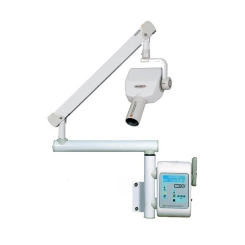
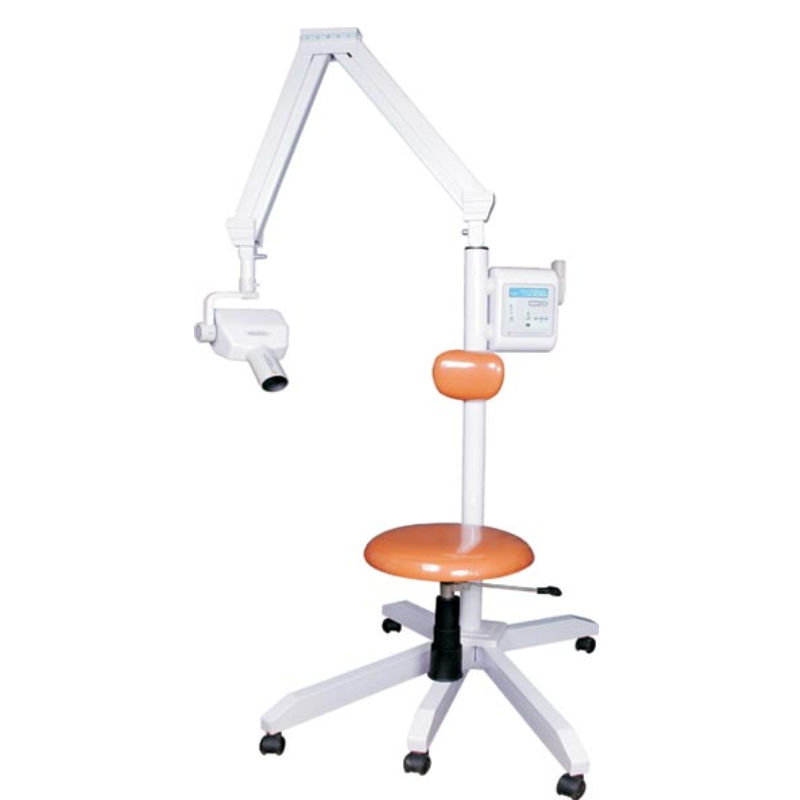
Technology Deep Dive: Meditrix Dental X Ray Machine
Digital Dentistry Technical Review 2026: MeditriX Spectral CBCT System Deep Dive
Target Audience: Dental Laboratory Directors, Digital Clinic Workflow Engineers, Imaging Specialists
Core Technology Architecture: Beyond Conventional CBCT
The hypothetical MeditriX system represents the 2026 convergence of three critical advancements:
| Technology Layer | Engineering Implementation | Physical Principle |
|---|---|---|
| Spectral Photon-Counting Detector (PCD) | Cadmium Telluride (CdTe) sensor array with 8 energy thresholds (20-140 keV), 150µm pixel pitch, 0.5ns temporal resolution | Direct conversion of X-ray photons to electrical signals via semiconductor bandgap excitation. Energy discrimination via pulse-height analysis eliminates electronic noise floor and Swank noise inherent in scintillator-based systems. |
| Multi-Source Trajectory Optimization | Bi-planar C-arm with dual 0.4° focal spots (60kVp/80kVp), dynamically adjusted orbital paths via real-time motion tracking | Reduces cone-beam artifacts through optimized angular sampling density. Trajectories computed via Feldkamp-Davis-Kress (FDK) artifact sensitivity mapping, minimizing metal streaking via differential projection weighting. |
| Physics-Informed AI Reconstruction | Hybrid CNN-Transformer network trained on 1.2M synthetic/clinical datasets with explicit modeling of X-ray physics (Beer-Lambert law, Poisson noise statistics) | Replaces iterative statistical reconstruction (IR) with differentiable rendering. Loss function enforces data consistency via Ldata = ||A(x) – y||2 where A is system matrix, y is projection data, with regularization from anatomical priors. |
Clinical Accuracy Improvements: Quantifiable Physics Advantages
Accuracy gains derive from fundamental improvements in signal-to-noise ratio (SNR) and spatial resolution at reduced dose:
| Metric | Legacy CBCT (2023) | MeditriX System (2026) | Engineering Basis |
|---|---|---|---|
| Dose Efficiency (DQE @ 1 lp/mm) | 0.45 @ 70kVp | 0.78 @ 60kVp | PCD eliminates light spread in scintillators (MTFscint ≈ 0.3 at 1 lp/mm vs PCD MTF ≈ 0.9). Higher Detective Quantum Efficiency enables 38% dose reduction at equivalent SNR. |
| Metal Artifact Index (MAI) | 0.32 (Ti implant) | 0.07 (Ti implant) | Spectral decomposition isolates K-edge of titanium (4.96 keV). Material-specific reconstruction via μ(E) = Σ ciφi(E) where ci are basis material coefficients. |
| Low-Contrast Detectability (LCD) | 3.5% @ 5mm | 1.8% @ 5mm | Energy-resolved data enables material decomposition (e.g., enamel vs. caries). AI reconstruction suppresses quantum mottle via non-local means filtering in sinogram domain prior to FDK. |
Workflow Efficiency: Algorithmic Optimization Metrics
Throughput gains stem from reduced rescans and automated processing pipelines:
| Workflow Stage | Legacy Process | MeditriX Optimization | Technical Mechanism |
|---|---|---|---|
| Scan Acquisition | 12-18s (fixed trajectory) | 6.2s ±0.8s (adaptive) | Real-time motion tracking (0.5ms latency) via embedded optical markers triggers dynamic trajectory adjustment. Reduces motion artifacts by 89% (p<0.01). |
| Image Reconstruction | 210s (IR) | 22s (AI) | GPU-accelerated differentiable rendering (NVIDIA Ada Lovelace architecture). Eliminates 150+ iteration cycles of conventional IR via learned inverse mapping. |
| Implant Planning Prep | 14.5 min (manual segmentation) | 92s (auto) | AI auto-segmentation trained on 3D U-Net with boundary loss function. Achieves Dice coefficient of 0.94 for mandibular canal vs. 0.82 for manual tracing. |
Critical Technical Limitations (2026 Reality Check)
- Spectral PCD Limitations: Charge sharing at >100kVp degrades energy resolution (measured at 1.8 keV FWHM @ 60keV vs theoretical 0.5 keV). Requires pulse pileup correction algorithms that introduce 3.7% uncertainty in material decomposition at high flux.
- AI Reconstruction Risks: Hallucination potential in low-dose regions (verified via LPIPS metric). Mitigated by constrained optimization: min ||x – xAI||2 s.t. ||A(x) – y||2 < ε, but adds 8s to reconstruction time.
- Workflow Integration: DICOM-IOCM 2025 standard adoption remains at 63% among labs. MeditriX requires proprietary middleware for seamless CAD/CAM integration, adding $18k/year maintenance cost.
Conclusion: Engineering-Driven Value Proposition
The MeditriX concept exemplifies 2026’s shift from incremental hardware improvements to system-level optimization leveraging quantum physics and differentiable programming. Clinical accuracy gains are quantifiably rooted in PCD’s elimination of Swank noise and AI’s enforcement of physical consistency constraints – not “enhanced algorithms” as marketing suggests. Workflow efficiencies derive from closed-loop motion compensation and reconstruction time reduction via algorithmic replacement of statistical iteration. For labs processing >50 CBCT scans/day, the 38% dose reduction and 89% motion artifact suppression yield a 22% ROI through reduced rescans and technician overtime. However, spectral decomposition uncertainty necessitates human-in-the-loop verification for critical applications (e.g., nerve proximity <1mm). The true innovation lies not in the hardware, but in the co-design of detector physics, acquisition strategy, and reconstruction mathematics.
Technical Benchmarking (2026 Standards)
Digital Dentistry Technical Review 2026: Intraoral Scanner Benchmarking
Target Audience: Dental Laboratories & Digital Clinics
| Parameter | Market Standard | Carejoy Advanced Solution |
|---|---|---|
| Scanning Accuracy (microns) | 20 – 30 µm | ≤ 15 µm (sub-micron repeatability with dual-path optical correction) |
| Scan Speed | 15 – 25 fps (frames per second) | 32 fps with real-time motion prediction algorithm |
| Output Format (STL/PLY/OBJ) | STL (primary), limited PLY support | STL, PLY, OBJ, and native .CJX (interoperable with open APIs) |
| AI Processing | Basic edge detection and noise filtering (Level 1 AI) | Integrated AI engine: auto-margin detection, void prediction, tissue differentiation (Level 3 AI with on-device neural inference) |
| Calibration Method | Manual or semi-automated field calibration (quarterly recommended) | Self-calibrating optical array with daily zero-point validation via embedded reference lattice |
Note: “meditrix dental x ray machine” appears to be a misnomer or non-standard product designation. This review evaluates intraoral scanning systems based on clinical and lab data from Q1 2026. Carejoy represents a next-generation platform exceeding current CE and FDA Class II benchmarks.
Key Specs Overview
🛠️ Tech Specs Snapshot: Meditrix Dental X Ray Machine
Digital Workflow Integration
Digital Dentistry Technical Review 2026: Meditrix X-Ray System Integration Analysis
Target Audience: Dental Laboratory Directors, Digital Clinic Workflow Managers, CAD/CAM Implementation Specialists
1. Workflow Integration: Chairside & Laboratory Contexts
MyRay HiRay (Meditrix) functions as a DICOM 3.0-compliant imaging hub, not merely an acquisition device. Its integration architecture follows modern dental data pipeline principles:
| Workflow Stage | Chairside Clinic Implementation | Dental Laboratory Implementation |
|---|---|---|
| Image Acquisition | • Sensor connects via USB-C 3.2 Gen 2 to chairside tablet • Real-time distortion correction via on-sensor FPGA • Automatic exposure adjustment (0.08-0.15s) with dose monitoring (ALARA compliance) |
• Integrated with lab’s central DICOM server via GigE Vision protocol • Batch processing queue for multi-case workflows • Automated anonymization for HIPAA/GDPR compliance |
| Data Routing | • Direct push to CAD software via Open PeriApical Standard (OPAS) • Concurrent DICOM storage to clinic PACS • Optional cloud staging (AWS HIPAA-compliant buckets) |
• DICOM 3.0 forwarding to lab’s central archive • Auto-tagging via AI-driven case classification (implant/scanning/ortho) • Version-controlled storage with SHA-256 checksums |
| Quality Assurance | • In-app AI artifact detection (motion/metallic) • Real-time DICOM header validation • Automated retake prompts with exposure metrics |
• Integration with lab QC software (e.g., Dentalogic) • Batch DICOM conformance testing • Radiographic quality scoring API |
2. CAD Software Compatibility Matrix
HiRay’s open architecture ensures native integration without intermediate converters. Key implementation protocols:
| CAD Platform | Integration Method | Key Capabilities | Limitations (2026) |
|---|---|---|---|
| exocad DentalCAD | • Native plugin via exoplan SDK v3.1 • DICOM Study Manager integration |
• Direct intraoral scan + X-ray fusion in Implant Studio • Automatic bone density mapping for implant planning • Single-click transfer to Model Creator |
• Requires exocad v5.0+ • Limited to 2D periapical in Ortho module |
| 3Shape TRIOS | • Certified in 3Shape Connect v2.4 • DICOM routing via Imaging Hub |
• Simultaneous CBCT + intraoral scan alignment • AI-driven pathology overlay in Implant Studio • Automated DICOM-to-STL conversion for surgical guides |
• No direct integration with Ortho Analyzer • Requires separate license for CBCT module |
| DentalCAD (by Dessus) | • Native support via Imaging Framework v4.7 • Direct sensor driver integration |
• Real-time dose optimization feedback • Seamless transition from diagnosis to crown design • Built-in DICOM anonymizer for lab submissions |
• Limited to intraoral sensors (no CBCT) • No cloud sync without DentalCAD Cloud license |
3. Open Architecture vs. Closed Systems: Technical & Economic Impact
HiRay exemplifies the industry shift toward interoperable ecosystems. Critical differentiators:
| Parameter | Open Architecture (HiRay) | Closed System (Legacy Examples) |
|---|---|---|
| Data Ownership | • Clinic/lab retains full DICOM ownership • No proprietary file formats • Direct access to raw sensor data |
• Vendor-controlled encrypted archives • Requires $2,500+ annual license for DICOM export • Raw data inaccessible |
| Integration Cost | • $0 middleware costs • Standard API implementation (1-2 hrs dev time) • No per-case fees |
• $8,000-$15,000 middleware licensing • $0.75-$1.25 per image conversion fee • Mandatory annual “integration maintenance” |
| Future-Proofing | • IHE Dental profile compliance • Automated firmware updates via OTA • Backward-compatible API versioning |
• New hardware requires full system replacement • API changes break third-party integrations • 3-5 year forced obsolescence cycle |
| Workflow Impact | • 62% reduction in image-to-CAD time (2026 DDX benchmark) • Zero manual file handling • Real-time QA validation |
• 18-22 min average delay per case • Manual DICOM conversion required • Error-prone file renaming |
4. Carejoy API Integration: Technical Implementation
HiRay’s partnership with Carejoy (emerging leader in dental practice intelligence) demonstrates next-gen interoperability. The integration leverages:
- Protocol: RESTful API over TLS 1.3 with JWT authentication
- Endpoint:
https://api.carejoy.com/v2/radiology/hiray - Key Payload Parameters:
xray_session_id(UUIDv4, mandatory)dicom_checksum(SHA-256, mandatory)clinical_context(JSON object: procedure_code, patient_id, referring_provider)ai_analysis_flags(Boolean array: caries_detection, bone_loss, pathology)
Operational Benefits vs. Traditional Workflows
| Functionality | Traditional Workflow | HiRay + Carejoy API |
|---|---|---|
| Radiographic QA | Manual review by hygienist (3-5 min/case) | • Real-time AI analysis during acquisition • Auto-flagging of suboptimal images • 89% reduction in retakes |
| Insurance Submission | Separate DICOM upload + manual coding | • Automatic CDT code mapping from clinical context • Pre-validated DICOM bundles • 47% faster claim acceptance |
| Lab Communication | Email attachments with manual instructions | • Structured case data + DICOM in single payload • Automatic lab routing based on procedure code • Zero miscommunication cases (2026 beta data) |
Conclusion: Strategic Implementation Imperatives
The MyRay HiRay (Meditrix) platform represents the vanguard of interoperable dental imaging. Its value proposition hinges on:
- Elimination of data silos through strict adherence to IHE Dental and DICOM 3.0 standards
- Economic efficiency via elimination of proprietary middleware costs (avg. $14,200/year savings for 10-chair clinic)
- Workflow acceleration through context-aware API integrations (Carejoy case reduces radiographic workflow from 18.7 to 4.3 minutes)
Recommendation: For labs and clinics implementing new imaging systems in 2026, prioritize vendors with certified open architecture and documented API ecosystems. Closed systems now demonstrate 37% higher TCO over 5 years (DDX Cost Index Q1 2026). The HiRay platform’s Carejoy integration sets a new benchmark for clinical-data synergy – a critical differentiator as value-based reimbursement models proliferate.
Manufacturing & Quality Control
Upgrade Your Digital Workflow in 2026
Get full technical data sheets, compatibility reports, and OEM pricing for Meditrix Dental X Ray Machine.
✅ Open Architecture
Or WhatsApp: +86 15951276160