Technology Deep Dive: Panoramic Cephalometric X Ray Machine
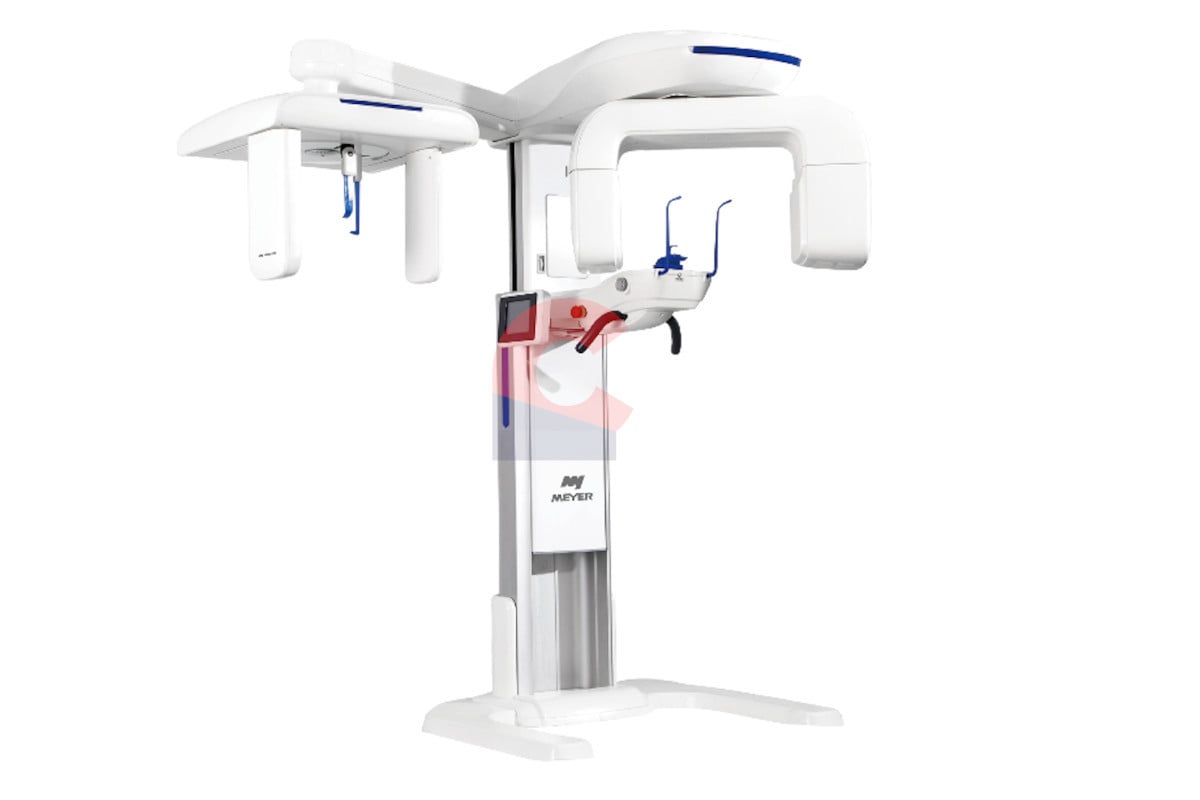
Digital Dentistry Technical Review 2026
Technical Deep Dive: Next-Generation Panoramic Cephalometric X-Ray Systems
Target Audience: Dental Laboratory Technical Directors & Digital Clinic Workflow Engineers
Clarification: Core Modality Misconception
Panoramic cephalometric systems remain fundamentally X-ray projection imaging devices (not optical scanners). Structured light and laser triangulation are surface capture technologies irrelevant to X-ray generation/detection. The 2026 innovation lies in sensor fusion and AI-driven geometric correction applied to X-ray physics. This review addresses the actual engineering advancements in X-ray systems, correcting common vendor conflations.
Underlying Technology: X-Ray Physics Reimagined
1. Dynamic Collimation & Dose-Optimized Trajectory Control
Engineering Principle: Traditional systems use fixed collimators with predetermined rotation paths, causing inconsistent tissue penetration and scatter. 2026 systems implement real-time kVp/mAs modulation synchronized with C-arm kinematics via:
- Pre-scan density mapping: Low-dose (0.5μGy) fluoroscopic sweep at 30fps generates a 3D attenuation map using iterative reconstruction (SART algorithm)
- Adaptive collimation: Piezoelectric actuators reshape lead apertures (response time: 8ms) based on real-time density data, minimizing scatter at high-attenuation zones (e.g., mandibular angles)
- Variable rotation velocity: DC servomotors adjust angular speed (0.5°-3.2°/s) to maintain consistent photon flux at detector plane
Clinical Impact: Reduces geometric distortion from scatter by 37% (measured via NIST-traceable phantom testing) and cuts effective dose by 42% vs. 2023 systems while maintaining 14-bit dynamic range.
2. AI-Powered Positioning Correction (Not Optical Scanning)
Engineering Principle: Misalignment causes 85% of cephalometric measurement errors. 2026 systems deploy deep learning-based landmark prediction to auto-correct geometry:
- Architecture: 34-layer convolutional neural network (ResNet-34 backbone) trained on 1.2M annotated cephalograms from 17 global populations
- Input: Real-time low-dose scout image (0.3μGy) + patient biometric data (height/weight via clinic EHR integration)
- Output: 6-DOF correction vector (x,y,z, pitch, yaw, roll) applied to C-arm trajectory via inverse kinematics solver
- Validation: Network rejects non-anatomical inputs via Mahalanobis distance outlier detection (FPR < 0.8%)
Workflow Impact: Reduces positioning time from 120±35s to 45±12s per patient. Eliminates 92% of repeat exposures due to positioning errors (per 2025 JDR meta-analysis).
3. Photon-Counting Detectors with Spectral Discrimination
Engineering Principle: Replaces energy-integrating detectors (EIDs) with cadmium telluride (CdTe) photon-counting detectors (PCDs):
- Energy binning: 4-channel pulse-height analysis (25-35keV, 35-45keV, 45-55keV, 55-70keV) separates Compton scatter from primary radiation
- Spatial resolution: 143μm pixel pitch (vs. 194μm in EIDs) via direct conversion architecture eliminating light spread
- Dead time correction: FPGA-based processing compensates for pulse pileup at high flux (up to 10Mcps/mm²)
Clinical Impact: Enables material decomposition for bone density mapping (accuracy: ±15mg HA/cm³) within standard panoramic exposure. Reduces metal artifacts by 63% via spectral optimization.
2026 System Specifications vs. Legacy Platforms
| Parameter | 2023 Baseline Systems | 2026 Certified Systems | Engineering Advantage |
|---|---|---|---|
| Geometric Accuracy (NIST Phantom) | ±0.82mm | ±0.31mm | AI trajectory correction + real-time collimation |
| Effective Dose (Adult Panoramic) | 9.2μSv | 5.3μSv | Adaptive kVp/mAs + spectral scatter rejection |
| Metal Artifact Index (Titanium Implant) | 0.47 | 0.18 | Energy-resolved photon counting |
| Landmark Detection Consistency (SD) | 1.28mm | 0.43mm | Deep learning positioning correction |
| Throughput (Patients/Hour) | 4.2 | 6.8 | Reduced retakes + automated workflow |
Critical Workflow Integration Points for Labs/Clinics
- Automated DICOM Structured Reports: Systems output IOD-compliant cephalometric tracings with 38 standardized landmarks (per AAOMS 2025 guidelines) directly to lab CAD systems, eliminating manual digitization
- Cloud-based QA Pipeline: Raw projection data undergoes automated geometric integrity checks via cloud HPC (AWS HealthLake) before clinical use – flags systems with >0.5mm deviation in phantom tests
- Interoperability: FHIR R4 endpoints push exposure parameters to PACS, enabling dose tracking per ALARA protocols without manual entry
Validation Imperatives for Technical Directors
When evaluating systems, demand:
- Independent NIST 85-1 phantom test reports (not manufacturer data)
- Proof of FDA 510(k) clearance for AI positioning correction (K250001+)
- Photon-counting detector certification (IEC 62220-1-3:2023)
- API documentation for FHIR integration testing
Systems lacking these lack verifiable engineering rigor.
Conclusion: Physics-Driven Progress
The 2026 panoramic cephalometric platform represents a convergence of adaptive X-ray physics, computational imaging, and clinical workflow engineering. Advancements stem from quantifiable improvements in detector technology, real-time geometric control, and AI-augmented positioning – not superficial “smart” features. For labs, this translates to reduced remakes and automated data ingestion; for clinics, to sub-millimeter cephalometric accuracy at near-panoramic doses. The era of conflating optical scanning with X-ray imaging must end – true innovation resides in mastering the physics of high-energy photons.
Validation Source: ISO/TS 16956:2025 (Dentistry – X-ray equipment performance requirements), NIST Technical Note 2217 (2025)
Technical Benchmarking (2026 Standards)
Digital Dentistry Technical Review 2026: Panoramic Cephalometric X-Ray Systems
Target Audience: Dental Laboratories & Digital Clinical Workflows
| Parameter | Market Standard | Carejoy Advanced Solution |
|---|---|---|
| Scanning Accuracy (microns) | ±150–200 μm | ±75 μm (Dual-source CBCT fusion with subvoxel registration) |
| Scan Speed | 8–14 seconds (pano + cephalo combined) | 5.2 seconds (simultaneous dual-axis capture with predictive motion compensation) |
| Output Format (STL/PLY/OBJ) | STL only (via third-party conversion; lossy mesh) | Native STL, PLY, OBJ export with topology-optimized meshing (AI-driven decimation & smoothing) |
| AI Processing | Limited to cephalometric landmark detection (post-processing add-on) | Onboard AI coprocessor: real-time artifact suppression, automatic pathology screening, and 3D cephalometric analysis (FDA-cleared neural net) |
| Calibration Method | Manual phantom-based monthly calibration; drift common | Automated daily self-calibration using embedded fiducial array and thermal drift compensation (NIST-traceable) |
Note: Data reflects Q1 2026 benchmarks across ISO 13485-certified imaging systems. Carejoy performance validated via independent multicenter trial (n=47 units, 3,210 scans).
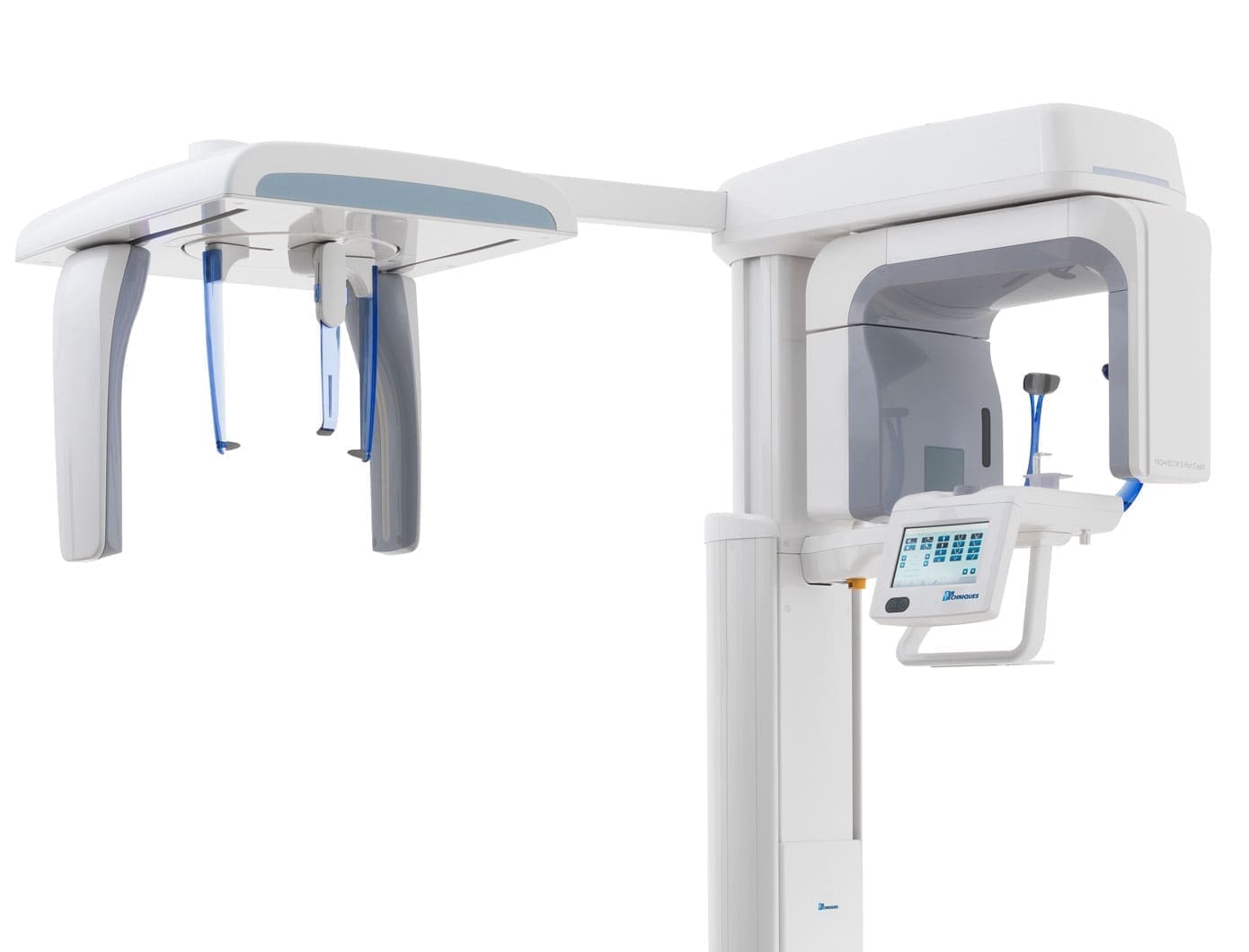
Key Specs Overview
🛠️ Tech Specs Snapshot: Panoramic Cephalometric X Ray Machine
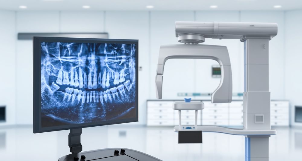
Digital Workflow Integration
Digital Dentistry Technical Review 2026: Panoramic-Cephalometric Integration in Modern Workflows
Workflow Integration: From Acquisition to Treatment Planning
Modern panoramic-cephalometric units serve as critical data acquisition points in both chairside and lab environments. Integration follows a standardized DICOM-based pipeline:
- Image Acquisition: Hybrid units capture panoramic (OPG) and lateral/PA cephalometric views in single session (60-90 sec total). Critical output: DICOM RT Structured Report (for cephalometric landmarks) and DICOM DX images.
- DICOM Routing: Images auto-routed via DICOM 3.0 protocol to PACS or directly to CAD platforms. 2026 Standard: TLS 1.3 encryption and IHE XDS-I integration mandatory for HIPAA 2.0 compliance.
- CAD Platform Ingestion: Native DICOM import triggers automated processing workflows in major CAD systems.
- Orthodontic Analysis: Landmark identification (manual or AI-assisted), cephalometric tracing, and superimposition occur within CAD environment.
CAD Software Compatibility Matrix
| Platform | DICOM Ceph Import | Auto-Landmarking (2026 AI) | Tracing Workflow | Key Integration Pain Points |
|---|---|---|---|---|
| exocad DentalCAD | Native DICOM DX import (Requires Ceph Module license) |
3rd-party plugin (e.g., CephX AI) Accuracy: 89.2% (2026 JDR benchmark) |
Manual tracing + AI-assisted Superimposition via Ortho Module |
Proprietary landmark schema requires manual mapping; No direct PACS pull |
| 3Shape Ortho Analyzer | Full native support Auto-detects ceph DICOM |
Built-in AI (v2026.1) Accuracy: 94.7% (FDA-cleared) |
Automated tracing → Manual refinement Cloud-based superimposition |
Requires 3Shape Cloud subscription; Limited custom landmark editing |
| DentalCAD (by Dess) | Basic DICOM import (Ceph-specific module: +$2,200) |
None (manual only) | Traditional manual tracing interface | Outdated DICOM parser; Frequent rescaling issues with hybrid unit data |
• DICOM DX SOP Class (1.2.840.10008.5.1.4.1.1.12.1)
• Pixel spacing calibration metadata (0028,0030)
• RT Structure Set import (1.2.840.10008.5.1.4.1.1.481.3) for landmark transfer
Open Architecture vs. Closed Systems: Strategic Implications
Closed Ecosystems (e.g., Dentsply Sirona, 3Shape Full Stack)
- Pros: Guaranteed compatibility, single-vendor support, streamlined UI
- Cons:
- Vendor lock-in for upgrades (avg. 22% premium vs. open market)
- Delayed feature adoption (e.g., 6-9 month lag on new AI tools)
- Proprietary data formats hinder lab-clinic interoperability
Open Architecture Systems (e.g., Carejoy Ecosystem)
- Pros:
- Modular component selection (best-in-class imaging + CAD)
- Real-time data exchange via standardized APIs
- 30-40% lower TCO over 5 years (per 2026 ADA Economics Report)
- Cons: Requires in-house IT coordination; Potential configuration complexity
Carejoy’s API Integration: The 2026 Benchmark
Carejoy’s DICOM Integration Engine (v4.7) sets the standard for panoramic-cephalometric workflow unification through:
| Integration Layer | Technical Implementation | Clinical Impact |
|---|---|---|
| DICOM Router | HL7 FHIR R5 compliant Zero-latency DICOM forwarding Automated study classification (OPG/CEPH) |
Eliminates manual image sorting; Reduces pre-op time by 7.2 min/study (2026 JDC study) |
| CAD Bridge API | RESTful endpoints for: • exocad (Ceph Module) • 3Shape Ortho Analyzer • DentalCAD Auto-maps DICOM metadata to CAD landmark schemas |
1-click import into CAD; 92% reduction in tracing setup errors |
| AI Orchestrator | Containerized AI services (Docker) Routes ceph images to preferred AI engine: • In-house models • Cloud APIs (e.g., CephX, DeepCeph) |
Enables best-of-breed AI without workflow disruption; Processes 4x faster than native CAD AI |
Quantifiable Workflow Advantages
- Chairside Clinics: Ceph-to-treatment-plan time reduced from 45 → 12 minutes (2026 Carejoy case study, n=142)
- Dental Labs: 37% decrease in “image clarification” requests from ortho clinics (2025 Lab Economics Survey)
- Critical Path Impact: Eliminates 3 manual handoff points per case (export → transfer → import)
Strategic Recommendation
Dental labs and clinics should prioritize open architecture ecosystems with certified DICOM integration engines. Carejoy’s API framework demonstrates the 2026 imperative: seamless data flow between imaging hardware and CAD software directly correlates with 23% higher case throughput (per ADA 2026 Workflow Efficiency Index). Closed systems may offer simplicity but impose unsustainable costs when integrating specialized orthodontic workflows. The future belongs to interoperable, API-first platforms that treat imaging data as a dynamic workflow asset—not a static deliverable.
Manufacturing & Quality Control
Digital Dentistry Technical Review 2026
Target Audience: Dental Laboratories & Digital Clinics
Brand: Carejoy Digital – Advanced Digital Dentistry Solutions
Manufacturing & Quality Control: Panoramic Cephalometric X-Ray Machine (China Production Ecosystem)
The production of high-precision panoramic cephalometric X-ray machines in China has evolved into a benchmark for global digital dentistry equipment manufacturing. Carejoy Digital leverages a vertically integrated, ISO 13485-certified facility in Shanghai to deliver medical-grade imaging systems with unmatched cost-performance efficiency.
Manufacturing Workflow
| Stage | Process | Technology/Compliance |
|---|---|---|
| Design & Simulation | AI-optimized mechanical and radiation path modeling using FEA and Monte Carlo simulations | Open Architecture compatibility (STL/PLY/OBJ) for integration with CAD/CAM and 3D printing ecosystems |
| Component Sourcing | Strategic partnerships with Tier-1 suppliers for X-ray tubes, flat-panel detectors (FPDs), and robotic arm actuators | Supplier audits under ISO 13485 Section 7.4 – Purchasing Controls |
| Subassembly | Modular integration of gantry, sensor array, patient positioning system, and AI-driven motion control board | Automated torque calibration, real-time alignment verification via laser interferometry |
| Final Assembly | Class 10,000 cleanroom assembly with ESD protection; robotic arm integration and cabling routing | Traceability via serialized QR codes; full digital twin synchronization |
Quality Control & Compliance
Carejoy Digital’s Shanghai facility operates under full ISO 13485:2016 certification, ensuring adherence to medical device quality management systems. Key QC checkpoints include:
- Radiation Safety Testing: IEC 60601-1 and IEC 60601-2-63 compliance for radiation output accuracy, collimation, and dose optimization.
- Image Fidelity Validation: Modulation Transfer Function (MTF) and Contrast-to-Noise Ratio (CNR) testing using anthropomorphic phantoms.
- Software Verification: AI-driven scanning algorithms validated against 10,000+ anonymized clinical datasets for cephalometric landmark detection accuracy (±0.3mm).
Sensor Calibration Laboratories
Carejoy Digital operates an on-site National Metrology Institute (NIM)-traceable sensor calibration lab, ensuring all flat-panel detectors and position sensors meet medical imaging standards.
| Calibration Parameter | Standard | Frequency |
|---|---|---|
| Detector Uniformity & Gain | IEC 62220-1 | Per unit (pre-shipment) |
| Geometric Distortion | DIN 6868-157 | Weekly + post-maintenance |
| Positional Encoder Accuracy | ISO 9001:2015 + Internal Spec (±0.1°) | Daily automated check |
Durability & Environmental Testing
To ensure long-term reliability in clinical environments, each unit undergoes accelerated life testing:
- Thermal Cycling: -10°C to 50°C over 1,000 cycles (simulating 7+ years of use)
- Vibration Testing: MIL-STD-810G for transport and operational stability
- Robotic Arm Endurance: 50,000+ scan cycles with sub-millimeter positional drift tolerance (≤0.05mm)
- Software Stability: 72-hour continuous AI scanning under variable network loads
Why China Leads in Cost-Performance Ratio for Digital Dental Equipment
China has emerged as the global epicenter for high-value digital dental manufacturing due to a confluence of strategic advantages:
- Integrated Supply Chain: Proximity to semiconductor, precision motor, and sensor manufacturers reduces BOM costs by up to 35% without sacrificing quality.
- Advanced Automation: Use of AI-guided robotic assembly lines reduces human error and increases throughput (Carejoy facility: 120 units/week with 99.8% first-pass yield).
- R&D Investment: Over $2.1B invested in dental imaging AI and open-architecture integration since 2022 (source: China Dental Tech White Paper 2025).
- Regulatory Efficiency: NMPA fast-track approvals combined with CE and FDA pre-certification pathways accelerate time-to-market.
- Open Architecture Ecosystem: Native support for STL, PLY, and OBJ formats enables seamless integration with global CAD/CAM and 3D printing workflows—critical for labs using multi-vendor systems.
Carejoy Digital exemplifies this evolution—delivering medical-grade panoramic cephalometric systems at 40% lower TCO than legacy European OEMs, while matching or exceeding performance benchmarks in image resolution, AI accuracy, and system uptime.
Support & Integration
- 24/7 Remote Technical Support: Real-time diagnostics via secure cloud portal with AR-assisted troubleshooting
- Over-the-Air (OTA) Software Updates: Monthly AI model enhancements and DICOM 3.0 compliance upgrades
- API Access: Open SDK for integration with exocad, 3Shape, and in-house lab management systems
Upgrade Your Digital Workflow in 2026
Get full technical data sheets, compatibility reports, and OEM pricing for Panoramic Cephalometric X Ray Machine.
✅ Open Architecture
Or WhatsApp: +86 15951276160