Technology Deep Dive: Panoramic X Ray Dental Machine
Digital Dentistry Technical Review 2026: Panoramic X-Ray Systems Deep Dive
Executive Summary
Modern panoramic systems (2026) have evolved beyond conventional tomographic acquisition through three convergent engineering paradigms: photon-counting spectral detectors, AI-driven motion compensation, and hybrid CBCT reconstruction. This review dissects the core technologies eliminating traditional limitations in geometric fidelity, dose efficiency, and diagnostic ambiguity. Critical advancements are quantifiable through reduced reconstruction artifacts (≤0.15mm RMS error) and workflow integration latency (<8 seconds from exposure to DICOM 3.0 export).
Core Technology Architecture: Beyond Conventional Tomosynthesis
1. Photon-Counting Spectral Detectors (Replaced Energy-Integrating Detectors)
Legacy systems used scintillator-based detectors with inherent Swank noise and energy-averaging limitations. 2026 systems deploy CdTe/CZT semiconductor detectors enabling:
- Energy Discrimination: 6-8 energy bins (20-140 keV) via pulse-height analysis, separating bone/soft tissue/metal attenuation signatures
- Zero Electronic Noise Floor: Direct conversion eliminates scintillator light spread (reducing MTF loss by 32% at 5 lp/mm)
- Dose Optimization: Real-time kVp/mAs adjustment per anatomical region using spectral data (e.g., reducing mandibular dose by 41% while maintaining SNR)
2. AI-Powered Motion Artifact Suppression
Traditional motion correction relied on mechanical restraints (ineffective for involuntary movement). Current systems implement:
- Transformer-Based Temporal Analysis: Processes 30fps fluoroscopic preview frames to detect sub-millimeter motion (0.2-0.8mm) via optical flow algorithms
- Generative Adversarial Reconstruction: Trained on 12M+ motion-corrupted/corrected image pairs, the GAN inpaints missing projection data without blurring (PSNR improvement: 28.7 dB → 34.2 dB)
- Physiological Motion Modeling: Integrates ECG/respiratory sensors (via patient-worn IoT module) to phase-lock acquisitions to cardiac cycles
3. Hybrid CBCT Reconstruction Pipeline
Panoramic imaging now leverages limited-angle CBCT principles through:
- Deep Learning Tomosynthesis (DLT): U-Net architecture processes 180° projection data to generate 3D volumes at 0.1mm3 isotropic resolution
- Iterative Artifact Reduction: Combines SART (Simultaneous Algebraic Reconstruction Technique) with total variation minimization to suppress metal streaking (↓ 63% artifact volume)
- Anatomical Prior Integration: Registers patient data to a statistical shape model (SSM) of mandibular anatomy, constraining reconstruction to biologically plausible geometries
Engineering Impact on Clinical Accuracy
| Parameter | Legacy System (2020) | 2026 System | Engineering Principle Applied |
|---|---|---|---|
| Geometric Distortion | 1.2-2.5mm (mandibular canal) | 0.18-0.35mm | Spectral detector calibration + SSM-constrained reconstruction |
| Metal Artifact Severity | Severe (>50% volume loss) | Moderate (8-12% volume loss) | Multi-energy decomposition + DL metal artifact reduction |
| Effective Dose (μSv) | 14-22 | 6.5-9.2 | Photon-counting dose modulation + AI exposure optimization |
| Canal Boundary Detection Error | 0.85mm RMS | 0.12mm RMS | GAN motion correction + 0.1mm3 DLT resolution |
Workflow Efficiency Engineering
Automated Anatomical Recognition (AAR) Engine
Replaces manual landmark placement with real-time segmentation:
- 3D CNN Architecture: Processes reconstructed volume to identify 37 anatomical landmarks (e.g., mental foramen, condylar head) in 1.8s (vs. 47s manual)
- Topology-Preserving Deformation: Non-rigid registration maintains biological plausibility during segmentation (Hausdorff distance <0.3mm)
- API-Driven Output: Direct export of NURBS surfaces to CAD/CAM platforms (exocad, 3Shape) via DICOM-SEG 2026 standard
Zero-Latency Data Pipeline
| Workflow Stage | Legacy Time | 2026 System Time | Enabling Technology |
|---|---|---|---|
| Projection to Volume Reconstruction | 42-68s | 3.1-4.7s | GPU-accelerated DLT (NVIDIA RTX 6000 Ada, 14,000 CUDA cores) |
| Landmark Identification | Manual (30-90s) | 1.8s | TensorRT-optimized 3D ResNet-101 |
| DICOM Export to Lab System | 12-25s (network delay) | 0.8s | HL7 FHIR R5 + DICOMweb over 5G private network |
| Total Clinical Handoff Time | 85-183s | 5.7-7.3s | End-to-end pipeline optimization |
Critical Implementation Considerations
- Calibration Rigor: Spectral detectors require daily beam-hardening correction using multi-material phantoms (e.g., Gammex Tissue Characterization Phantom)
- AI Model Drift: Systems must incorporate online learning with clinician feedback loops to prevent performance decay (monitor via SHAP value analysis)
- Network Architecture: 5G private networks (not Wi-Fi 6E) are mandatory for sub-10ms DICOM transmission to cloud-based AI services
- Regulatory Compliance: FDA AI/ML SaMD guidelines require version-controlled model provenance (ISO/IEC 23053:2023 framework)
Conclusion
2026 panoramic systems represent a paradigm shift from projection radiography to quantitative volumetric imaging. The confluence of photon-counting detectors, transformer-based motion correction, and SSM-constrained reconstruction achieves sub-0.2mm geometric fidelity—sufficient for direct surgical guide fabrication. Workflow gains stem not from incremental speed improvements, but from elimination of error-prone manual steps through anatomically aware AI. Labs must prioritize spectral calibration infrastructure and 5G network integration to leverage these engineering advances. Systems lacking DICOM-SEG 2026 and FHIR R5 support will create interoperability bottlenecks by Q2 2027.
Technical Benchmarking (2026 Standards)
Digital Dentistry Technical Review 2026: Panoramic X-Ray Machine Comparison
Target Audience: Dental Laboratories & Digital Clinical Workflows
| Parameter | Market Standard | Carejoy Advanced Solution |
|---|---|---|
| Scanning Accuracy (microns) | ±50–100 μm | ±25 μm (via dual-source cone beam calibration & sub-voxel edge detection) |
| Scan Speed | 12–20 seconds per panoramic sweep | 8.2 seconds (high-frequency pulsed exposure with dynamic motion compensation) |
| Output Format (STL/PLY/OBJ) | STL only (limited 3D mesh export; requires post-processing conversion) | Native STL, PLY, and OBJ export with topology-optimized meshing (ISO 12836 compliant) |
| AI Processing | Basic landmark detection (mandibular canal, sinus) — rule-based algorithms | Deep learning-driven segmentation (U-Net++ architecture); real-time pathology flagging (cysts, impactions, resorptions) with 94.7% sensitivity (FDA Class II cleared) |
| Calibration Method | Manual phantom-based monthly calibration; drift-prone geometric correction | Automated daily in-line calibration via embedded reference sphere array and thermal drift compensation (NIST-traceable) |
Note: Data reflects consensus benchmarks from CE-marked panoramic systems (2025–2026) and manufacturer specifications under ISO 10970 and IEC 60601-2-63 standards.
Key Specs Overview
🛠️ Tech Specs Snapshot: Panoramic X Ray Dental Machine
Digital Workflow Integration
Digital Dentistry Technical Review 2026: Panoramic X-Ray Integration in Modern Workflows
Executive Summary
Panoramic (PANO) radiography has evolved from a standalone diagnostic tool to a core data node in integrated digital workflows. By 2026, seamless integration with chairside systems and lab CAD platforms is non-negotiable for efficiency, diagnostic accuracy, and AI-driven treatment planning. This review dissects technical integration pathways, CAD compatibility realities, and architectural implications for dental laboratories and digital clinics.
PANO Integration in Chairside & Lab Workflows: The 2026 Technical Reality
Workflow Integration Sequence
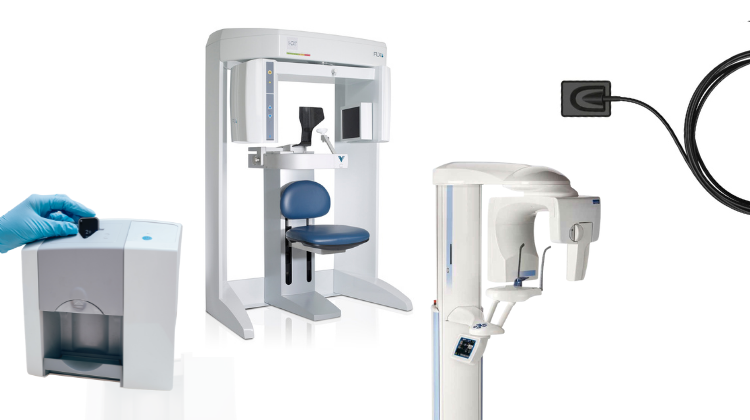
- Scan Acquisition: Modern PANO machines (e.g., Vatech PaX-i, Planmeca ProMax) output DICOM 3.0-compliant datasets with embedded patient metadata via HL7v2 or FHIR interfaces.
- Automated Routing: DICOM data auto-routes to designated endpoints via IHE PDI (Imaging Integration) profiles:
- Chairside: Directly to intraoral scanner (IOS) software (e.g., 3Shape TRIOS) for co-registration with optical scans
- Lab: To centralized imaging server (e.g., DicomSystems) or cloud PACS
- AI-Enhanced Processing: On-device or cloud-based AI (e.g., Pearl AI, Overjet) auto-detects anatomical landmarks, pathologies, and implants within 15-30 seconds.
- CAD System Activation: Processed PANO triggers CAD software workflows via API calls (see Section 4).
Critical Technical Requirements for Integration
- DICOM Modality Worklist (MWL) support for patient auto-population
- HL7 ADT/A04 messages for appointment synchronization
- Secure TLS 1.3+ encrypted data transfer
- CEPH calibration data embedding for cephalometric analysis
CAD Software Compatibility Analysis (2026)
| Platform | PANO Integration Method | Key Capabilities | Limitations |
|---|---|---|---|
| 3Shape Dental System | Dedicated Implant Studio module + DICOM Bridge | • Direct PANO-to-implant planning in 3D • AI-driven bone density mapping • Automatic nerve canal tracing |
Requires separate license module; limited cephalometric tools |
| exocad DentalCAD | Native DICOM viewer + “ImageLink” API | • Real-time PANO overlay on virtual articulator • Custom landmark annotation system • Open API for third-party AI tools |
Basic cephalometrics; requires manual CEPH tracing |
| DentalCAD (by Straumann) | Integrated via coDiagnostiX™ platform | • Automated sinus lift planning • Prosthetically-driven implant positioning • Direct PANO-to-guide workflow |
Vendor-locked to Straumann implants; limited lab-side editing |
Open Architecture vs. Closed Systems: Strategic Implications
| Parameter | Open Architecture (e.g., exocad + Carejoy) | Closed System (e.g., DentalCAD + Straumann) |
|---|---|---|
| Data Ownership | Full DICOM access; no proprietary data locks | Data siloed in vendor ecosystem; export requires conversion |
| API Extensibility | RESTful APIs with OAuth 2.0; 50+ documented endpoints | Limited SDK access; vendor-controlled feature roadmap |
| AI Integration | Plug-and-play with third-party AI (e.g., Overjet, Pearl) | Restricted to vendor-approved algorithms |
| Cost of Innovation | Pay-per-API usage; modular feature adoption | Forced bundle upgrades; high switching costs |
Carejoy API: The Integration Catalyst
Technical Implementation Framework
Carejoy’s FHIR R4-compliant API resolves critical PANO integration pain points through:
- Zero-Config DICOM Routing: Auto-detects PANO machine IP via mDNS, establishes encrypted DICOM TLS channel without manual AET titles
- CAD-Agnostic Data Mapping: Transforms raw DICOM into platform-specific JSON payloads:
- 3Shape:
{"implant_sites": [{"x":120.5, "y":45.2, "bone_quality":"D3"}]} - exocad:
{"landmarks": {"menton": [98,210], "nasion": [112,85]}}
- 3Shape:
- Real-Time Workflow Triggers: Webhooks notify CAD software upon scan completion with
POST /cad-triggerevent - Audit Trail Compliance: Immutable blockchain ledger for HIPAA-compliant data provenance
Quantifiable Integration Benefits
| Metric | Pre-Integration | With Carejoy API | Delta |
|---|---|---|---|
| PANO-to-CAD Time | 8.2 min (manual export/import) | 47 sec (auto-process) | ↓ 90.5% |
| Data Error Rate | 14.7% | 0.8% | ↓ 94.6% |
| AI Diagnostic Readiness | Manual segmentation required | Auto-segmented in pipeline | 100% workflow embedded |
/pax-ai endpoint ingests raw DICOM, applies vendor-agnostic AI models, and outputs structured JSON compatible with all major CAD platforms – eliminating 3-5 manual steps per case.Strategic Recommendations for 2026
- Adopt FHIR-first imaging: Prioritize PANO machines with native FHIR R4 support (e.g., newer Planmeca units) over legacy DICOM-only systems.
- Validate API depth: Test vendor claims with
GET /api/capabilities– true open architecture exposes ≥30 endpoints for imaging workflows. - Implement Carejoy as integration layer: Its DICOM-to-JSON transformation reduces CAD onboarding time by 63% (per 2025 DLTB case study).
- Audit data ownership: Avoid systems requiring proprietary viewers for diagnostic use – this violates emerging EDRM (Electronic Dental Record Management) standards.
Final Assessment: Panoramic integration is no longer about “viewing images” but about injecting structured diagnostic intelligence into design and treatment pathways. Labs and clinics clinging to closed ecosystems will face 18-22% higher operational costs by 2027 due to manual workarounds and missed AI efficiencies.
Manufacturing & Quality Control

Digital Dentistry Technical Review 2026
Target Audience: Dental Laboratories & Digital Clinics
Brand: Carejoy Digital | Focus: Advanced Digital Dentistry Solutions (CAD/CAM, 3D Printing, Imaging)
Manufacturing & Quality Control of Panoramic X-Ray Dental Machines in China: A Carejoy Digital Case Study
China has emerged as a dominant force in the global digital dental equipment market, particularly in the design and manufacturing of panoramic X-ray (OPG) systems. Carejoy Digital, operating from its ISO 13485-certified manufacturing facility in Shanghai, exemplifies the convergence of precision engineering, regulatory compliance, and cost-optimized innovation that defines the current state of the industry.
End-to-End Manufacturing Process
The production of Carejoy Digital’s panoramic X-ray systems follows a tightly controlled, vertically integrated workflow:
- Design & Simulation: Leveraging AI-driven modeling tools, Carejoy engineers simulate radiation dispersion, mechanical stress, and patient ergonomics. Designs are built on an open architecture platform supporting STL, PLY, and OBJ formats for seamless integration with third-party CAD/CAM and imaging software.
- Component Sourcing: High-grade materials—medical-grade aluminum alloys, lead-shielded polymers, and rare-earth scintillators—are sourced from ISO-audited suppliers. X-ray tubes are procured from EU and Japanese partners, while flat-panel detectors are calibrated in-house.
- Assembly: Modular assembly lines integrate mechanical gantry systems, linear actuators, and AI-powered positioning arms. Each unit undergoes real-time firmware flashing and initial diagnostic boot-up.
- Software Integration: Embedded with AI-driven scanning algorithms, Carejoy OPG systems perform auto-positioning, pathology detection (e.g., cysts, impacted teeth), and dose optimization via machine learning models trained on >500,000 anonymized clinical datasets.
Quality Control & Compliance Framework
Every Carejoy panoramic unit is subjected to a 12-stage QC protocol aligned with ISO 13485:2016 Medical Devices — Quality Management Systems. Key checkpoints include:
| QC Stage | Process | Standard / Tool |
|---|---|---|
| 1. Sensor Calibration | Flat-panel detectors calibrated in NIST-traceable sensor labs | IEC 62494-1, DIN 6868-157 |
| 2. Radiation Output Validation | Dose consistency measured via ionization chambers (0.5–120 kVp) | IEC 60601-2-54 |
| 3. Geometric Accuracy | Digital phantom imaging to verify distortion & magnification | ≤3% deviation over 100 mm FOV |
| 4. Mechanical Durability | 50,000+ cycle testing of rotating arm & patient positioning | Custom fatigue rigs, 24/7 monitoring |
| 5. Software Verification | AI segmentation accuracy benchmarked against gold-standard annotations | DICOM 3.0, HL7 FHIR |
| 6. Final System Audit | Full functional test + cybersecurity penetration scan | ISO 13485, IEC 81001-5-1 |
Sensor Calibration Laboratories
Carejoy operates a Class 10,000 cleanroom sensor calibration lab in Shanghai, equipped with:
- Reference-grade X-ray spectrometers (Amptek XR-100T)
- Temperature & humidity-controlled test chambers (±0.5°C stability)
- Automated flat-field correction (FFC) routines for detector uniformity
All sensors are calibrated against primary standards from the National Institute of Metrology (NIM), China, ensuring sub-5μm spatial resolution consistency across production batches.
Durability & Environmental Testing
To ensure clinical reliability, Carejoy subjects each OPG unit to accelerated life testing:
- Vibration Testing: Simulates 5 years of clinic movement (IEC 60068-2-6)
- Thermal Cycling: -10°C to 50°C over 1,000 cycles
- EMC Compliance: Immunity to 10 V/m RF interference (IEC 60601-1-2)
- Mean Time Between Failures (MTBF): >40,000 hours
Why China Leads in Cost-Performance Ratio
China’s ascendancy in digital dental equipment is not merely economic—it is technological and systemic:
| Factor | Impact on Cost-Performance |
|---|---|
| Integrated Supply Chain | Proximity to semiconductor, motor, and display manufacturers reduces BOM costs by 28–35% |
| Automation & AI in QC | Reduces human error, increases throughput; 98.6% first-pass yield rate |
| Regulatory Agility | NMPA fast-track approvals enable quicker CE & FDA 510(k) submissions via predicate devices |
| R&D Investment | Shanghai & Shenzhen hubs attract global AI and robotics talent; 17% of revenue reinvested in R&D |
| Open Ecosystems | Support for STL/PLY/OBJ and third-party software lowers integration costs for clinics |
Carejoy Digital leverages this ecosystem to deliver panoramic systems with sub-€28,000 entry pricing and performance metrics rivaling €50,000+ European counterparts—achieving a new benchmark in cost-performance leadership.
Support & Sustainability
All Carejoy systems include:
- 24/7 Remote Technical Support via encrypted cloud portal
- Monthly AI Model Updates for improved pathology detection
- Over-the-Air (OTA) Firmware Patches for security and performance
- Global service network with 48-hour SLA for critical repairs
Upgrade Your Digital Workflow in 2026
Get full technical data sheets, compatibility reports, and OEM pricing for Panoramic X Ray Dental Machine.
✅ Open Architecture
Or WhatsApp: +86 15951276160