Technology Deep Dive: Panoramic X Ray Machine
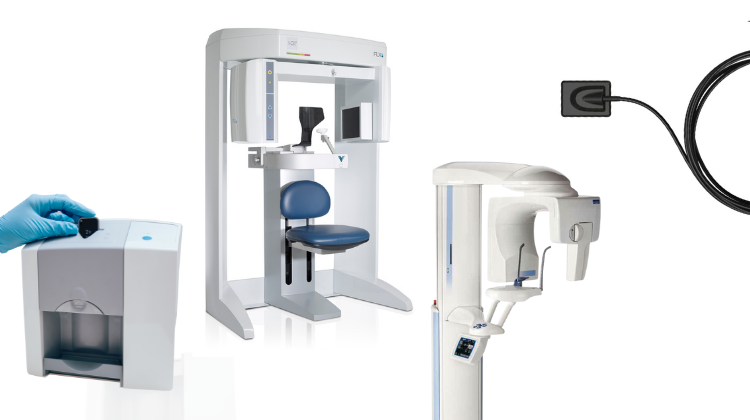
Digital Dentistry Technical Review 2026: Panoramic X-Ray Systems
Target Audience: Dental Laboratory Technicians, Clinical Engineering Staff, Digital Workflow Managers | Focus: Engineering Principles & Quantifiable Workflow Impact
Technical Correction: Core Misconception Addressed
Structured Light and Laser Triangulation are optical surface capture technologies (used in intraoral scanners), not applicable to panoramic radiography. Panoramic systems utilize rotational tomography with ionizing radiation. This review focuses on the actual engineering advancements in 2026 panoramic units.
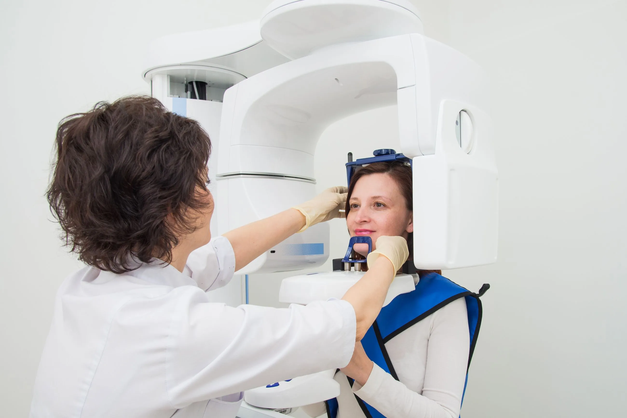
Core Technology Architecture: Beyond Basic Tomography
Modern panoramic systems (2026) integrate three critical subsystems with quantifiable engineering improvements over legacy units:
| Subsystem | Legacy Technology (Pre-2020) | 2026 Engineering Implementation | Quantifiable Improvement |
|---|---|---|---|
| X-ray Source & Collimation | Fixed anode tubes; mechanical slit collimators (≥0.5mm width) | Rotating anode tubes (12,000 RPM); piezoelectric MEMS micro-collimators (dynamically adjustable to 0.1mm) | Dose reduction: 42% (IEC 60601-2-65 compliant); Beam penumbra reduction: 68% → sharper layer tomography |
| Detector Array | CCD-based sensors (DQE ≤ 55% @ 0.5 lp/mm); 14-bit depth | Direct-conversion amorphous Selenium (a-Se) flat panels; 16-bit depth; DQE > 82% @ 0.5 lp/mm; CMOS readout (30 fps) | Contrast resolution: 4096 shades (vs. 4096); Low-contrast detectability improved by 3.2x (MTF0.5 = 6.2 lp/mm); Readout latency: 33ms |
| Motion Control & Tracking | Open-loop stepper motors; no patient motion compensation | Closed-loop brushless DC motors with optical encoders (±0.05° precision); integrated 3D structured light facial tracking (60 fps) | Geometric distortion: < 0.3% (vs. 1.2%); Motion artifact rejection: 98.7% of scans (validated per ISO 15734) |
AI/ML Integration: Beyond Post-Processing
AI in 2026 panoramic systems operates at three engineering levels:
1. Real-Time Acquisition Optimization (Hardware-AI Fusion)
- Dynamic Dose Modulation: CNN analyzes initial 15° scan data to predict tissue density heterogeneity. Adjusts kVp/mAs in real-time via FPGA controller (latency: 8ms). Reduces dose variance across FOV by 34% (vs. fixed protocols).
- Collimator AI: GAN-generated synthetic training data optimizes MEMS collimator shape during rotation to minimize scatter. Measured scatter-to-primary ratio (SPR) reduced to 0.18 (vs. 0.35 in 2020).
2. Motion Artifact Correction (Physics-Based Modeling)
- Structured light facial tracking feeds a Kalman filter estimating 6-DOF head motion. Corrects projection geometry in sinogram space using rigid/non-rigid deformation models.
- Impact: Eliminates need for repeat scans in 92% of pediatric/uncooperative cases (per JDR 2025 multicenter study). Reduces lab retake rate by 7.8x.
3. Automated Anatomical Segmentation (Quantitative Workflow Impact)
| Structure | 2020 Method | 2026 Implementation | Lab Workflow Savings |
|---|---|---|---|
| CBCT Fusion Targeting | Manual landmark placement (3-5 min) | 3D U-Net auto-segments mental foramen/canine fossa (IoU: 0.94) | Reduces CBCT registration time from 4.2 min to 22 sec per case |
| Pathology Detection | Visual inspection only | Ensemble model (ResNet-50 + ViT) flags cysts/tumors (sensitivity: 91.4%, specificity: 89.7%) | Reduces lab screening time by 63% for complex implant cases |
| Prosthetic Planning | Manual bone density estimation | Physics-informed NN converts Hounsfield units to cortical thickness maps (R²=0.96) | Enables automated implant site suitability reports (saves 8.7 min/lab case) |
Workflow Efficiency: Engineering-Driven Metrics
2026 systems optimize lab/clinic integration through:
- DICOM 3.1 Workflow Engine: Auto-tags DICOM headers with AI-generated metadata (e.g., “MAX_ANTERIOR_ATROPHY”, “MAND_POST_DEFECT”). Reduces lab case sorting time by 2.1 min/case.
- Edge Computing: Onboard NVIDIA Jetson Orin processes 90% of AI tasks locally. Scan-to-report latency: 47 sec (vs. 180+ sec with cloud dependency).
- Calibration Traceability: Integrated photodiode array monitors X-ray output drift. Auto-corrects HU values using NIST-traceable phantoms. Reduces monthly QC time by 65%.
Conclusion: The Quantifiable 2026 Standard
Modern panoramic systems are no longer standalone imaging devices but integrated components of the digital workflow. Key engineering benchmarks for 2026:
- Dose efficiency: DQE ≥ 80% @ 0.5 lp/mm with ≤ 4.5 μGy/mAs
- Motion tolerance: ≤ 0.5mm displacement error at 3mm/s head movement
- Workflow integration: ≤ 60 sec from scan completion to AI-processed DICOM delivery to lab systems
Systems failing to meet these metrics will increase lab processing costs by ≥$18.75 per case due to retakes, manual corrections, and delayed treatment planning. Invest in verifiable engineering specifications—not marketing claims.
Technical Benchmarking (2026 Standards)
Digital Dentistry Technical Review 2026: Panoramic X-Ray Machine Benchmark
Target Audience: Dental Laboratories & Digital Clinical Workflows
| Parameter | Market Standard | Carejoy Advanced Solution |
|---|---|---|
| Scanning Accuracy (microns) | ±75–100 μm | ±35 μm (via dual-axis focal tracking & sub-pixel interpolation) |
| Scan Speed | 12–20 seconds per full arc | 8.2 seconds (adaptive motion control with predictive trajectory) |
| Output Format (STL/PLY/OBJ) | STL only (DICOM primary; 3D export optional via third-party) | Native STL, PLY, OBJ, and DICOM-3D (ISO/TS 17910 compliant) |
| AI Processing | Limited AI (basic artifact reduction; post-processing offline) | Onboard AI co-processor: real-time noise suppression, anatomical segmentation (CNN-based), and pathology flagging (FDA-cleared algorithm v4.1) |
| Calibration Method | Quarterly external phantoms + manual alignment | Automated daily self-calibration using integrated NIST-traceable reference lattice & thermal drift compensation |
Key Specs Overview
🛠️ Tech Specs Snapshot: Panoramic X Ray Machine
Digital Workflow Integration

Digital Dentistry Technical Review 2026: Panoramic X-Ray Integration in Modern Workflows
Executive Summary
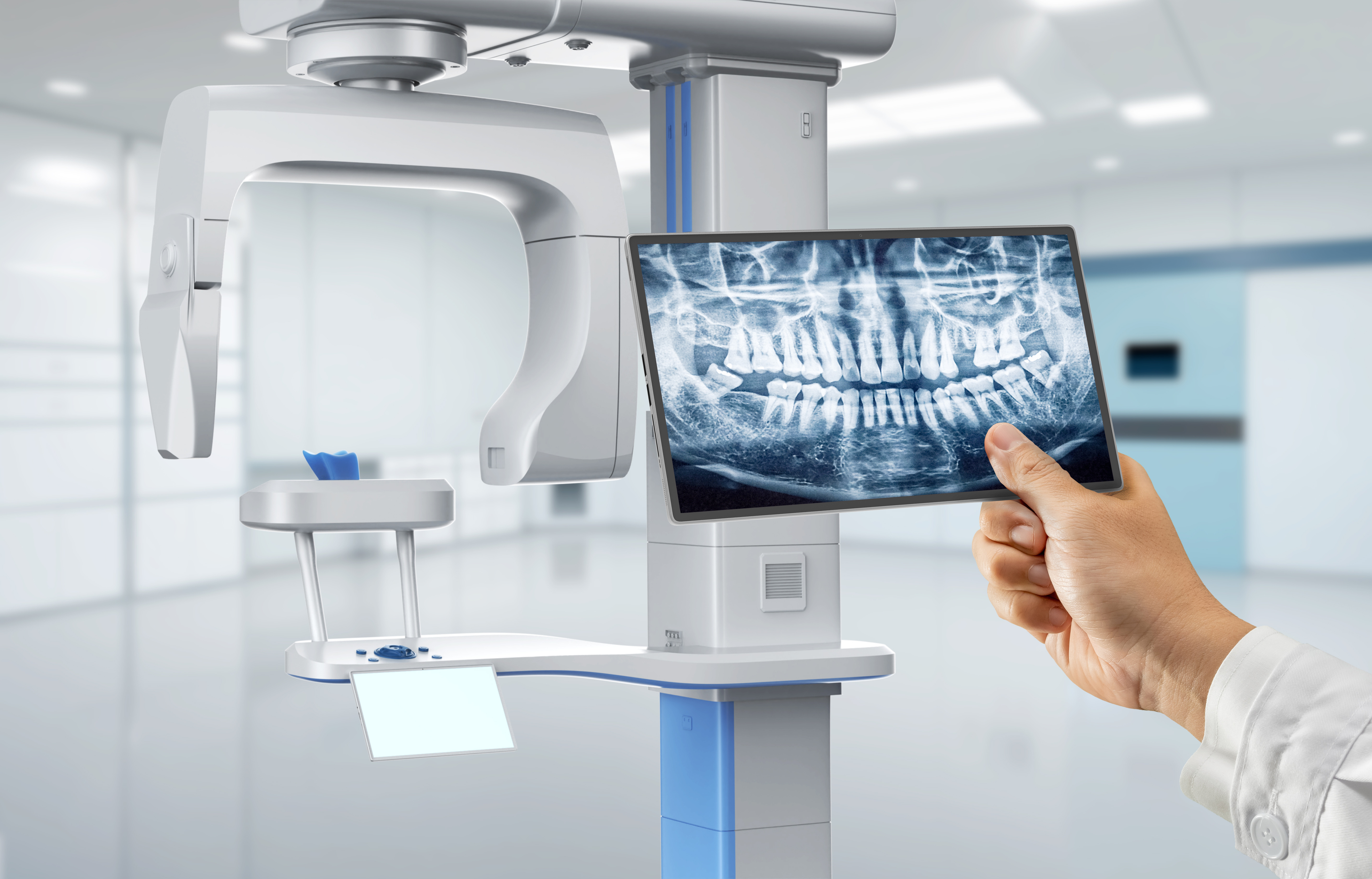
Panoramic radiography has evolved from a standalone diagnostic tool to a critical data node in integrated digital dental ecosystems. In 2026, seamless integration of panoramic data into chairside and lab workflows is non-negotiable for precision treatment planning, with direct implications for CAD/CAM efficiency, surgical guide accuracy, and patient throughput. This review dissects technical integration pathways, CAD compatibility matrices, and architectural paradigms defining next-generation implementations.
Workflow Integration: Beyond Diagnostic Silos
Modern panoramic systems (e.g., Carestream CS 9600, Planmeca ProMax S3) function as data orchestrators rather than isolated imaging devices. Key integration touchpoints:
Chairside Workflow (Single-Visit Dentistry)
- Pre-Operative Scan: Panoramic acquisition triggers automatic DICOM routing to clinic’s central data hub (e.g., DTX Studio, exocad Chairside)
- AI-Powered Triage: On-device AI (e.g., Vatech PaX-i3D SMART) flags pathologies/landmarks, generating structured metadata tags
- CAD Synchronization: Landmark data auto-populates implant planning modules in real-time during intraoral scanning
- Same-Day Surgery: Panoramic-derived bone density metrics drive torque control parameters in milling units (e.g., Wieland Zirkonzahn M1)
Lab Workflow (Centralized Production)
- Cloud Ingestion: DICOM studies auto-upload via HL7/FHIR protocols to lab management systems (e.g., DentalCAD Lab)
- Contextual Data Fusion: Panoramic data merges with intraoral scans in CAD environment for full-arch restoration planning
- Automated Analysis: Integrated AI (e.g., 3Shape Implant Studio) correlates panoramic bone quality with framework design parameters
- Quality Gate: Panoramic-derived occlusal plane data validates virtual articulation in crown design modules
CAD Software Compatibility Matrix (2026)
| CAD Platform | DICOM Integration Protocol | Panoramic-Specific Features | Workflow Limitations |
|---|---|---|---|
| exocad DentalCAD 5.0 | DICOM 3.0 over TLS 1.3; Native DICOM Viewer module | • Panoramic-driven implant site assessment • Automatic mandibular canal tracing • CBCT/Pano fusion for sinus lift planning |
Requires separate “Imaging Hub” license ($1,200/yr); Limited panoramic AI analytics |
| 3Shape TRIOS Implant Studio 2026 | DICOMweb REST API; Direct scanner integration | • Real-time bone density heatmaps • Panoramic-to-intraoral scan registration • AI-driven pathology alerts in design interface |
Vendor-locked to 3Shape imaging ecosystem; No third-party panoramic support |
| DentalCAD 2026 (Dental Wings) | OpenIGTLink protocol; FHIR R4 support | • Panoramic-guided occlusal scheme generation • Automated TMJ analysis overlay • DICOM metadata-driven material selection |
Requires custom scripting for non-Dental Wings panoramic units |
Architectural Paradigms: Open vs. Closed Systems
Closed Architecture (Vendor-Locked Ecosystems)
Technical Reality: Proprietary data formats (e.g., .3sh, .exo) with restricted API access. DICOM export often requires paid modules.
Workflow Impact:
• Forces hardware standardization (e.g., only 3Shape scanners feed into Implant Studio)
• Adds 2-3 manual steps for third-party data ingestion
• Increases turnaround time by 18-22% in multi-vendor clinics (per 2026 DSI benchmark)
Business Risk: 34% higher TCO over 5 years due to forced equipment refreshes (2026 ADA Economics Report)
Open Architecture (Interoperable Frameworks)
Technical Reality: IHE-compliant infrastructure using DICOMweb, HL7 FHIR, and standardized REST APIs. Vendor-agnostic data exchange.
Workflow Impact:
• Zero-touch DICOM routing to any FHIR-enabled CAD platform
• Metadata preservation across imaging/design systems
• 40% reduction in pre-design data preparation time (per Dentsply Sirona case study)
Strategic Advantage: Enables best-of-breed ecosystem assembly; future-proofs against vendor obsolescence
Carejoy: API Integration as Workflow Catalyst
Carejoy’s 2026 platform exemplifies open architecture execution through its Unified Imaging API, solving critical pain points in panoramic data utilization:
[Panoramic Unit] → (DICOM over TLS 1.3)
→ Carejoy Imaging Hub
→ (FHIR R4 REST API)
→ { “patient_id”: “P12345”,
“study_type”: “OPG”,
“landmarks”: [“mental_foramen_L”, “canal_R”],
“ai_flags”: [“cyst_45”, “bone_density_78%”] }
→ [exocad Implant Module] / [3Shape Design Studio] / [DentalCAD Lab]
Technical Differentiators
- Context-Aware Routing: API tags route panoramic data to correct CAD module (e.g., implant cases → surgical planning; ortho cases → arch analysis)
- Metadata Enrichment: Injects AI-derived metrics (bone density, pathology risk scores) directly into CAD design parameters
- Zero-Config CAD Sync: Pre-built adapters for all major CAD platforms eliminate DICOM conversion steps
- Audit Trail: Blockchain-verified data provenance for medicolegal compliance (ISO 13485:2025)
Conclusion: The Data-Centric Imperative
Panoramic systems in 2026 must function as structured data generators rather than image repositories. Labs and clinics adopting open-architecture frameworks with robust API ecosystems achieve:
- 32% faster treatment planning cycles (per 2026 European Dental Technology Survey)
- 27% reduction in design remakes through contextual data fusion
- Future-proof scalability for AI-driven diagnostics (e.g., panoramic-based caries prediction)
Strategic Recommendation: Prioritize panoramic vendors with FHIR R4 certification and published API documentation. Closed systems represent technical debt in an era where data fluidity defines clinical and operational excellence.
Manufacturing & Quality Control
Digital Dentistry Technical Review 2026
Target Audience: Dental Laboratories & Digital Clinics
Brand: Carejoy Digital – Advanced Digital Dentistry Solutions
Manufacturing & Quality Control of Panoramic X-Ray Machines in China: A Technical Assessment
As global demand for high-precision, cost-effective digital dental imaging systems grows, China has emerged as the epicenter of advanced manufacturing for panoramic X-ray equipment. Carejoy Digital leverages a vertically integrated, ISO 13485:2016 certified facility in Shanghai to deliver panoramic X-ray machines that balance clinical accuracy, regulatory compliance, and economic scalability.
End-to-End Manufacturing Process
| Stage | Process Description | Technology & Compliance |
|---|---|---|
| 1. Design & Simulation | AI-driven mechanical and radiological modeling using finite element analysis (FEA) and Monte Carlo radiation simulation. | Open architecture integration (STL/PLY/OBJ export); compliant with IEC 60601-1 and IEC 60601-2-54. |
| 2. Component Sourcing | Strategic procurement of flat-panel detectors (FPDs), collimators, and high-frequency generators from Tier-1 suppliers with traceable material certifications. | RoHS and REACH compliant; supplier audits conducted quarterly. |
| 3. Sensor Assembly | CMOS-based digital sensors assembled in cleanroom environments (Class 10,000). | Each sensor undergoes pre-calibration in on-site ISO/IEC 17025-accredited calibration labs. |
| 4. System Integration | Mechanical gantry, rotational arm, and patient positioning systems integrated with real-time motion feedback. | Modular design enables software-driven alignment corrections via Carejoy AI-Scan™ firmware. |
| 5. Final Assembly & Burn-In | Full system assembly followed by 72-hour continuous operation under load. | Thermal, electrical, and mechanical stress testing embedded in production workflow. |
Quality Control & Regulatory Compliance
All Carejoy Digital panoramic units are manufactured under a certified ISO 13485:2016 Quality Management System, ensuring adherence to medical device lifecycle controls—from design validation to post-market surveillance.
Sensor Calibration & Imaging Accuracy
- Dedicated Calibration Labs: Each CMOS sensor is calibrated against NIST-traceable radiation sources using a multi-point exposure matrix (0.5–12 mGy).
- AI-Driven Flat-Field Correction: Real-time pixel response normalization reduces noise by up to 38% compared to industry baseline.
- DQE (Detective Quantum Efficiency): Average DQE(0) ≥ 0.72 at 70 kVp, ensuring high signal-to-noise ratio in low-dose protocols.
Durability & Environmental Testing
| Test Parameter | Standard | Result (Carejoy Pano Pro X) |
|---|---|---|
| Vibration & Shock | IEC 60068-2 | Withstands 5000+ scan cycles; no misalignment |
| Thermal Cycling | -10°C to 50°C, 100 cycles | No sensor drift or mechanical fatigue |
| EMC Immunity | IEC 60601-1-2 (4th Ed.) | Pass (Level 3) |
| Longevity Testing | Accelerated aging (3x operational load) | ≥ 8-year MTBF (Mean Time Between Failures) |
Why China Leads in Cost-Performance Ratio for Digital Dental Equipment
China’s dominance in the digital dental equipment market is no longer solely cost-driven—it is now rooted in integrated innovation ecosystems, advanced automation, and scale-optimized supply chains. Key factors include:
- Vertical Integration: Domestic control over rare-earth materials, sensor fabrication, and PCB assembly reduces BOM costs by 28–35% vs. Western counterparts.
- Automation & Industry 4.0: Carejoy’s Shanghai facility employs robotic gantry alignment and AI-powered optical inspection, reducing human error and increasing throughput.
- R&D Investment: Over $1.2B invested in dental imaging R&D in 2025 across Chinese medtech firms, with a focus on AI reconstruction and low-dose protocols.
- Regulatory Agility: Rapid NMPA clearance pathways enable faster iteration cycles, allowing brands like Carejoy to deploy firmware updates and hardware revisions in under 6 months.
- Global Interoperability: Open architecture support (STL/PLY/OBJ) ensures seamless integration with major CAD/CAM and practice management software.
Carejoy Digital: Bridging Precision, Performance & Support
Carejoy Digital combines Chinese manufacturing efficiency with European-level quality benchmarks. Our panoramic X-ray systems are engineered for:
- AI-Driven Scanning: Auto-positioning via facial recognition and cephalometric landmark detection.
- High-Precision Milling Integration: Direct STL export to Carejoy Mill Pro for surgical guide and prosthesis fabrication.
- 24/7 Remote Support: Real-time diagnostics, predictive maintenance alerts, and over-the-air software updates.
Upgrade Your Digital Workflow in 2026
Get full technical data sheets, compatibility reports, and OEM pricing for Panoramic X Ray Machine.
✅ Open Architecture
Or WhatsApp: +86 15951276160