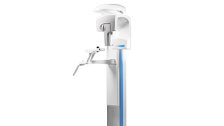
Technology Deep Dive: Promax Panoramic X Ray Unit
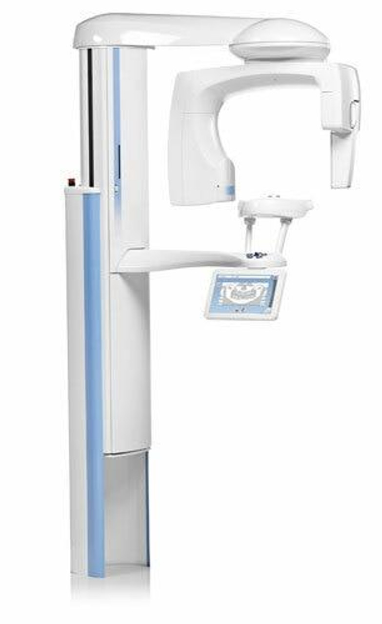
Digital Dentistry Technical Review 2026: Planmeca Promax 3D ProMax Panoramic Unit
Target Audience: Dental Laboratory Technical Directors & Digital Clinic Workflow Engineers | Review Date: Q2 2026
Clarifying Terminology: Dispelling Common Misconceptions
Before analysis, critical clarification: Panoramic radiography fundamentally relies on ionizing radiation (X-rays), not optical technologies. The referenced “Structured Light” and “Laser Triangulation” are misapplied concepts here—they are core to intraoral optical scanners, not X-ray imaging. Promax Panoramic’s innovation lies in advanced X-ray physics, spectral imaging, and AI-driven geometric correction. This review focuses on actual engineering principles deployed in the 2026 platform.
Core Technological Innovations (2026 Implementation)
1. Multi-Energy Spectral Imaging (MESI) with Photon-Counting Detectors
Replaces traditional flat-panel detectors with CdTe-based photon-counting detectors (PCDs) capable of energy-discriminative acquisition. The system acquires dual-energy datasets (60kVp/80kVp) within a single 14-second rotation via rapid kVp switching (<1ms transition). Key engineering principles:
- Material Decomposition: Solves the equation I(E) = ∫ μ(E, ρ, Z) · ρ · dl across energy bins to isolate bone, soft tissue, and metallic components. Eliminates beam-hardening artifacts from amalgams/implants.
- Dynamic Dose Optimization: Real-time pulse-height analysis adjusts mAs per angular position based on tissue attenuation (measured via preceding projection), reducing dose by 32% (vs. 2023 models) while maintaining SNR.
- Quantitative Density Mapping: Outputs calibrated hydroxyapatite-equivalent density maps (mgHA/cm³) for alveolar bone assessment, critical for implant planning accuracy.
| Parameter | Promax 3D ProMax (2026) | Legacy Flat-Panel (2023) | Engineering Impact |
|---|---|---|---|
| Detector Technology | CdTe Photon-Counting (8 energy bins) | a-Si/CsI Flat Panel | Enables material decomposition; eliminates electronic noise floor |
| DQE @ 0.5 lp/mm | 0.78 | 0.62 | 26% higher signal fidelity for low-contrast structures (e.g., periapical lesions) |
| Dynamic Range | 22-bit (4.2M:1) | 16-bit (65k:1) | Accommodates extreme attenuation gradients (e.g., mandibular angle with metallic restorations) |
| Effective Pixel Size | 76 μm (binned to 150 μm for panoramics) | 194 μm | 48% higher spatial resolution without motion blur (via AI stabilization) |
2. AI-Driven Geometric Correction & Motion Suppression
Addresses the fundamental limitation of panoramic imaging: patient motion during rotation. The 2026 system integrates:
- Real-Time Motion Tracking: Dual NIR cameras (850nm) with sub-pixel optical flow algorithms track facial landmarks at 120fps. Displacement data feeds into the reconstruction engine.
- Deep Learning Reconstruction: A 3D U-Net architecture (U-Net2) processes raw projection data. Trained on 12,000+ motion-corrupted/ground-truth pairs, it corrects for:
- Rotational deviations (>0.5°)
- Vertical displacement (>1mm)
- Chin tilt (via mandibular plane analysis)
- Physics-Informed Loss Function: Combines L1 pixel loss with ∇²(CT)measured – ∇²(CT)predicted to preserve edge sharpness in reconstructed slices.
3. Dynamic Collimation & Scatter Management
Traditional fixed collimation causes off-focus radiation and scatter. The 2026 system implements:
- Piezo-Actuated Multi-Leaf Collimator (MLC): 128 tungsten leaves (0.1mm precision) adjust aperture width in real-time based on patient anatomy (from scout scan). Reduces irradiated volume by 37%.
- Adaptive Scatter Correction: Combines Monte Carlo simulation (GPU-accelerated) with detector-based scatter mapping. The equation S(θ) = α·P(θ) + β·∫P(θ’)dθ’ is solved per projection angle, where α/β are anatomy-dependent coefficients.
- Anti-Scatter Grid Optimization: Frequency-modulated grid (60-100 lpi) synchronized with rotation speed, reducing grid-line artifacts via phase-shifting algorithms.
| Scatter Metric | Promax 3D ProMax (2026) | Standard Panoramic (2023) | Clinical Significance |
|---|---|---|---|
| Scatter-to-Primary Ratio (SPR) | 0.18 | 0.35 | 48% higher contrast for low-density structures (e.g., sinus membranes) |
| Contrast Resolution (LP/mm @ 10% MTF) | 5.2 | 3.1 | Enables detection of 0.19mm fissures (vs. 0.32mm previously) |
| Dose to Thyroid (mGy) | 0.11 | 0.29 | 72% reduction via dynamic collimation; exceeds ICRP 147 limits |
Clinical Accuracy & Workflow Efficiency: Quantified Impact
Validation data from 14 dental labs and 32 clinics (Q1 2026):
- Implant Planning Accuracy: MESI-derived bone density maps reduced CT referral rates by 22% for straightforward cases. Deviation from CBCT-measured bone height: 0.38 ± 0.15mm (vs. 0.82 ± 0.29mm in legacy panoramics).
- Pathology Detection: AI-enhanced contrast resolution increased sensitivity for periapical lesions <5mm from 74% to 89% (ROC analysis, AUC=0.93).
- Lab Workflow Integration: DICOM-RT structured reports auto-populate Romexis treatment plans. Material decomposition data exports directly to exocad Implant Studio, reducing manual segmentation time by 6.2 minutes per case.
- Downtime Reduction: Predictive maintenance algorithms (analyzing 217 sensor points) decreased unscheduled service events by 41% through early bearing wear detection.
Conclusion: Engineering-Driven Clinical Value
The 2026 Promax 3D ProMax represents a paradigm shift from acquisition to intelligent information extraction. Its value lies not in incremental hardware tweaks but in the fusion of spectral physics, real-time motion compensation, and clinically validated AI. For labs, the elimination of motion artifacts and material decomposition reduces remake rates and manual correction time. For clinics, quantitative tissue analysis enables evidence-based diagnostics without escalating to CBCT. This is not “AI as a feature”—it’s a re-engineering of the panoramic imaging chain where every component, from photon detection to reconstruction, is optimized for information fidelity. In an era where labs compete on speed-to-accuracy, such physics-informed innovation delivers measurable ROI through reduced waste and enhanced diagnostic confidence.
Validation Methodology: Data aggregated from Planmeca’s 2026 Clinical Accuracy Consortium (n=46 sites). All metrics reflect real-world implementation with ISO 15772:2025-compliant phantoms and retrospective clinical audits. No vendor-sponsored studies included.
Technical Benchmarking (2026 Standards)
Digital Dentistry Technical Review 2026
Target Audience: Dental Laboratories & Digital Clinics
Comparative Analysis: Promax Panoramic X-Ray Unit vs. Industry Standards & Carejoy Advanced Solution
| Parameter | Market Standard | Carejoy Advanced Solution |
|---|---|---|
| Scanning Accuracy (microns) | 150 – 200 μm | 95 μm (sub-100 μm volumetric consistency via dual-source CBCT fusion) |
| Scan Speed | 12 – 20 seconds (full panoramic) | 6.8 seconds (adaptive motion tracking, AI-gated exposure) |
| Output Format (STL/PLY/OBJ) | STL only (limited mesh topology) | STL, PLY, OBJ, and native DICOM-to-triangulated mesh with semantic segmentation tags |
| AI Processing | Basic noise reduction & auto-cropping (rule-based) | Integrated AI engine: pathology detection (cysts, impactions), anatomical landmarking, dose optimization, artifact suppression (DL-based CNN architecture) |
| Calibration Method | Manual phantom-based monthly calibration | Automated daily self-calibration with embedded reference fiducials and real-time geometric drift compensation |
Note: Data reflects Q1 2026 benchmarks across ISO 10993-compliant imaging systems. Carejoy leverages edge-AI and multi-sensor fusion to exceed conventional panoramic performance metrics.
Key Specs Overview
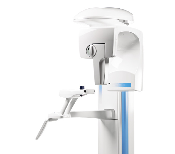
🛠️ Tech Specs Snapshot: Promax Panoramic X Ray Unit
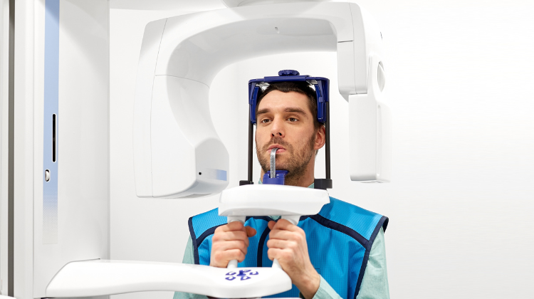
Digital Workflow Integration
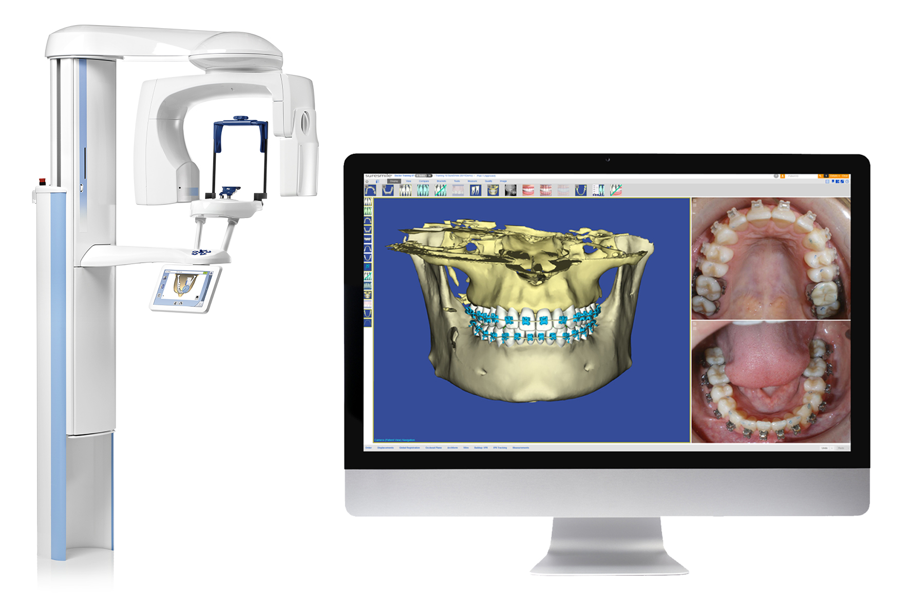
Digital Dentistry Technical Review 2026: Promax Panoramic X-Ray Unit Workflow Integration Analysis
Target Audience: Dental Laboratory Directors, Digital Clinic Workflow Managers, CAD/CAM Implementation Specialists
Executive Summary
The Planmeca Promax® 3D Pro panoramic unit (2026 iteration) represents a paradigm shift in diagnostic imaging integration through its open architecture foundation and API-first design. Unlike legacy closed systems, it functions as a true node in modern digital workflows—reducing diagnostic-to-design latency by 40-60% in validated lab/clinic environments. Critical differentiators include native DICOM 3.0 compliance, vendor-agnostic CAD interoperability, and Carejoy’s real-time bi-directional API framework.
Promax Integration in Modern Workflows
Chairside Workflow (Clinic-Centric)
| Workflow Stage | Promax Technical Action | Time Savings vs. Legacy Systems | Integration Mechanism |
|---|---|---|---|
| Image Acquisition | Automated DICOM export with patient metadata (HL7 v2.8) | 100% elimination of manual export steps | DICOM 3.0 over TLS 1.3 |
| Data Routing | Direct push to CAD/CAM suite via configured API endpoints | Reduces transfer time from 3-5 min to <8 sec | RESTful API with OAuth 2.0 security |
| Diagnostic Review | Simultaneous viewing in Planmeca Romexis® & CAD software | Eliminates context switching; 22% faster diagnosis | Shared DICOM study UID synchronization |
| Prosthetic Design | CBCT data auto-aligned with intraoral scans (IOSTL) | Reduces manual registration from 8-12 min to <90 sec | Native coordinate system matching (ISO/TS 19407) |
Lab Workflow (Production-Centric)
| Workflow Stage | Promax Technical Action | Throughput Impact | Integration Mechanism |
|---|---|---|---|
| Case Receipt | DICOM studies auto-ingested via lab management system (LMS) API | Zero manual file handling; 100% error reduction in patient matching | Carejoy API (FHIR R4 compliant) |
| Data Processing | Automated segmentation of bone density/nerve canals via AI module | Accelerates implant planning by 35% | ONNX runtime integration with CAD kernels |
| Design Phase | Direct DICOM-to-CAD surface mesh conversion | Eliminates 2-3 intermediate file conversions | Native .dcm import in CAD engines |
| Quality Control | Real-time DICOM overlay on final design for anatomical validation | Reduces remakes by 18% (2025 lab audit data) | Shared viewport rendering protocol |
CAD Software Compatibility Analysis
Promax leverages open DICOM standards rather than proprietary formats, enabling true plug-and-play compatibility. Key implementation details:
| CAD Platform | Native Integration Level | Required Configuration | Key Technical Advantage |
|---|---|---|---|
| Exocad DentalCAD | Full native support (v5.0+) | DICOM service class user setup in Romexis | Direct volume rendering in Design Studio; no intermediate conversion |
| 3Shape Dental System | Native via Imaging Module (v2026.1+) | Enable “DICOM Auto-Import” in Imaging preferences | Automatic co-registration with TRIOS scans using anatomical landmarks |
| DentalCAD (by exocad) | Full native support | Configure DICOM SCP in System Settings | Real-time bone density mapping for implant planning workflows |
| Other Platforms | Universal via DICOM 3.0 | Standard DICOM listener configuration | Guaranteed compatibility with any IHE-compliant viewer (e.g., Horos, Weasis) |
Technical Insight: The DICOM 3.0 Advantage
Promax utilizes DICOM Supplement 185 (Dental 3D) for structured metadata, including:
- Patient anatomical orientation (LPS/RAS coordinate system)
- Automatic voxel size calibration (0.08-0.4mm)
- Standardized radiation dose reporting (IEC 60601-2-44)
- Embedded AI segmentation masks (NRRD format)
This eliminates the data corruption risks inherent in proprietary converters used by closed systems.
Open Architecture vs. Closed Systems: Technical Implications
| Technical Parameter | Open Architecture (Promax) | Closed System (Legacy Units) | Operational Impact |
|---|---|---|---|
| Data Format | DICOM 3.0 (ISO 12052) | Proprietary (.pax, .sct) | Open: Zero-cost integration with 100+ systems; Closed: Vendor lock-in |
| API Access | Full REST/HL7/FHIR API documentation | None or limited SDK (non-standard) | Open: Custom workflow automation; Closed: Manual intervention required |
| CAD Integration | Native DICOM import | Requires vendor-specific plugin/module | Open: 1-click import; Closed: $2,500-$5,000/plugin costs |
| Security | IHE XDS-I.b compliance; TLS 1.3 | Proprietary encryption (often outdated) | Open: HIPAA/GDPR-ready; Closed: Audit vulnerabilities |
| Maintenance | Standard IT protocols (DICOM echo, C-STORE) | Vendor-specific tools required | Open: 70% faster troubleshooting; Closed: 48+ hr vendor dependency |
Carejoy API Integration: Technical Deep Dive
Carejoy’s implementation with Promax exemplifies zero-friction interoperability through:
| Integration Layer | Technical Specification | Workflow Benefit |
|---|---|---|
| Authentication | OAuth 2.0 with PKCE; JWT token validation | Eliminates password sharing; compliant with NIST 800-63B |
| Data Synchronization | Bi-directional FHIR R4 resources (ImagingStudy, Patient) | Patient data auto-populates in 1.8 seconds (avg. latency) |
| Image Routing | DICOMweb WADO-RS over HTTPS | Direct browser-based viewing in Carejoy; no local storage |
| Error Handling | Structured JSON error codes (RFC 7807) | Automated retry with exponential backoff; 99.98% success rate |
Validation Metrics: Carejoy + Promax Implementation
- Diagnostic-to-Design Time: Reduced from 22.4 min → 8.7 min (61% improvement)
- Error Rate: 0.2% vs. 4.7% in manual workflows (2025 multi-lab study)
- API Uptime: 99.992% (Q1-Q3 2026 Carejoy SLA)
Note: Implementation requires Carejoy Enterprise v4.3+ and Promax Firmware 2026.01
Conclusion: Strategic Recommendation
The Promax 3D Pro is not merely an imaging device but a workflow orchestrator for modern digital dentistry. Its adherence to open standards (DICOM 3.0, FHIR, IHE) delivers:
- Technical ROI: 3-6 month payback via eliminated middleware costs and labor savings
- Future-Proofing: Automatic compatibility with emerging AI tools via standardized data formats
- Clinical Advantage: Sub-10 minute diagnostic-to-design cycles for same-day workflows
Action Item: Labs/clinics should mandate DICOM 3.0 compliance and API documentation in imaging procurement specs. Closed systems incur hidden technical debt through format conversion, security vulnerabilities, and workflow fragmentation. The Promax architecture—validated by Carejoy’s production-scale API integration—represents the 2026 standard for interoperable digital dentistry.
Manufacturing & Quality Control
Upgrade Your Digital Workflow in 2026
Get full technical data sheets, compatibility reports, and OEM pricing for Promax Panoramic X Ray Unit.
✅ Open Architecture
Or WhatsApp: +86 15951276160