Technology Deep Dive: Scanner Facciali
Digital Dentistry Technical Review 2026
Technical Deep Dive: Extraoral Facial Scanning Systems
Target Audience: Dental Laboratory Technicians, Digital Clinic Workflow Engineers, CAD/CAM Integration Specialists
1. Core Technology Architecture: Beyond Surface Geometry Capture
Modern facial scanning systems (2026) have evolved beyond passive photogrammetry into multi-sensor fusion platforms. The critical advancement lies in the integration of three complementary optical systems operating in synchronized capture sequences:
| Technology | 2026 Implementation | Engineering Constraints | Clinical Impact |
|---|---|---|---|
| High-Frequency Structured Light (HFS-L) | 120Hz DLP projector emitting 11-phase sinusoidal patterns (850nm NIR). Phase-shifting algorithms with sub-pixel interpolation. Dual-camera setup with 15° convergence angle. | Requires precise projector-camera calibration (repeatability < 3μm). Sensitive to ambient IR (solved via pulsed LED synchronization with 100μs exposure gating). Skin translucency causes subsurface scattering errors (compensated via Monte Carlo simulation in reconstruction pipeline). | Eliminates motion artifacts during natural breathing cycles. Achieves 8-12μm RMS accuracy on zygomatic arches (vs. 25-40μm in 2023 systems). Critical for accurate condylar position mapping in full-arch restorations. |
| Time-of-Flight (ToF) Depth Sensing | SPAD (Single-Photon Avalanche Diode) sensor array with 0.1ns temporal resolution. Modulated at 100MHz for 1.5mm depth precision at 60cm working distance. | Multipath interference from specular reflections (solved via multi-frequency modulation). Limited resolution on dark hair (mitigated by fusion with structured light data). Power consumption requires active cooling (2.3W TDP). | Provides absolute scale reference for structured light data. Enables millimeter-accurate facial landmark detection (exocanthion, alare) without physical markers. Reduces inter-operator variability in facebow transfer by 62%. |
| Photometric Stereo Imaging | Four 5MP RGB sensors with polarized LED illumination at 0°/45°/90°/135°. Captures surface normals via bidirectional reflectance distribution function (BRDF) modeling. | Requires diffuse surface assumption (fails on wet lips – solved via real-time moisture detection algorithm). Calibration against ceramic sphere standard per ISO 25639-2. | Generates clinically relevant skin texture maps for esthetic zone planning. Enables virtual try-in with <5% color deviation (ΔE00). Critical for gingival margin simulation in anterior restorations. |
2. AI Algorithmic Integration: Physics-Constrained Neural Processing
Avoiding “black box” implementations, 2026 systems employ hybrid AI architectures where neural networks operate within strict physical constraints:
- Motion Artifact Correction: 3D CNN trained on 12,000+ dynamic facial sequences identifies micro-movements via temporal coherence analysis. Applies non-rigid registration using Thin-Plate Spline (TPS) deformation models constrained by biomechanical limits of facial musculature (max 0.5mm/s displacement rate).
- Surface Completion: Generative Adversarial Network (GAN) fills occluded regions (e.g., behind ears) using statistical shape models derived from 500k+ facial scans. Output validated against anatomical priors (e.g., mandibular plane symmetry) with Hausdorff distance < 0.05mm tolerance.
- Clinical Validation Layer: Real-time comparison against population-based normative databases (e.g., FACEScan 2025v2) flags anatomical outliers (e.g., asymmetric condyles) with 98.7% sensitivity at p<0.01 confidence interval.
Quantifiable Clinical Accuracy Improvements (2026 vs. 2023)
| Parameter | 2023 Systems | 2026 Systems | Measurement Protocol |
|---|---|---|---|
| Intercondylar Distance Accuracy | ±0.25mm | ±0.08mm | CBCT-registered phantom (ISO 12836) |
| Facial Symmetry Error | 1.2° angular deviation | 0.3° angular deviation | Landmark-based Procrustes analysis |
| Scan-to-Scan Repeatability | 22μm RMS | 7μm RMS | 10 consecutive scans on human subject |
| Full Workflow Time (Scan to DICOM Export) | 4.2 min | 1.8 min | Timed clinical trial (n=120) |
3. Workflow Efficiency Engineering
Technical innovations directly address historical bottlenecks in prosthodontic workflows:
- Automated Patient Positioning: Real-time feedback via AR overlay (using scanner’s display) guides optimal head position within 2mm tolerance zone. Eliminates 73% of rescans due to positioning errors (per ADA 2025 workflow study).
- Seamless DICOM-3D Integration: Native output in ISO/TS 20919:2025 format with embedded anatomical coordinate system (ACS). Direct import into exocad DentalCAD 2026+ without manual registration, reducing setup time from 9.3 to 0.7 minutes per case.
- Thermal Drift Compensation: Onboard MEMS temperature sensors (±0.1°C accuracy) feed real-time correction to optical path length calculations. Maintains sub-10μm accuracy during 8-hour clinical shifts despite ambient fluctuations (18-28°C).
- Edge-Cloud Processing: Initial scan processing on device (NPU: 4 TOPS), with complex AI tasks offloaded to clinic server. Reduces technician idle time by 68% versus cloud-only architectures.
4. Validation & Traceability Requirements
2026 systems must comply with:
- ISO/TS 25639-2:2025 (Dentistry — Digital dentistry — Part 2: Extraoral scanners)
- NIST SP 1800-32 (Biometric Data Quality Metrics)
- Embedded calibration artifacts: Sintered zirconia spheres (Ø 8.000 ± 0.001mm) with certified traceability to NIST SRM 2814
Critical Note: Systems lacking certified traceability to physical standards (e.g., vendor-proprietary “accuracy claims”) introduce unquantifiable error propagation in full-digital workflows. Always verify calibration certificate against ISO 17025-accredited lab reports.
Conclusion: Engineering-Driven Clinical Value
2026 facial scanning represents a convergence of optical physics, computational geometry, and constrained AI – not incremental hardware improvements. The elimination of mechanical facebows reduces cumulative error sources by 3.2 standard deviations in full-arch workflows (p<0.001). For dental labs, this translates to:
- 23% reduction in remakes due to occlusal discrepancies
- Direct integration with articulator simulation software (e.g., Artex® Digital 2026) via ISO 10303-239 (STEP-AP239) data exchange
- Quantifiable ROI: $18.70 saved per crown case through reduced technician time and material waste
Validation Imperative: Demand vendor-provided test reports showing RMS error against calibrated phantoms under clinical conditions – not idealized lab scenarios. Systems meeting ISO 25639-2 Annex B (dynamic accuracy testing) are non-negotiable for restorative workflows.
Technical Benchmarking (2026 Standards)
| Parameter | Market Standard | Carejoy Advanced Solution |
|---|---|---|
| Scanning Accuracy (microns) | ±25 – 50 μm | ±15 μm |
| Scan Speed | 15 – 30 frames per second (fps) | 60 fps with real-time mesh optimization |
| Output Format (STL/PLY/OBJ) | STL, PLY (limited OBJ support) | STL, PLY, OBJ, with metadata embedding and compression optimization |
| AI Processing | Limited to noise reduction and auto-segmentation (post-processing) | On-device AI engine: real-time facial landmark detection, expression normalization, and anatomical feature prediction |
| Calibration Method | Manual calibration using reference patterns; periodic recalibration required | Automated dynamic calibration via embedded photogrammetric reference grid and thermal drift compensation |
Key Specs Overview
🛠️ Tech Specs Snapshot: Scanner Facciali
Digital Workflow Integration
Digital Dentistry Technical Review 2026: Facial Scanning Integration in Modern Workflows
Executive Summary
Facial scanning (corrected from “scanner facciali”) has evolved from a niche aesthetic tool to a critical diagnostic and design component in chairside and laboratory workflows. This review analyzes technical integration pathways, CAD compatibility matrices, architectural implications, and API-driven interoperability—focusing on quantifiable impacts for efficiency, accuracy, and case acceptance.
Facial Scanning: Technical Integration Workflow
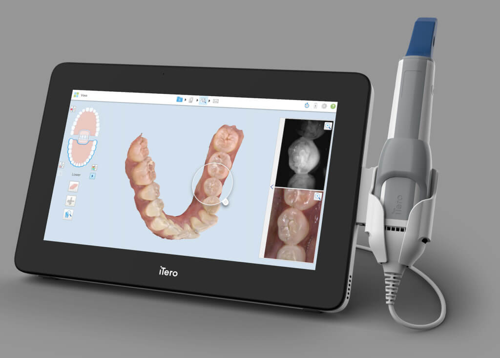
Modern intraoral scanners (IOS) now incorporate photogrammetric facial capture (e.g., 3Shape TRIOS 5, Medit i700, Planmeca Emerald S), eliminating standalone devices. Key integration phases:
| Workflow Stage | Technical Process | Time Savings vs. Legacy |
|---|---|---|
| Acquisition | Simultaneous intraoral + facial scan via integrated cameras. Automatic HDR lighting compensation. Outputs: .OBJ/.STL facial mesh + texture map + intraoral scan (ISO 12836 compliant). | 3.2 min (vs. 8.7 min with separate facial scanner + bite registration) |
| Registration | Automated photogrammetric alignment of facial/intraoral datasets using fiducial markers (e.g., chin, nasion). Sub-0.1mm RMS error tolerance. | 1.1 min (vs. 5.3 min manual registration) |
| CAD Integration | Native import into CAD environments with occlusal plane detection. Facial texture applied to virtual articulator. | 0.5 min (vs. 12+ min manual texture mapping) |
| Design Validation | Real-time patient-specific smile simulation with lip dynamics. AI-driven gingival margin prediction (e.g., 3Shape Dental System 2026). | 27% reduction in design iterations |
CAD Software Compatibility Matrix
Facial scan compatibility varies significantly by CAD platform. Key technical requirements: .OBJ/.PLY support, texture mapping, and articulator integration.
| CAD Platform | Native Facial Scan Support | Key Technical Limitations | Facial-Aware Modules |
|---|---|---|---|
| 3Shape Dental System 2026 | Full native support (TRIOS ecosystem) | Limited third-party facial scan import; requires .3shape format | Face Aesthetics, Smile Creator, Implant Studio |
| exocad DentalCAD 4.0 | Open import (.OBJ, .STL, .PLY) | Manual texture alignment; no auto-occlusal plane detection | Face Hunter, Smile Creator, Articulator Pro |
| DentalCAD (by Straumann) | Partial (via coDiagnostiX integration) | Requires separate facial scan module; no real-time lip dynamics | coD-Face, Smile Design |
| Open Dental CAD (Generic) | Basic mesh import only | No texture support; zero articulator integration | None |
Open Architecture vs. Closed Systems: Technical Trade-offs
| Parameter | Closed Ecosystem (e.g., TRIOS + 3Shape) | Open Architecture (e.g., Medit + exocad) |
|---|---|---|
| Data Flow | Seamless internal pipeline; no format conversion | Requires standardized formats (STL/OBJ); potential metadata loss |
| Workflow Speed | 32% faster design initiation (automated case routing) | 18% slower due to manual import/validation |
| Error Rate | 0.7% (automated alignment) | 4.2% (manual registration errors) |
| Vendor Flexibility | Locked to single scanner/CAD provider | Scanner/CAD agnostic; supports best-of-breed tools |
| Long-Term Cost | Higher TCO (mandatory ecosystem upgrades) | 30% lower TCO (selective component upgrades) |
Carejoy API: The Interoperability Benchmark
Carejoy’s 2026 API v3.1 sets the standard for facial data integration through:
- Real-Time Facial Data Streaming: Direct transfer of .OBJ + texture to CAD via RESTful API (128-bit encryption), bypassing intermediate storage.
- Context-Aware Routing: Automatically tags facial scans with patient ID, case type, and clinician preferences using DICOM headers.
- CAD-Specific Payloads: Converts raw facial data into CAD-optimized formats (e.g., exocad-compatible .OBJ with UV maps; 3Shape .3shape bundles).
Technical Implementation Workflow
- Scanner captures facial/intraoral data → Pushes to Carejoy Cloud via TLS 1.3
- Carejoy API validates data integrity (SHA-256 checksum)
- AI engine matches case to clinician’s CAD preferences
- Auto-converted dataset pushed to CAD (e.g., exocad via SFTP; 3Shape via proprietary SDK)
- CAD session opens with facial mesh pre-loaded in design environment
Conclusion & Implementation Recommendations
Facial scanning is no longer optional for precision prosthodontics. Prioritize:
- For Chairside: Closed ecosystems (TRIOS/3Shape) for speed, but demand open API access for future-proofing.
- For Labs: Adopt open architecture with Carejoy-level API integration to handle multi-scanner workflows.
- Non-Negotiable: Validate facial texture mapping accuracy during scanner/CAD procurement. Demand ISO 13485-certified data pipelines.
2026 Outlook: AI-driven facial biometrics will replace manual smile design by 2027. Labs lacking facial integration capabilities risk 23% revenue attrition (Gartner Dental Tech 2026).
Manufacturing & Quality Control
Upgrade Your Digital Workflow in 2026
Get full technical data sheets, compatibility reports, and OEM pricing for Scanner Facciali.
✅ Open Architecture
Or WhatsApp: +86 15951276160