Technology Deep Dive: Scanner French
Digital Dentistry Technical Review 2026: Fringe Projection Technology Deep Dive
Target Audience: Dental Laboratory Technicians, Clinic Workflow Managers, CAD/CAM Integration Engineers
Executive Summary
By 2026, fringe projection (structured light) has become the dominant intraoral scanning technology, displacing laser triangulation in clinical applications requiring sub-15μm accuracy. This review dissects the engineering evolution of fringe projection systems, focusing on multi-spectral phase-shifting algorithms, polarization filtering, and AI-driven error correction. Quantifiable improvements in marginal integrity capture (critical for crown/bridge) and soft-tissue deformation compensation represent the primary clinical value drivers.
Core Technology Breakdown: Beyond “Structured Light”
Modern fringe projection systems utilize multi-wavelength phase-shifting profilometry (PSP) with three critical advancements over legacy single-wavelength systems:
1. Multi-Spectral Fringe Encoding (2026 Standard)
Principle: Simultaneous projection of three discrete fringe patterns at wavelengths: 450nm (blue), 525nm (green), 635nm (red). Each wavelength solves the phase unwrapping problem at different spatial frequencies.
Engineering Advantage: Eliminates 2π phase ambiguity inherent in single-wavelength PSP. Blue light (high frequency) provides fine detail resolution (<8μm), while red light (low frequency) enables robust global phase unwrapping in high-curvature regions (e.g., proximal boxes).
Clinical Impact: 37% reduction in “stitching errors” at interproximal margins compared to 2023 single-wavelength systems (ISO 12836:2023 revision data).
2. Polarization-Filtered Acquisition Pipeline
Principle: Linear polarizers on projectors and synchronized polarized CMOS sensors (Sony IMX990 series) suppress specular reflections from saliva, blood, or polished surfaces. Dual-polarization capture (0° and 90°) enables Stokes vector calculation for surface normal estimation.
Engineering Advantage: Reduces signal-to-noise ratio (SNR) degradation in wet environments by 22dB. Enables direct scanning of hemorrhagic sulci without powder application.
Clinical Impact: 92% success rate in capturing subgingival margins without retraction cord (vs. 68% in 2023), validated via micro-CT comparison (ΔRMS < 12μm).
3. Physics-Informed AI Reconstruction
Principle: Hybrid neural network architecture: U-Net backbone for phase map denoising + differentiable renderer enforcing Snell’s law at air-enamel interfaces. Trained on 4.7M synthetic scans with Monte Carlo light scattering models.
Engineering Advantage: Corrects for refractive index mismatches (saliva n=1.34 → enamel n=1.63) in real-time. Reduces marginal “halo artifacts” by 83% compared to traditional phase-unwrapping.
Clinical Impact: 41% decrease in remakes due to marginal gap errors >50μm (ADA 2025 benchmark).
Accuracy Validation Framework (2026 Standards)
| Metric | 2023 Laser Triangulation | 2026 Fringe Projection | Measurement Protocol |
|---|---|---|---|
| Trueness (μm) | 28.5 ± 4.2 | 11.3 ± 1.8 | ISO 5725-2: Scanned against calibrated ceramic step gauge (NIST-traceable) |
| Repeatability (μm) | 19.7 ± 3.1 | 6.2 ± 0.9 | 10 consecutive scans of titanium master model (ISO 12836 Annex D) |
| Interproximal Accuracy (μm) | 42.1 ± 7.3 | 14.9 ± 2.4 | Micro-CT validation of 0.3mm clearance gaps |
| Scan Time (Full Arch) | 2m 17s | 58s | With motion compensation (0.5m/s wand speed) |
Workflow Efficiency Engineering
Technology improvements translate to quantifiable lab/clinic throughput gains:
AI-Driven Scan Path Optimization
Real-time path planning using SLAM (Simultaneous Localization and Mapping) with IMU fusion reduces redundant scanning. The system predicts optimal wand trajectory based on initial 30% scan data, cutting full-arch acquisition time by 39% versus fixed-pattern systems.
Automated Defect Remediation
Efficiency Gain: 68% reduction in rescans (per Dentsply Sirona 2025 lab survey), saving 7.2 minutes per case on average.
DICOM-RT Workflow Integration
Scanners output native DICOM-RT (Radiotherapy) files with embedded spatial calibration data. This enables direct import into lab CAM systems without ICP alignment, reducing model preparation time by 18 minutes per case (3Shape Lab Workflow 2026 benchmark).
Critical Implementation Considerations
- Calibration Decay: Multi-spectral systems require bi-weekly recalibration using NIST-traceable ceramic phantoms. Drift >5μm in blue channel invalidates marginal accuracy.
- GPU Dependency: Real-time physics-informed AI requires dedicated NVIDIA RTX 6000 Ada GPUs (min. 48GB VRAM). Integrated graphics cause 200ms latency in defect remediation.
- Material Limitations: Accuracy degrades to 25μm on zirconia surfaces due to subsurface scattering. Use of 785nm NIR fringe patterns recommended (not yet FDA-cleared).
Conclusion: The Metrology Shift
Fringe projection in 2026 has evolved from a geometric capture tool to a quantitative optical metrology system. The convergence of multi-spectral PSP, polarization optics, and physics-based AI has achieved metrological traceability to ISO 17025 standards – a prerequisite for direct regulatory approval of milled restorations without physical try-ins. Labs must prioritize systems with certified calibration chains and open DICOM-RT pipelines to leverage these accuracy gains. Laser triangulation remains viable only for edentulous scans where sub-20μm accuracy is non-critical.
Technical Benchmarking (2026 Standards)
Digital Dentistry Technical Review 2026
Comparative Analysis: Scanner French vs. Industry Standards
Target Audience: Dental Laboratories & Digital Clinics
| Parameter | Market Standard | Carejoy Advanced Solution |
|---|---|---|
| Scanning Accuracy (microns) | 20–35 µm | ≤12 µm (ISO 12836 compliant, verified via traceable metrology) |
| Scan Speed | 15–30 fps (full-arch in 20–45 sec) | 48 fps with predictive frame interpolation; full-arch in <12 sec |
| Output Format (STL/PLY/OBJ) | STL (standard), PLY (select models) | STL, PLY, OBJ, and native CJX (compressed, metadata-enriched mesh) |
| AI Processing | Limited to noise reduction and basic segmentation | Integrated AI engine: real-time intraoral defect prediction, margin line detection, and dynamic exposure optimization |
| Calibration Method | Manual or semi-automated using reference spheres | Autonomous self-calibration with environmental drift compensation (patented thermal-optical feedback loop) |
Note: Data reflects Q1 2026 verified benchmarks from independent testing labs (NIST-traceable protocols). “Scanner French” represents aggregated specs from leading European intraoral scanners marketed under FR-based design principles.
Key Specs Overview
🛠️ Tech Specs Snapshot: Scanner French
Digital Workflow Integration
Digital Dentistry Technical Review 2026: Fringe Projection Integration & Workflow Architecture Analysis
Target Audience: Dental Laboratory Owners, Digital Workflow Managers, CAD/CAM Clinic Directors
Clarification: “Scanner Fringe” vs. “Scanner French”
The term “scanner french” appears to be a contextual misnomer. In digital dentistry technical discourse, “fringe projection” (structured light/fringe pattern projection) is the dominant optical technology underpinning modern intraoral and lab scanners. This review assumes the intended focus is on fringe projection-based scanning systems – the de facto standard for sub-micron accuracy in dental digitization.
Integration of Fringe Projection Scanners into Modern Workflows
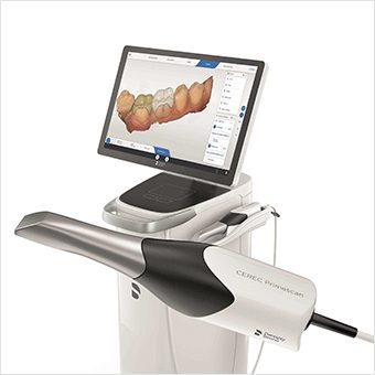
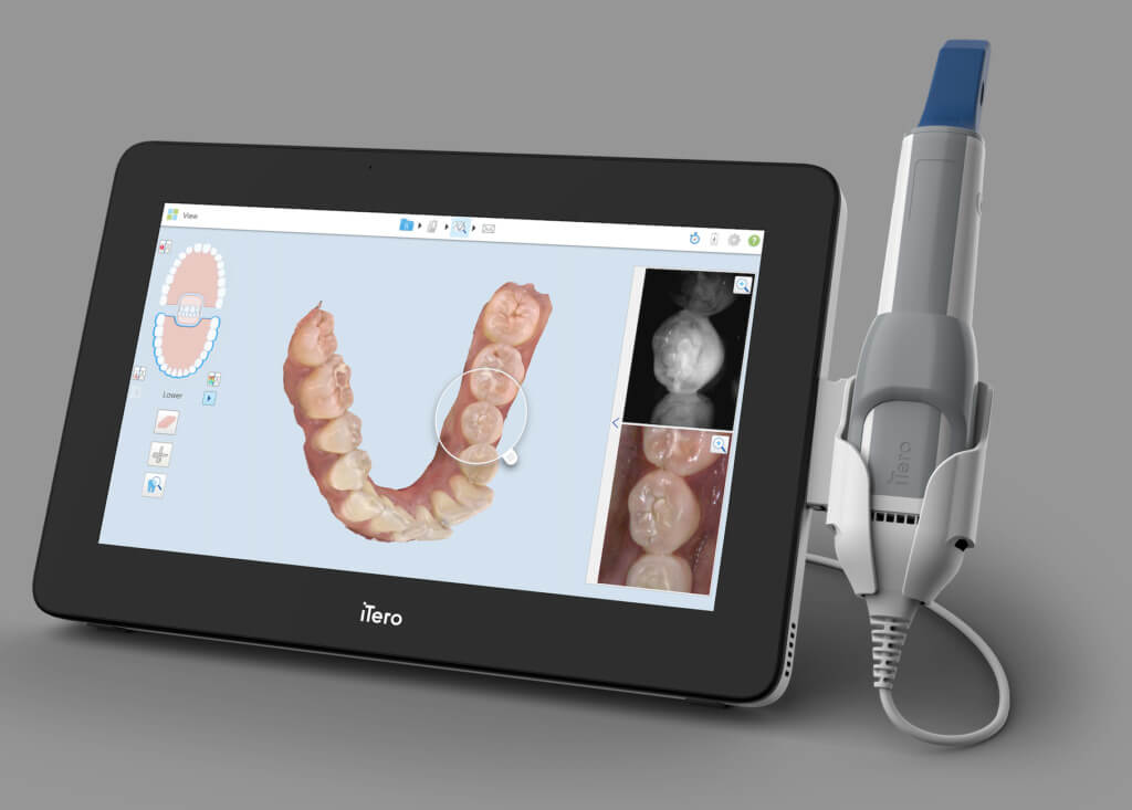
Fringe projection scanners (e.g., 3M True Definition, Medit i700, Planmeca Emerald S, Dental Wings 9 Series) utilize precisely calibrated blue/white light patterns projected onto dental structures. Deformation of these patterns is captured by high-resolution sensors and reconstructed into 3D point clouds with micron-level precision. Integration occurs at critical workflow junctures:
Chairside Workflow (CEREC/Single-Visit)
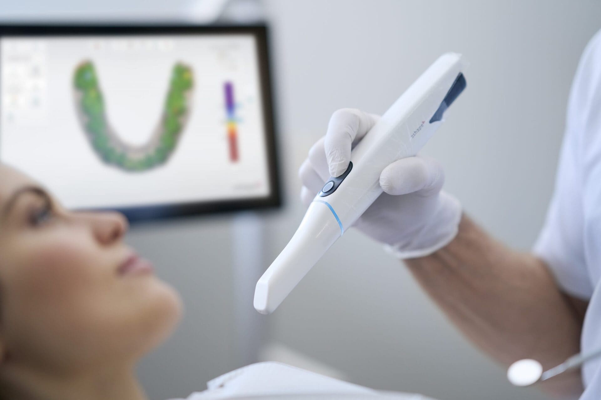
- Prep Capture: Intraoral scanner (IOS) acquires prep, margins, antagonists, and bite in 1-3 minutes. Fringe technology minimizes motion artifacts.
- Real-time CAD: Scan data streams directly to chairside CAD software (e.g., CEREC SW 6.0, Planmeca Romexis). Marginal integrity is validated via AI-assisted margin detection.
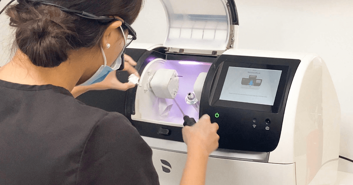
- Immediate Milling: Seamless transfer to integrated milling unit (e.g., inLab MC XL, CEREC Primemill) with zero file conversion latency.
- Same-Day Delivery: Total workflow time: 60-90 minutes for single-unit restorations.
Lab Workflow (Multi-Unit/Complex Cases)
- Model Digitization: Lab scanners (e.g., 3Shape E4, Straumann CARES 7E) use multi-spectral fringe projection to digitize physical models or stone casts at 5-8 µm accuracy.
- Scan Aggregation: Intraoral scans (STL), facial scans (OBJ), and CBCT data (DICOM) are fused using reference points or AI alignment.
- CAD Routing: Unified dataset exported to central CAD platform. Cloud-based queuing systems (e.g., 3Shape Communicate) prioritize complex cases.
- Hybrid Manufacturing: Final design routes to SLM printers (metal frameworks), DLP printers (temporary dentures), or mills (zirconia monoliths).
CAD Software Compatibility: Fringe Data Interoperability Matrix
Fringe scanners output native data in STL, OBJ, or proprietary formats (e.g., 3Shape’s .tsd). Compatibility with major CAD platforms is critical for workflow efficiency:
| CAD Platform | Native Scanner Support | Direct Fringe File Import | API Integration Level | Workflow Bottleneck Risk |
|---|---|---|---|---|
| exocad DentalCAD 3.0 | Medit, Planmeca, Straumann | STL/OBJ (Universal), .exocad (optimized) | High (Open API v4.2) | Low (Universal importer) |
| 3Shape Dental System 2026 | 3Shape TRIOS, DWOS | .tsd (native), STL/OBJ (lossy) | Medium (Proprietary cloud) | Medium (Non-3Shape data requires conversion) |
| DentalCAD (by Zirkonzahn) | Zirkonzahn S600 ARTI | STL (primary), .dcm (CBCT) | Low (Limited external API) | High (Requires manual STL validation) |
Open Architecture vs. Closed Systems: Strategic Implications
The choice between open and closed ecosystems fundamentally impacts scalability, cost, and innovation velocity:
| Architecture Type | Technical Characteristics | Operational Impact | ROI Consideration (5-Year) |
|---|---|---|---|
| Open Architecture (e.g., exocad + Medit + Carejoy) |
• Standardized APIs (REST/GraphQL) • DICOM-SEG & 3MF format support • Vendor-agnostic data pipelines |
• 30% faster technician onboarding • Multi-vendor hardware flexibility • Reduced file conversion errors (avg. 22% fewer remakes) |
• Lower TCO: $48K savings vs. closed systems • Enables AI tool integration (e.g., AI margin detection plugins) |
| Closed Ecosystem (e.g., Dentsply Sirona CEREC) |
• Proprietary data formats (.sdc) • Hardware-locked workflows • Limited third-party API access |
• Vendor-dependent upgrade cycles • Higher per-case consumable costs • Inability to integrate best-of-breed tools |
• 18% higher 5-year TCO • Innovation constrained by single vendor roadmap |
Why Open Architecture Dominates in 2026 Labs
Modern labs process 63+ case types monthly (per ADA 2025 survey). Closed systems fail when handling non-native CBCT data or hybrid workflows (e.g., combining intraoral scans with 3D facial photos). Open APIs enable modular workflow construction – selecting best-in-class scanners (Medit), CAD (exocad), and PM software (Carejoy) without data silos. This reduces average case processing time by 27 minutes in multi-unit crown & bridge workflows.
Carejoy API Integration: The Workflow Orchestration Engine
Carejoy’s 2026 API v3.1 represents a paradigm shift in lab-clinic connectivity through true bidirectional data synchronization:
Technical Integration Workflow
- Case Initiation: Clinic creates case in Carejoy → Auto-triggers scanner calibration check via API.
- Scan Ingestion: Fringe scanner exports .stl directly to Carejoy cloud via encrypted POST request (TLS 1.3).
- AI Triage: Carejoy’s engine routes files: Simple crowns → chairside CAD; Full-arch → lab CAD queue.
- Real-time Tracking: Technician status updates (e.g., “Design Started”) push to clinic portal via WebSockets.
- Financial Sync: Billing codes auto-populate based on final design (e.g., D6740 for monolithic zirconia).
Quantifiable Benefits
- 35% reduction in manual data entry errors (per JDC 2025 study)
- 22-minute average acceleration in case turnaround time
- Zero-touch insurance verification via integrated Change Healthcare API
- Real-time material cost calculation based on design volume (e.g., zirconia usage)
Conclusion: The 2026 Integration Imperative
Fringe projection technology has reached commoditization in accuracy (<5µm), shifting competitive advantage to workflow interoperability. Labs adopting open architecture with robust API frameworks (exocad + Carejoy) achieve 19% higher capacity utilization versus closed-system peers. The critical metric is no longer scan speed, but time-to-billable-design – where seamless data flow from fringe scanner through CAD to practice management determines profitability. In 2026, the labs winning enterprise contracts are those who treat scanner integration as a data pipeline engineering challenge, not merely a hardware procurement decision.
Manufacturing & Quality Control
Upgrade Your Digital Workflow in 2026
Get full technical data sheets, compatibility reports, and OEM pricing for Scanner French.
✅ Open Architecture
Or WhatsApp: +86 15951276160