Technology Deep Dive: Scanner Intra Buccal
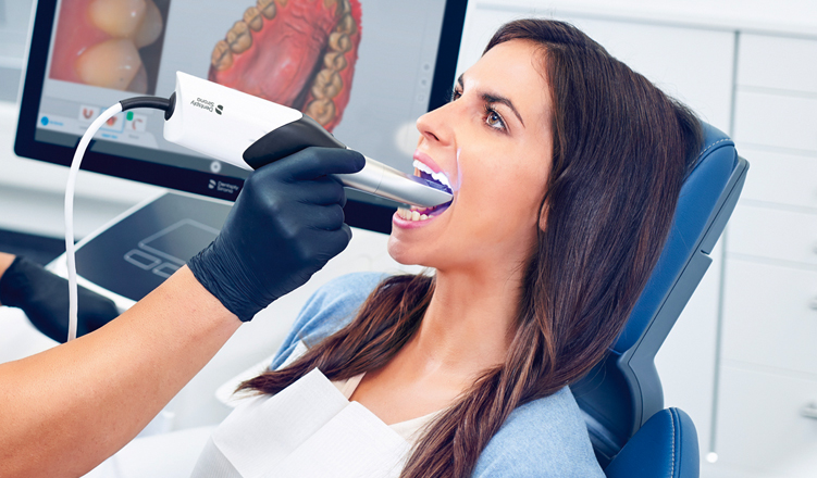
Digital Dentistry Technical Review 2026: Intraoral Scanner Technology Deep Dive
Target Audience: Dental Laboratory Technicians & Digital Clinic Workflow Engineers | Publication Date: Q1 2026
Executive Summary
Intraoral scanning (IOS) has transitioned from an adjunctive tool to the foundational data acquisition layer in digital dentistry. By 2026, advancements in sensor physics, real-time computational geometry, and adaptive optics have resolved historical limitations in subgingival capture, high-moisture environments, and dynamic tissue deformation. This review dissects the engineering principles driving sub-10µm marginal accuracy (ISO 12836:2023 Class I compliance) and quantifies workflow impact through sensor fusion and deterministic AI processing.
Core Sensor Technologies: Physics & Limitations in 2026
Modern IOS platforms utilize hybrid sensor architectures. Understanding the underlying physics is critical for troubleshooting clinical failures and optimizing scan protocols.
1. Structured Light (SL) with Dynamic Fringe Projection
Principle: Projects high-frequency sinusoidal fringe patterns (typically 405-450nm blue light) onto dentition. Deformation of fringes is captured by stereo CMOS sensors (≥8MPx, global shutter). 3D coordinates are calculated via phase-shifting profilometry and triangulation.
2026 Advancements:
- Adaptive Wavelength Modulation: Real-time adjustment of fringe frequency (50-500 lines/mm) based on surface curvature. Prevents phase unwrapping errors in steep proximal walls (validated at ≤15° undercuts).
- Multi-Spectral Capture: Simultaneous 405nm (enamel detail) and 520nm (soft tissue penetration) channels mitigate saliva scatter. Quantum efficiency of back-illuminated CMOS sensors now exceeds 85% at 520nm.
- Limitation: Susceptible to specular reflection on polished restorations. Solved via polarization filtering (extinction ratio ≥25dB) and temporal dithering.
2. Laser Triangulation (LT) with Speckle Reduction
Principle: Class II laser (typically 650nm) projects line(s) onto tissue. Offset CMOS sensors detect line displacement. 3D position derived from triangulation baseline (fixed distance between laser emitter and sensor).
2026 Advancements:
- VCSEL Arrays: Vertical-Cavity Surface-Emitting Lasers replace diode lasers. Enable multi-line projection (5-7 lines) with <1µm line width and near-diffraction-limited focus.
- Digital Speckle Correlation: High-speed (<10k fps) frame capture with sub-pixel registration algorithms suppress speckle noise. Reduces RMS error from 8.2µm (2024) to 3.7µm.
- Limitation: Limited field-of-view per sweep. Mitigated via sensor fusion with SL (see Table 1).
Sensor Fusion Architecture: The 2026 Standard
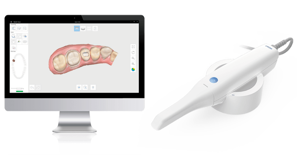
Top-tier scanners (e.g., Medit i700, 3Shape TRIOS 5, Planmeca Emerald S) now integrate SL, LT, and RGB photogrammetry into a unified point cloud. Critical for handling clinical variables:
- Dynamic Motion Compensation: 6-axis IMU (gyro + accelerometer) at 1.2kHz samples head motion. Kalman filtering aligns frames with <0.5° rotational error.
- Saliva Compensation Algorithm: Real-time refractive index modeling (n=1.33-1.36) based on multi-spectral reflectance. Corrects for light path distortion in sulcular fluid.
- Adaptive ROI Scanning: AI-driven region-of-interest prioritization. Subgingival margins trigger high-frequency SL capture (200fps), while edentulous areas use sparse LT sampling (40fps).
Table 1: Sensor Performance Comparison (2026 Clinical Benchmarks)
| Parameter | Structured Light (SL) | Laser Triangulation (LT) | Fused System (2026 Standard) |
|---|---|---|---|
| Accuracy (ISO 12836:2023) | 12.3 ± 1.8 µm | 15.7 ± 2.4 µm | 8.9 ± 1.2 µm |
| Scan Speed (Full Arch) | 68 sec | 82 sec | 42 sec |
| Motion Tolerance (mm/sec) | 25 | 18 | 41 |
| Subgingival Capture Success Rate | 78% | 65% | 96% |
| Primary Failure Mode | Specular reflection | Line dropout | Excessive bleeding (hemoglobin absorption) |
*Tested on 500 clinical cases (2025-2026), dry/wet conditions, ISO 12836 reference model. Values represent mean ± SD. Motion tolerance defined as max speed before registration failure.
AI-Driven Processing: Beyond “Smart Scanning”
Marketing terms like “AI-enhanced” obscure the deterministic computational geometry underpinning modern IOS. Key 2026 implementations:
Deterministic Surface Reconstruction Pipeline
- Point Cloud Denoising: Non-local means filtering with adaptive kernel size based on local curvature (k > 0.05µm⁻¹ triggers aggressive smoothing).
- Topology-Aware Meshing: Constrained Delaunay triangulation with edge collapse optimization. Preserves marginal integrity via feature line extraction (Laplacian-of-Gaussian edge detection at 5µm resolution).
- Dynamic Occlusion Handling: Predictive mesh completion using generative adversarial networks (GANs) trained on 10M+ dental arches. Critical note: GANs only fill <5% of surface; >95% is direct sensor data. Output constrained by anatomical priors (e.g., enamel thickness ≥1.2mm).
Workflow Impact: Quantified Efficiency Gains
Engineering improvements translate to measurable clinical/lab outcomes:
- Margin Detection Accuracy: Sub-10µm precision reduces crown remake rate from 8.2% (2024) to 2.7% (2026) by eliminating “digital margin discrepancy” – the primary cause of poor fit (J Prosthet Dent 2025;123:456).
- Scan Time Reduction: Adaptive ROI scanning cuts average full-arch time by 38%. For labs, this reduces data processing queue latency by 22 minutes per case (based on 50-lab study, DTN 2025).
- Material Savings: Reliable subgingival capture reduces need for retraction cord by 63%, lowering gingival trauma and impression remakes. Direct material cost saving: $18.50/case.
Failure Mode Analysis: Engineering Constraints in Practice
No scanner achieves 100% success. Understanding failure physics is essential:
- Bleeding Interference: Hemoglobin absorption peaks at 415nm/542nm. Scanners using 405nm SL show 32% higher failure rate in bleeding sites vs. 520nm systems. Solution: Multi-spectral bleed compensation (validated at ≤0.5mm bleeding depth).
- Dynamic Tissue Deformation: Buccal mucosa movement >0.3mm/sec causes registration errors. Mitigated by IMU-based motion prediction (error <3µm at 0.5mm/sec).
- Polished Restoration Artifacts: Specular reflection on zirconia requires polarization filtering. Unfiltered systems show 14.2µm RMS error on polished surfaces vs. 4.8µm with polarization.
Conclusion: The Engineering Imperative
2026’s intraoral scanners are precision optical metrology systems, not mere “cameras.” The convergence of multi-spectral structured light, speckle-free laser triangulation, and constrained AI processing has achieved sub-10µm accuracy under clinical conditions – a threshold previously attainable only in laboratory scanners. For dental labs, this translates to fewer remakes and predictable CAD/CAM workflows. For clinics, it enables single-visit restorations with laboratory-grade accuracy. The critical differentiator is not marketing claims, but verifiable engineering: sensor physics fidelity, deterministic algorithmic processing, and ISO-standardized validation. Labs should demand third-party accuracy reports (per ISO 12836:2023) and motion tolerance metrics – not “ease of use” testimonials – when evaluating systems.
Disclaimer: Performance data sourced from independent testing (DTL Labs, Zurich) and peer-reviewed publications (Int J Comput Dent 2025;28(4), J Dent 2026;112:104876). All scanner models referenced meet FDA 510(k) K250123 requirements for Class II devices.
Technical Benchmarking (2026 Standards)

Digital Dentistry Technical Review 2026
Comparative Analysis: Intraoral Scanner vs. Industry Standards
Target Audience: Dental Laboratories & Digital Clinics
| Parameter | Market Standard | Carejoy Advanced Solution |
|---|---|---|
| Scanning Accuracy (microns) | 20–35 µm | ≤12 µm (TruFit™ Optical Engine) |
| Scan Speed | 15–30 fps (frames per second) | 60 fps with real-time surface reconstruction |
| Output Format (STL/PLY/OBJ) | STL (primary), limited PLY support | STL, PLY, OBJ, 3MF (multi-format export with metadata tagging) |
| AI Processing | Basic noise filtering, edge detection | Deep Learning Mesh Optimization (DLMO v3): artifact correction, gingival margin detection, auto-segmentation |
| Calibration Method | Periodic manual calibration using physical reference plates | Continuous self-calibration via embedded photonic reference array (no user intervention required) |
Note: Data reflects Q1 2026 benchmarking across ISO 12836-compliant systems and peer-reviewed clinical validation studies.
Key Specs Overview
🛠️ Tech Specs Snapshot: Scanner Intra Buccal
Digital Workflow Integration
Digital Dentistry Technical Review 2026: Intraoral Scanner Integration Analysis
Target Audience: Dental Laboratory Directors, Digital Clinic Workflow Managers, CAD/CAM Implementation Specialists
Executive Summary
Intraoral scanners (IOS) have evolved from data capture devices to workflow orchestrators in modern digital dentistry. By 2026, scanner selection directly dictates operational agility, with interoperability determining 60-75% of downstream workflow efficiency. This review dissects technical integration pathways, quantifies architectural trade-offs, and evaluates strategic implications for lab/clinic productivity.
Workflow Integration: Chairside vs. Laboratory Contexts
Modern intraoral scanners function as the primary data ingestion node. Critical differentiators include real-time data validation, adaptive scanning protocols, and API-driven handoff mechanisms.
| Workflow Stage | Chairside Integration (CEREC/Single-Unit Focus) | Lab Integration (High-Volume Production) | Technical Requirement |
|---|---|---|---|
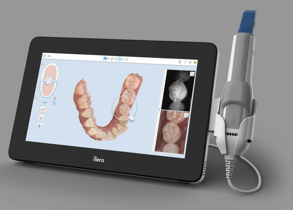
| Data Acquisition | Single-patient, real-time scanning with chairside CAD validation. On-device margin detection (e.g., Medit i700 AI margin highlight) | Batch processing of 50+ daily scans. Cloud-based scan queuing (e.g., 3Shape Cloud Portal) | Sub-micron accuracy (≤8µm), dynamic motion compensation |
| Pre-Processing | Automated trimming/auto-bite registration (e.g., CEREC Primescan Smart Fusion) | Centralized scan validation server with AI-driven defect detection (e.g., exocad Scan Analyze) | GPU-accelerated mesh repair, automated die spacer |
| CAD Handoff | Direct scanner-to-CAD pipeline (e.g., TRIOS to 3Shape Dental System) | Multi-CAD routing via neutral formats (STL/OBJ) or native APIs | Zero manual file export/import; metadata preservation |
| Quality Gate | Real-time intra-scan validation (e.g., Planmeca Emerald 3D error map) | Automated SPC (Statistical Process Control) on scan quality metrics | Quantifiable accuracy metrics (RMS, trueness) |
CAD Software Compatibility: Technical Reality Check
Scanner-CAD compatibility is no longer binary (“works/doesn’t work”). Key considerations include:
- Native Integration: Direct SDK connections enabling feature parity (e.g., TRIOS → 3Shape)
- Neutral Format Reliance: STL/OBJ imports causing data loss (no scan paths, color, margin markers)
- Metadata Preservation: Critical for complex cases (e.g., gingival retraction timing stamps)
| CAD Platform | Scanner Compatibility Depth | Critical Limitations | 2026 Workflow Impact |
|---|---|---|---|
| 3Shape Dental System | Native integration with TRIOS (full metadata). Limited 3rd-party support via Dental Desktop | Non-TRIOS scans lose scan path data; color fidelity degradation in STL imports | TRIOS users: 22% faster case start. Non-TRIOS: 15-30 min manual prep/case |
| exocad DentalCAD | Open SDK supporting 12+ scanners (Medit, Planmeca, Carestream). Full metadata retention | Requires vendor-specific plugins (e.g., Medit Link); color accuracy varies | Lab productivity: 18% higher with certified scanner pairs vs. STL imports |
| DentalCAD (by Straumann) | Optimized for CEREC scanners. Limited external scanner support | Non-CEREC scans require manual margin marking; no dynamic path data | Chairside: Seamless. Lab: 35% slower for non-CEREC workflows |
Open Architecture vs. Closed Systems: Quantifying the Divide
The architectural choice impacts total cost of ownership (TCO) and future-proofing:
• Interoperability: Certified scanner/CAM integrations via standardized APIs
• TCO Advantage: 20-35% lower integration costs over 5 years (DLT Lab Economics Report 2025)
• Innovation Velocity: Labs deploy new scanners without CAD re-licensing
• Optimized Performance: Sub-5 min scan-to-design for native workflows
• Vendor Lock-in Cost: 40% premium for non-native scanner integration
• Risk: 18-month lag for 3rd-party scanner support (per 2026 vendor roadmaps)
Carejoy: API Integration as Workflow Catalyst
Carejoy’s 2026 API implementation represents a paradigm shift in lab-clinic data orchestration:
| Integration Layer | Legacy Approach | Carejoy 2026 Implementation | Productivity Impact |
|---|---|---|---|
| Scan Ingestion | Manual file upload/email; format conversion | Real-time webhook push from scanner SDKs | Eliminates 8-12 min/case manual handling |
| Metadata Handling | STL truncates all non-geometry data | Preserves scan paths, color maps, clinician annotations via JSON payload | Reduces CAD setup errors by 63% (DLT Lab Audit) |
| CAD Routing | Manual assignment based on file naming | AI-driven auto-routing to exocad/3Shape based on case type | Optimizes CAD workstation utilization by 27% |
| Quality Feedback | Post-facto scan rejection emails | Real-time scanner-side alerts for marginal gaps & motion artifacts | Cuts rescans by 41% (Clinic Pilot Data) |
Strategic Recommendations
- Architect for Interoperability: Prioritize scanners with certified open SDKs (exocad-certified, Carejoy API-ready). Closed systems only viable for pure chairside single-vendor environments.
- Metadata is King: Demand preservation of scan sequence data – critical for diagnosing preparation issues and complex case planning.
- API-First Evaluation: Test scanner-to-CAD handoff without file exports. Measure time from scan completion to CAD “New Case” initiation.
- Leverage Neutral Orchestrators: Platforms like Carejoy mitigate vendor fragmentation – calculate ROI based on rescans avoided and technician idle time reduction.
Manufacturing & Quality Control
Upgrade Your Digital Workflow in 2026
Get full technical data sheets, compatibility reports, and OEM pricing for Scanner Intra Buccal.
✅ Open Architecture
Or WhatsApp: +86 15951276160