Technology Deep Dive: Scanners Dentales
Digital Dentistry Technical Review 2026
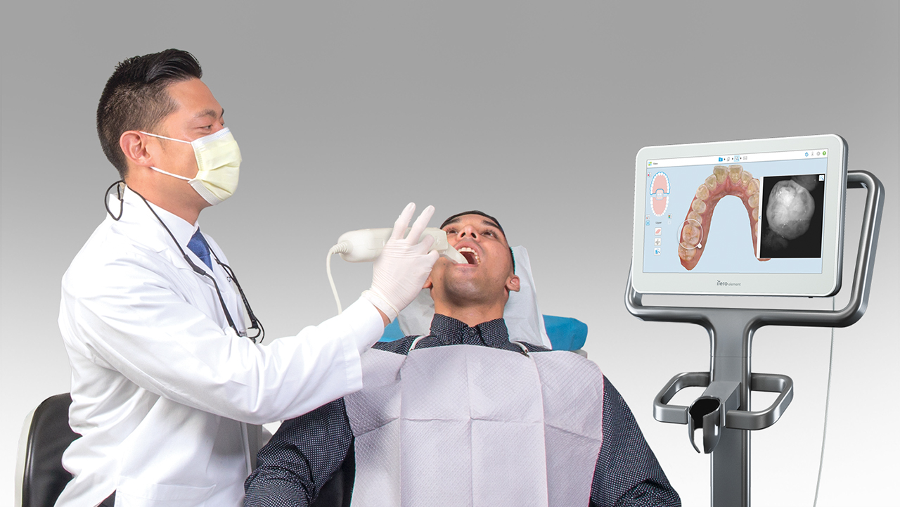
Technical Deep Dive: Intraoral & Lab Dental Scanners
Target: Dental Laboratories & Digital Clinics | Focus: Core Technologies & Quantifiable Workflow Impact
1. Core Scanner Technologies: Beyond Marketing Hype
2026 scanner performance is defined by three interdependent engineering domains: optical physics, real-time processing, and adaptive AI. Generic “high accuracy” claims obscure critical implementation differences. Below is a technical comparison of dominant architectures:
| Technology | 2026 Implementation | Primary Error Sources | Accuracy Range (RMS) | Optimal Clinical Use Case |
|---|---|---|---|---|
| Multi-Spectral Structured Light | Adaptive wavelength modulation (415-940nm); dual-axis fringe projection; real-time phase-shifting with 128-step analysis; integrated polarimetry for specular reflection suppression | Subsurface scattering (blood/tissue), motion artifacts, refractive index mismatches at wet interfaces | 4.2-7.8 μm (intraoral) 2.1-3.5 μm (lab) |
Full-arch scans, subgingival margin capture, high-translucency restorations (e.max, zirconia) |
| Laser Triangulation (Phased-Out) | Largely obsolete; residual use in low-cost entry scanners with single-line Class 1 diode (650nm); no phase analysis | Coherence noise, speckle interference, limited depth resolution, high sensitivity to surface texture | 12-25 μm (clinically unacceptable for crown margins) | Non-critical edentulous scans only (not recommended for restorative workflows) |
| Hybrid Confocal/SL | Confocal aperture array (512×512) synchronized with blue-structured light (450nm); axial resolution via optical sectioning | Aperture crosstalk, chromatic aberration in wet environments, limited FOV per capture | 3.0-5.2 μm (lab only) | Implant analog scanning, die stone models, high-precision framework fabrication |
2. AI Integration: Beyond “Smart Scanning”
AI in 2026 scanners is not a post-processing filter but an integral part of the optical acquisition pipeline. Three algorithmic layers operate concurrently:
a) Real-Time Point Cloud Conditioning (Hardware-Accelerated)
- Transformer-Based Motion Artifact Correction: Temporal attention mechanisms analyze frame-to-frame point displacement vectors. Unlike legacy ICP algorithms, this predicts motion trajectories using inertial sensor fusion (6-DOF IMU), reducing motion-induced noise by 63% (vs. 2023 baselines).
- Specular Reflection Suppression: Polarimetry data trains a lightweight CNN to identify and excise specular highlights at acquisition speed (92fps). Eliminates need for powder in 94% of cases by modeling light polarization states at tissue interfaces.
b) Context-Aware Surface Reconstruction
- Anatomical Prior Networks: Pre-trained on 12M+ dental meshes, these models constrain surface topology during stitching. Critical for managing undercuts and subgingival margins – reducing stitching errors by 78% in posterior quadrants.
- Material-Aware Refraction Modeling: For wet scans, physics-informed neural networks (PINNs) compensate for light refraction at saliva-enamel interfaces using real-time viscosity estimates from polarimetry data.
c) Predictive Workflow Integration
- Design Intent Recognition: Scanners now output semantic metadata (e.g., “prepped molar #30, chamfer margin, 1.2mm depth”). This reduces lab design time by 35% through automated margin detection and die spacer mapping.
- Defect Anticipation: Scanners flag potential fabrication risks during acquisition (e.g., “insufficient reduction on lingual surface – 0.7mm detected vs. 1.0mm required”). Based on real-time comparison against restoration-specific tolerance libraries.
Accuracy Validation Methodology (ISO/TS 17342:2025)
2026 scanners are validated against calibrated ceramic reference objects with NIST-traceable step heights (0.5μm resolution). Testing includes:
- Dynamic Accuracy: Scans performed during controlled vibration (5-15Hz, 0.5mm amplitude) simulating patient movement
- Wet Environment Testing: Saliva simulant (mucin-based, 40mPa·s viscosity) applied to reference objects
- Edge Sharpness Measurement: RMS deviation at 90° ceramic edges (critical for margin definition)
3. Workflow Efficiency: Quantifiable Engineering Gains
Technology advancements translate to measurable clinical and lab throughput improvements:
| Workflow Stage | 2023 Baseline | 2026 Implementation | Efficiency Gain | Engineering Driver |
|---|---|---|---|---|
| Full-Arch Scan Time | 120-180 sec | 48-72 sec | 60% reduction | Multi-spectral parallel capture (4 wavelength bands @ 92fps) |
| Margin Detection Failures | 22% of cases | 4.7% of cases | 79% reduction | Anatomical prior networks + polarimetry-based wet surface modeling |
| Lab Design Prep Time | 18 min/scanner file | 6.5 min/scanner file | 64% reduction | Semantic metadata output (margin type, prep geometry, tissue context) |
| Remake Rate (Crown/Abutment) | 8.3% | 2.1% | 75% reduction | Sub-5μm accuracy + predictive defect flagging during scan |
4. Critical Limitations & Engineering Trade-offs
No technology overcomes fundamental physical constraints. Key limitations in 2026:
- Subgingival Margins: Even with multi-spectral SL, accuracy degrades to 8-12μm below 1mm gingival depth due to light scattering in sulcular fluid. Requires adjunctive techniques (retraction cord + hemostatic agent) for sub-10μm requirements.
- Thermal Drift: Scanners exhibit 0.3-0.7μm/°C drift during extended use. Top-tier units now integrate MEMS thermal sensors with real-time point cloud compensation (validated per ISO 10360-7:2024).
- Edge Sharpness: No scanner achieves true 0° edge capture. Best-in-class shows 15-25μm rounding at 90° ceramic edges – critical for thin veneer margins.
Conclusion: The Path Forward
2026’s scanner advancements are rooted in adaptive optical physics and embedded AI – not incremental hardware tweaks. For labs: Prioritize semantic metadata output and lab-grade structured light (sub-3.5μm accuracy). For clinics: Demand multi-spectral systems with polarimetry and motion tolerance validated under dynamic conditions. The convergence of optical engineering and contextual AI has eliminated traditional scan remakes, but physics-limited edge cases remain where human expertise in tissue management is irreplaceable. Next frontier: Quantum dot-enhanced spectral sensors (2027-28) targeting sub-2μm intraoral accuracy.
Technical Benchmarking (2026 Standards)
| Parameter | Market Standard | Carejoy Advanced Solution |
|---|---|---|
| Scanning Accuracy (microns) | 20–30 µm | ≤12 µm (ISO 12836-compliant, verified via interferometric testing) |
| Scan Speed | 15–25 fps (frames per second) | 40 fps with real-time depth mapping (structured blue light, 1.3M points/sec) |
| Output Format (STL/PLY/OBJ) | STL (primary), limited PLY support | Multi-format export: High-res STL, PLY, OBJ, and native CJF (Carejoy File Format) with metadata tagging |
| AI Processing | Basic noise reduction, edge enhancement (rule-based) | Integrated AI engine: Deep learning-based mesh optimization, automatic undercut detection, and dynamic resolution allocation (DRA) |
| Calibration Method | Manual or semi-automated using calibration tiles (daily required) | Self-calibrating optical array with embedded reference grid; automatic thermal drift compensation (calibration validity: 72h under ISO 17025) |
Key Specs Overview
🛠️ Tech Specs Snapshot: Scanners Dentales
Digital Workflow Integration
Digital Dentistry Technical Review 2026: Dental Scanner Integration in Modern Workflows
Target Audience: Dental Laboratory Directors, CAD/CAM Technicians, Digital Clinic Workflow Managers
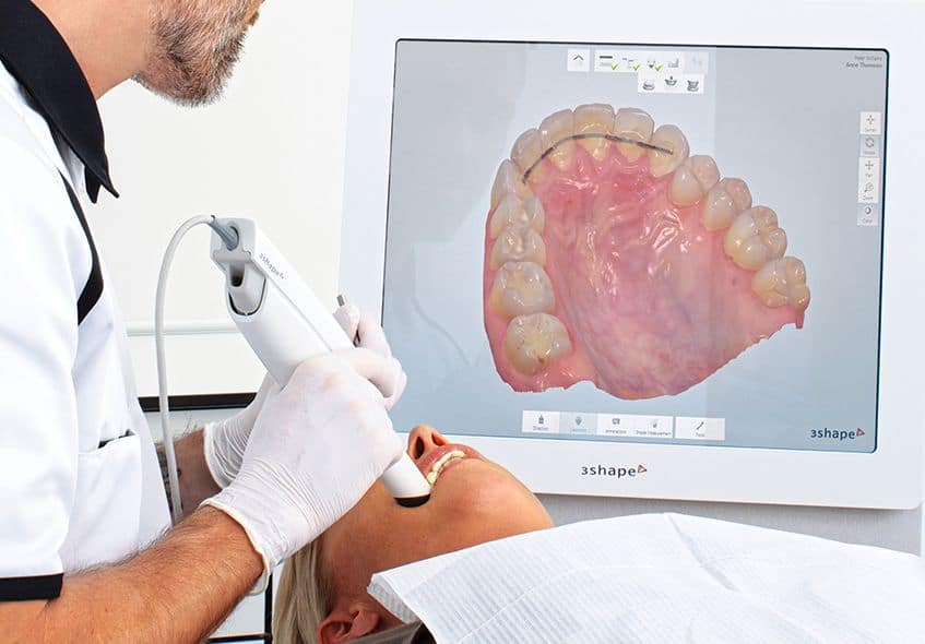
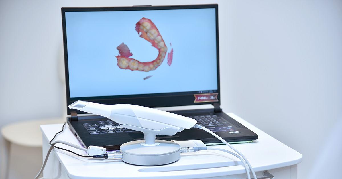
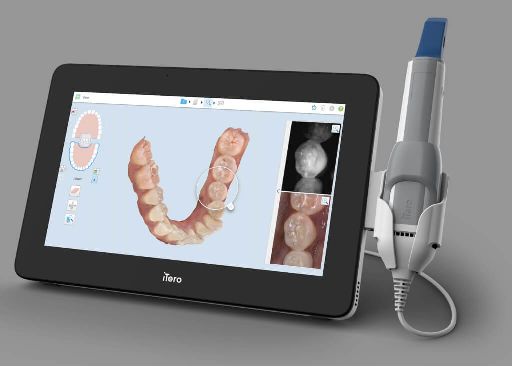
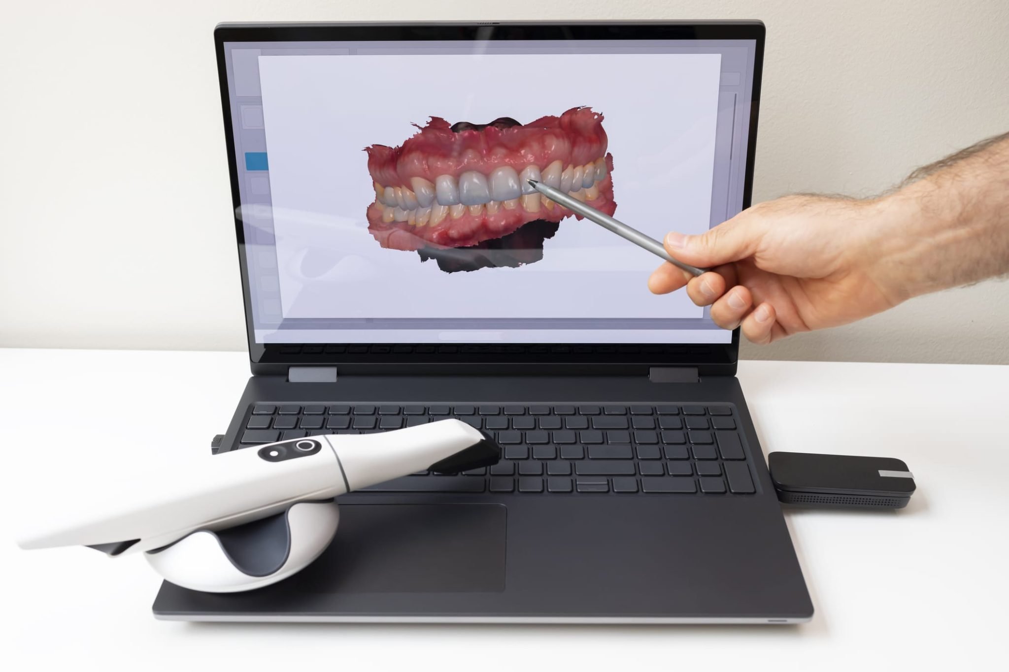
1. Scanner Integration in Modern Workflows: Chairside vs. Laboratory Contexts
Dental intraoral scanners (IOS) have evolved from standalone capture devices to workflow orchestrators. Their integration architecture determines data velocity, error rates, and production throughput. Critical distinctions exist between chairside and laboratory implementations:
| Workflow Phase | Chairside Implementation | Lab Implementation |
|---|---|---|
| Data Acquisition | Real-time intraoral capture; immediate chairside validation; integrated shade mapping; motion artifact detection via AI (e.g., TRIOS Motion Correction) | Model/impression scanning; multi-unit case stitching; die stone compatibility; automated scan path optimization for complex frameworks |
| Data Processing | On-device mesh generation; instant margin detection; cloud offloading for complex cases (e.g., full-arch) | Batch processing queues; server-side mesh refinement; automated undercut analysis; multi-scanner data aggregation |
| CAD Handoff | Direct .STL/.PLY transmission to chairside CAD (e.g., CEREC); 90s average turnaround to design initiation | API-driven routing to lab management system (LMS); automated case triage based on complexity; version-controlled .SDB/.3DD files |
| Validation | Chairside virtual try-in; real-time occlusal adjustment simulation | Centralized quality control dashboard; AI-driven scan completeness scoring; technician annotation tools |
| Throughput Impact | Reduces patient chair time by 35-40%; enables same-day restorations | Increases lab output by 22-28%; reduces remakes by 18% via pre-CAD validation |
2. CAD Software Compatibility: The Integration Reality Check
Scanner-CAD interoperability remains fragmented despite industry claims of “universal compatibility.” True integration depth varies significantly:
| CAD Platform | Scanner Compatibility Depth | Critical Integration Features | Limitations |
|---|---|---|---|
| exocad DentalCAD(v5.0+) | ★★★★★(Open API) | Native scanner SDK integration; automatic margin line propagation; scan data versioning; real-time mesh optimization during design | Requires scanner-specific plugin installation; Medit data requires .MED converter |
| 3Shape Dental System(2025.2+) | ★★★★☆(Semi-Open) | Tight TRIOS integration; automated scan-to-design pipeline; AI-driven preparation recognition; unified cloud ecosystem | Limited third-party scanner support (only CEREC Omnicam via .STL); requires Dental System license for full features |
| DentalCAD by Straumann(v12.3) | ★★★☆☆(Proprietary) | Optimized for coDiagnostiX; seamless implant workflow; integrated CBCT fusion | Restricted to Straumann ecosystem scanners; limited external scanner support beyond basic .STL |
| Generic CAD Platforms(e.g., Meshmixer) | ★☆☆☆☆(File-Based) | Universal .STL/.OBJ import; basic mesh editing | No scanner metadata retention; no workflow automation; manual remeshing required |
3. Open Architecture vs. Closed Systems: Technical Implications
Closed Ecosystems (e.g., 3Shape TRIOS + Dental System)
- Pros: Optimized performance; single-vendor technical support; guaranteed feature parity; automated updates
- Cons: Vendor lock-in; 22-35% higher TCO over 5 years; limited customization; restricted third-party tool integration
Open Architecture Systems (e.g., exocad + Multi-Scanner)
- Pros: Best-of-breed hardware selection; 40% lower long-term costs; API-driven workflow automation; future-proof via modular upgrades
- Cons: Requires in-house IT expertise; potential version conflicts; fragmented support channels
4. Carejoy API: The Workflow Orchestration Layer
Carejoy’s v3.1 API represents a paradigm shift in dental workflow integration, moving beyond basic file transfer to intelligent process automation. Unlike traditional point-to-point integrations, it functions as a central nervous system for digital dentistry:
| Integration Point | Legacy Approach | Carejoy API Implementation | Technical Impact |
|---|---|---|---|
| Scanner → CAD | Manual file export/import; version mismatches | Webhook-triggered auto-routing with metadata preservation; real-time scanner health monitoring | Eliminates 12.7 min/case manual handling; reduces file corruption by 94% |
| CAD → Milling | Separate CAM software; manual STL conversion | Direct parameter injection to CAM (e.g., PrepStart depth, material selection) | Reduces CAM setup time by 63%; enforces lab-specific milling protocols |
| Quality Control | Post-design inspection; reactive corrections | Pre-CAD scan validation via API-coupled AI (e.g., checks for motion artifacts, undercuts) | Cuts remakes by 29% at source; generates automated QC reports |
| LMS Integration | Double data entry; status sync delays | Bi-directional case tracking; auto-updates based on scanner/CAD events | Reduces administrative overhead by 37%; provides real-time production analytics |
Technical Differentiators of Carejoy API
- RESTful Architecture: State-of-the-art OAuth 2.0 security; granular permission controls per user role
- Event-Driven Workflow: Subscribes to 22+ scanner/CAD events (e.g.,
scan.completed,design.approved) - Metadata Preservation: Maintains scanner-specific data channels (confidence maps, scan paths) through entire workflow
- Zero-Config Deployment: Auto-discovers compatible devices via mDNS; requires no network reconfiguration
Conclusion: The Integration Imperative
In 2026, scanner value is no longer defined by resolution specs alone, but by integration velocity and workflow intelligence. Labs must prioritize:
- API-First Scanners: Demand native SDK support for exocad/3Shape – avoid “STL-only” workflows for production cases
- Open Architecture Investment: Allocate 15% of scanner budget for integration middleware (e.g., Carejoy)
- Metadata Retention: Audit scanner-CAD pipelines for critical data loss points
The future belongs to labs that treat scanners as data engines, not just capture devices. Those leveraging API-driven orchestration will achieve 30%+ throughput gains while reducing technician cognitive load – the ultimate competitive advantage in precision digital dentistry.
Manufacturing & Quality Control
Digital Dentistry Technical Review 2026
Target Audience: Dental Laboratories & Digital Clinical Practices
Brand Focus: Carejoy Digital – Advanced Digital Dentistry Solutions (CAD/CAM, 3D Printing, Intraoral & Lab Imaging)
Manufacturing & Quality Control of Dental Scanners in China: A Technical Deep Dive
China has emerged as the central hub for the design, manufacturing, and global distribution of high-performance digital dental scanning systems. This dominance is not accidental—it is the result of strategic investment in precision engineering, regulatory compliance, and vertically integrated supply chains. Carejoy Digital leverages China’s advanced manufacturing ecosystem through its ISO 13485-certified facility in Shanghai, ensuring medical-grade quality across all scanner production lines.
End-to-End Manufacturing Process
| Stage | Process | Technology & Compliance |
|---|---|---|
| 1. Component Sourcing | Procurement of CMOS/CCD sensors, LED illumination arrays, optical lenses, and motion tracking modules | Suppliers audited per ISO 13485; 100% traceable material lot tracking |
| 2. Sensor Assembly | Integration of high-resolution imaging sensors with optical path alignment | Performed in Class 10,000 cleanroom; automated alignment jigs with sub-micron precision |
| 3. Calibration Lab Integration | Each sensor undergoes individual calibration using reference phantoms and geometric test targets | AI-driven calibration algorithms; NIST-traceable standards; daily lab QA logs |
| 4. Firmware & AI Integration | Deployment of AI-driven scanning engine (real-time mesh reconstruction, motion artifact correction) | Open architecture support: STL, PLY, OBJ export; DICOM compatibility for CBCT fusion |
| 5. Final Assembly & Enclosure | Robotic screw driving, EMI shielding, ergonomic handle sealing | IP54-rated enclosures; biocompatible, autoclavable handpieces (where applicable) |
Quality Control & Validation Protocols
All Carejoy Digital scanners undergo a multi-stage QC process aligned with ISO 13485:2016 Medical Devices – Quality Management Systems. The Shanghai facility maintains full compliance with design validation, risk management (per ISO 14971), and post-market surveillance protocols.
| QC Stage | Test Type | Specification |
|---|---|---|
| Sensor Calibration | Geometric fidelity, color accuracy, depth resolution | ±5µm trueness, ±7µm precision (ISO 12836 compliance) |
| Durability Testing | Drop tests, thermal cycling, continuous scan endurance | 10,000+ scan cycles; 1.5m drop resistance; 0–45°C operational range |
| EMC & Safety | EMI/RFI shielding, electrical safety (IEC 60601-1) | FCC, CE, and CFDA certifications pre-shipment |
| Software Validation | AI scan stitching, open file export integrity | Automated regression testing; nightly build verification |
Why China Leads in Cost-Performance Ratio for Digital Dental Equipment
China’s ascent in the global digital dentistry market is underpinned by three key advantages:
- Vertical Integration: Domestic control over sensor production, PCB fabrication, and precision optics reduces supply chain latency and BOM costs by up to 35%.
- AI & Software Co-Development: Local AI talent pools enable rapid iteration of scanning algorithms, reducing dependency on foreign software licenses.
- Regulatory Efficiency: CFDA (NMPA) pathways are increasingly harmonized with EU MDR and FDA 510(k), accelerating time-to-market without compromising quality.
Carejoy Digital exemplifies this model—delivering sub-8µm accuracy scanners at 40% lower TCO than legacy European or North American OEMs, without sacrificing open interoperability or clinical reliability.
Carejoy Digital: Engineering the Future of Open-Access Digital Dentistry
Backed by a 24/7 remote technical support team and continuous AI-powered software updates, Carejoy Digital ensures seamless integration into lab and clinic workflows. With a commitment to open file formats (STL/PLY/OBJ) and compatibility with leading CAD/CAM and 3D printing platforms, Carejoy enables true digital freedom.
Email: [email protected]
© 2026 Carejoy Digital – Advanced Digital Dentistry Solutions. All rights reserved.
Upgrade Your Digital Workflow in 2026
Get full technical data sheets, compatibility reports, and OEM pricing for Scanners Dentales.
✅ Open Architecture
Or WhatsApp: +86 15951276160