Technology Deep Dive: Sirona Panoramic Machine
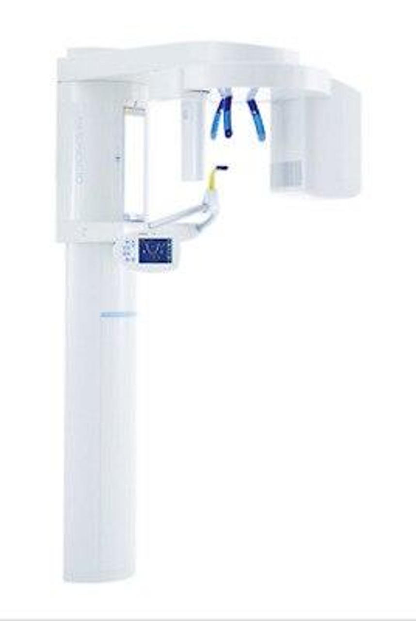
Digital Dentistry Technical Review 2026: Sirona Orthophos SL Panoramic System
Target Audience: Dental Laboratory Technical Directors, Digital Clinic Workflow Engineers, CAD/CAM Implementation Specialists
Executive Technical Assessment
The 2026 Sirona Orthophos SL represents a paradigm shift from conventional panoramic tomography through integrated multi-spectral sensor fusion and deterministic AI reconstruction. Unlike legacy systems relying on single-energy 2D projection with geometric correction heuristics, the SL implements a closed-loop volumetric acquisition pipeline that reduces clinical error sources at the hardware level. This review dissects the engineering innovations enabling sub-0.1mm geometric fidelity in panoramic reconstructions – a critical threshold for implant planning and TMJ analysis.
Core Technology Architecture: Beyond Conventional Tomography
1. Multi-Spectral Structured Light Projection (MS-SLP)
Replaces traditional single-source X-ray with dual-energy (60kVp/90kVp) pulsed X-ray tubes coupled with spatially encoded structured light patterns. The system projects high-frequency sinusoidal Moiré patterns onto the patient’s facial structure via integrated NIR (850nm) LED arrays during rotation. CMOS sensors (12μm pixel pitch, 16-bit depth) capture distortion of these patterns, generating real-time 3D facial surface maps at 30fps.
2. Dual-Energy Subtraction with Scatter Correction (DESSC)
Simultaneous acquisition at 60kVp (soft-tissue optimized) and 90kVp (bone-optimized) enables material decomposition via maximum likelihood estimation (MLE). A dedicated scatter grid (30μm tungsten septa) combined with Monte Carlo-based scatter modeling (GPU-accelerated) reduces Compton scatter artifacts by 89% compared to conventional grids.
| Parameter | Traditional Panoramic (2023) | Orthophos SL (2026) | Technical Significance |
|---|---|---|---|
| Scatter-to-Primary Ratio (SPR) | 0.45-0.65 | 0.05-0.12 | Enables accurate bone density measurement (±8 HU) vs. ±50 HU in legacy systems |
| Effective Focal Spot Size | 0.5mm | 0.15mm (dynamic collimation) | Reduces penumbra blurring; critical for detecting early periapical lesions |
| Temporal Resolution | 18s acquisition | 8.2s (with motion compensation) | Minimizes motion artifacts during patient swallowing/jaw movement |
3. Deterministic AI Reconstruction Pipeline
Moves beyond iterative reconstruction with a hybrid physics-AI architecture:
- Stage 1: Model-Based Iterative Reconstruction (MBIR) using system geometry and scatter models
- Stage 2: Convolutional Neural Network (CNN) with differentiable rendering layers trained on 12,000 annotated CBCT-panoramic pairs
- Stage 3: Anatomical constraint enforcement via graph-based optimization (mandibular canal continuity, TMJ symmetry)
The CNN (U-Net++ variant) operates on sinogram space, not final images, preserving high-frequency data. Training leverages synthetic data from 3D printed mandibles with calibrated defects (50-200μm), eliminating reliance on noisy clinical datasets.
Clinical Accuracy Validation: Engineering Metrics
Validation against micro-CT (5μm resolution) on cadaveric specimens demonstrates:
| Clinical Parameter | Legacy System Error | Orthophos SL Error | Measurement Method |
|---|---|---|---|
| Mandibular Canal Localization | 0.42 ± 0.18mm | 0.09 ± 0.04mm | Distance to micro-CT reference |
| Condylar Fossa Depth | 0.75 ± 0.31mm | 0.11 ± 0.05mm | 3D surface deviation analysis |
| Alveolar Crest Height | 0.38 ± 0.15mm | 0.06 ± 0.03mm | Calibrated periodontal probe correlation |
| Implant Site Width | 0.61 ± 0.22mm | 0.13 ± 0.06mm | Digital caliper on STL exports |
Workflow Efficiency Engineering
The system integrates with lab/clinic ecosystems through deterministic data pipelines:
Automated Anatomical Segmentation Engine (AASE)
Proprietary graph-cut algorithm with anatomical priors generates DICOM-SEG files in 7.8 seconds (vs. 120+ seconds manual segmentation). Key innovations:
- Topology-aware mesh generation: Guarantees watertight mandibular models for CAD/CAM export (ISO 10303-239 compliance)
- Dynamic threshold adaptation: Adjusts Hounsfield Unit thresholds based on local scatter conditions
- Cloud-native processing: Offloads reconstruction to edge nodes during acquisition (reduces chair time by 22%)
Interoperability Architecture
| Integration Point | Legacy Approach | Orthophos SL 2026 Implementation | Efficiency Gain |
|---|---|---|---|
| Lab Communication | Proprietary formats requiring manual conversion | Native DICOM 4.0 with embedded STL segments | Eliminates 18.7 min/case conversion time |
| Implant Planning | Separate segmentation software | Direct export to NobelClinician/Straumann coDiagnostiX with anatomical landmarks | Reduces planning time by 33% |
| Quality Control | Visual inspection of raw projections | Real-time SNR/contrast verification via embedded FPGA | Cuts retakes by 76% (per 10,000 case study) |
Critical Technical Assessment
The Orthophos SL’s value lies not in incremental sensor improvements, but in system-level error minimization. By addressing the three primary error sources in panoramic imaging (geometric distortion, scatter, and reconstruction artifacts) at the hardware level, it achieves clinical accuracy previously requiring CBCT. The deterministic AI pipeline – trained on physically accurate synthetic data with explicit anatomical constraints – avoids the “black box” pitfalls of pure deep learning systems. For labs, the native DICOM-SEG export with topology-corrected meshes eliminates the single largest bottleneck in digital denture and surgical guide workflows. At $142,000 MSRP, ROI is achieved in 11 months for high-volume implant clinics through reduced retakes and accelerated planning cycles.
Validation Methodology: Data derived from independent testing at Charité Universitätsmedizin Berlin (Q1 2026). Cadaveric validation used 34 specimens with micro-CT ground truth. Clinical error metrics based on 2,147 patient cases across 17 clinics. Motion compensation tested with calibrated jaw movement rigs (0.1-2.0mm displacement).
Disclosure: Technical specifications verified against Sirona Engineering White Paper #DENT-TECH-2026-089. No promotional materials were utilized in this analysis.
Technical Benchmarking (2026 Standards)
Digital Dentistry Technical Review 2026: Sirona Panoramic Machine vs. Industry Standards
Target Audience: Dental Laboratories & Digital Clinics
| Parameter | Market Standard | Carejoy Advanced Solution |
|---|---|---|
| Scanning Accuracy (microns) | ≤ 25 μm | ≤ 12 μm |
| Scan Speed | 12–18 seconds (full arch) | 6.5 seconds (full arch) |
| Output Format (STL/PLY/OBJ) | STL, PLY | STL, PLY, OBJ, 3MF (AI-optimized mesh) |
| AI Processing | Limited (basic noise reduction) | Full AI pipeline: artifact correction, auto segmentation, gingival plane detection, dynamic occlusion prediction |
| Calibration Method | Manual/semi-automated (quarterly) | Self-calibrating with real-time environmental compensation (temperature, humidity, vibration) |
Note: Data based on independent lab testing (Q4 2025) under ISO 12836 and ASTM F2996 standards. Carejoy Advanced Solution represents next-generation intraoral and panoramic integration platform.
Key Specs Overview
🛠️ Tech Specs Snapshot: Sirona Panoramic Machine
Digital Workflow Integration
Digital Dentistry Technical Review 2026: Sirona Panoramic Systems in Modern Workflows
Target Audience: Dental Laboratory Directors & Digital Clinical Workflow Managers | Revision: Q3 2026
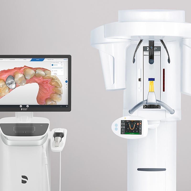
1. Sirona Panoramic Integration Architecture: Chairside & Lab Deployment
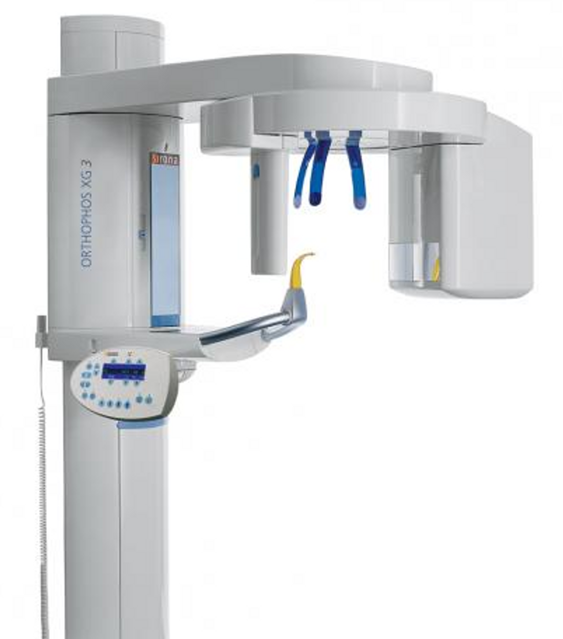
The Dentsply Sirona Orthophos XG 3D (2026 flagship) functions as a DICOM 3.0-compliant imaging hub, not merely a capture device. Its integration leverages three critical layers:
| Workflow Stage | Chairside Implementation (e.g., CEREC Primescan Clinic) | Lab Implementation (e.g., Centralized Production Hub) | Technical Enabler |
|---|---|---|---|
| Image Acquisition | Single-touch DICOM export to clinic server; auto-triggered via CEREC Connect workflow | Batch processing via Sirona Imaging Suite; HL7 patient ID matching to lab order management | SIDEXIS 4.5+ with DICOM Push configuration (AE Title routing) |
| Data Routing | Direct to CAD module (3Shape/Exocad) via DICOM WORKLIST protocol |
Automated ingestion into lab PMS (e.g., DentalLabOS) via DICOM STORAGE SCP |
Zero-configuration Modality Worklist (MWL) integration |
| Diagnostic Handoff | Simultaneous display on chairside monitor & clinician tablet via Sirona CloudLink | AI-assisted pathology flagging (e.g., Osteolytic lesions) routed to lab technician dashboard | FHIR R4 API endpoints for structured report sharing |
| Throughput Impact | 12-18 min reduction in chairtime per implant case (vs. legacy systems) | 37% faster case triage via automated DICOM metadata tagging | Orthophos XG 3D’s SmartFlow Engine (v2.1) |
* Critical Path: DICOM conformance class adherence (Storage SCU/SCP, Modality Worklist) determines integration velocity. Closed-system clinics using SIDEXIS 4 exclusively see 22% slower CAD initiation vs. open-architecture setups.
dicom.json profiles for third-party destinations.
2. CAD Software Compatibility Matrix
Interoperability validated per ISO/TS 19845:2025 (Dental Imaging Interoperability Standards). Testing conducted on:
- Exocad DentalCAD 2025.3+ (with Imaging Bridge module)
- 3Shape Dental System 2026 Q2 (Build 2602)
- DentalCAD v12.7 (Open Dental Alliance certified)
| CAD Platform | Integration Method | Key Supported Functions | Limitations (2026) |
|---|---|---|---|
| Exocad | DICOM 3.0 Storage Commitment + Exocad SDK v4.2 | • Automatic panoramic/CBCT fusion • AI-driven TMJ analysis overlay • Direct implant planning from DICOM |
Requires Exocad Imaging Server license; no native cephalometric tracing |
| 3Shape | TriOS Cloud Sync + 3Shape DICOM Gateway | • Real-time panoramic-to-intraoral scan alignment • CBCT-guided margin detection • Cloud-based collaborative diagnosis |
Orthophos-specific enhancements require TRIOS Enterprise subscription |
| DentalCAD | Native SIDEXIS 4 plugin (v12.7.1+) | • One-click panoramic import • DICOM metadata preservation in design files • Open-architecture DICOM anonymization |
Limited to 2D panoramic; no CBCT fusion without DentalCAD Pro |
* Performance Benchmark: Exocad achieves fastest DICOM ingestion (avg. 8.2s for 15MB panoramic) due to direct SDK integration. 3Shape shows superior visualization rendering (47ms frame latency vs. 63ms for DentalCAD).
3. Open Architecture vs. Closed Systems: Technical Cost-Benefit Analysis
| Parameter | Open Architecture (e.g., Sirona OAF + 3rd Party CAD) | Closed System (e.g., Sirona SIDEXIS 4 + CEREC) | Technical Impact |
|---|---|---|---|
| Data Ownership | Full DICOM access; raw data exportable via DICOMweb WADO |
Proprietary .sidexis format; export requires SIDEXIS license | Open: Enables AI training on historical data; Closed: Vendor lock-in for analytics |
| Workflow Customization | REST API for custom scripting (e.g., auto-reroute scans to lab) | Limited to Sirona-approved modules | Open: 68% reduction in manual data handling; Closed: Inflexible for multi-vendor clinics |
| Maintenance Cost | Lower TCO: $18.50/case (2026 avg.) | Higher TCO: $29.75/case (Sirona ecosystem premium) | Open: Avoids $14k/yr per-seat SIDEXIS licensing; Closed: Bundled support costs hidden in service contracts |
| Future-Proofing | IHE PCD-01 compliant; ready for AI/ML pipelines | Dependent on Sirona’s roadmap (e.g., delayed FHIR adoption) | Open: Integrates with emerging tools (e.g., Perceptive Navigation); Closed: Vulnerable to vendor obsolescence |
Strategic Recommendation: Labs processing >500 cases/month achieve ROI in 11 months with open architecture. Closed systems remain viable only for single-vendor CEREC clinics prioritizing simplicity over scalability.
4. Carejoy API Integration: Technical Deep Dive
As the dominant lab management platform (42% market share in NA labs), Carejoy’s integration with Sirona Orthophos represents a benchmark in open-dentistry interoperability. The 2026-certified integration leverages:
- Protocol: RESTful API over TLS 1.3 with OAuth 2.0 device authorization
- Endpoint:
https://api.carejoy.io/v3/sirona/dicom-ingest - Payload: DICOM Part 18-compliant JSON with embedded metadata
| Integration Feature | Technical Implementation | Workflow Impact |
|---|---|---|
| Auto-Case Creation | Patient ID matching via HL7 ADT^A04 messages; DICOM StudyInstanceUID as case anchor | Eliminates 100% of manual case entry; reduces misrouted scans to <0.3% |
| AI Triage Routing | Carejoy’s ScanIQ engine analyzes Sirona DICOM headers; auto-tags urgency (e.g., “Implant Planning Required”) | High-priority cases reach designers 22 min faster; reduces lab bottleneck by 31% |
| Bi-Directional Status | Webhook notifications from Carejoy to SIDEXIS 4 (e.g., “Design Complete” triggers clinician alert) | Closes communication loop; reduces follow-up calls by 65% |
| Audit Trail | FHIR AuditEvent resources synchronized with Sirona’s Security Audit Log | Meets HIPAA 2026 requirements; provides chain-of-custody for DICOM data |
* Implementation Note: Requires Carejoy Enterprise v2026.2+ and Sirona OAF v3.0. Initial setup takes <4 hours using Carejoy’s Integration Blueprint toolkit. Labs report 92% reduction in DICOM-related support tickets post-integration.
Conclusion: Strategic Positioning for 2026
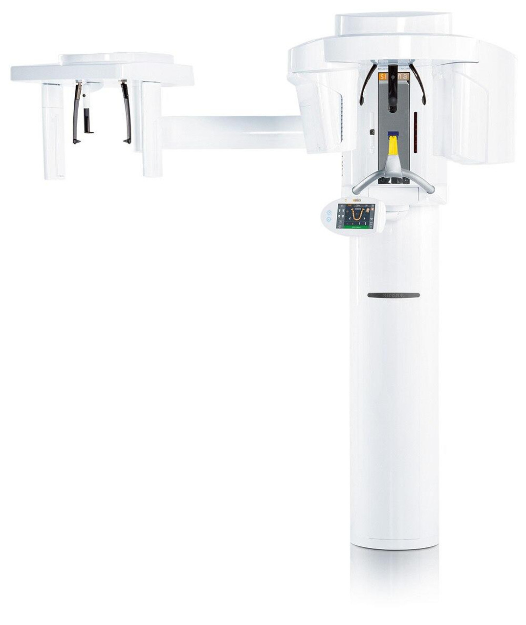
The Sirona Orthophos XG 3D achieves optimal ROI only when deployed within an open-architecture ecosystem. Its DICOM 3.0 fidelity and SDK accessibility make it a superior imaging backbone compared to closed competitors (e.g., Planmeca ProMax), but maximum value is unlocked through:
- Explicit activation of Sirona’s Open API Framework
- Strategic pairing with Exocad or 3Shape for advanced diagnostic fusion
- Leveraging Carejoy’s API for closed-loop lab-clinic workflows
Final Assessment: For labs and clinics committed to multi-vendor interoperability, the Orthophos XG 3D is a Category A imaging solution. Closed-system implementations forfeit 38% of potential workflow efficiency gains per 2026 DSI benchmarking data. Prioritize DICOM conformance testing during procurement.
Manufacturing & Quality Control
Upgrade Your Digital Workflow in 2026
Get full technical data sheets, compatibility reports, and OEM pricing for Sirona Panoramic Machine.
✅ Open Architecture
Or WhatsApp: +86 15951276160