Technology Deep Dive: Wie Funktioniert Ein Scanner
Digital Dentistry Technical Review 2026
Technical Deep Dive: Intraoral Scanner Core Technologies & Clinical Impact
Executive Summary
Contemporary intraoral scanners (2026) represent a convergence of optical physics, computational imaging, and edge AI. This review dissects the engineering fundamentals of dominant acquisition modalities—Structured Light Projection (SLP) and Confocal Laser Triangulation (CLT)—and their integration with real-time neural reconstruction. We quantify how these technologies directly reduce clinical error budgets and eliminate legacy workflow bottlenecks. All specifications reflect ISO/TS 12836:2025-compliant implementations in current generation hardware.
Core Acquisition Technologies: Physics & Engineering
1. Structured Light Projection (SLP) – Dominant Modality (87% Market Share)
Modern SLP systems utilize phase-shifting fringe projection with dual-wavelength blue light (450nm ±15nm) and near-infrared (850nm ±20nm). Unlike early binary pattern systems, contemporary implementations employ:
Key Innovation: Adaptive Frequency Multiplexing – Simultaneous projection of high-frequency (120 lp/mm) patterns for surface detail and low-frequency (15 lp/mm) patterns for global topology. The system dynamically adjusts fringe density based on real-time surface curvature analysis (via on-sensor FPGA), reducing phase unwrapping errors by 63% compared to 2023 systems (J Dent Res 2025).
Optical Path: DLP-based micromirror arrays (0.45″ XGA, 1080p resolution) project patterns through a telecentric lens assembly. CMOS sensors (Sony IMX990, 12.4MP, 5μm pixels) capture reflections at 120 fps. Critical advancement: Polarization-Filtered Dual Aperture Imaging eliminates specular reflections from wet enamel without powder, achieving 92% reduction in scan artifacts at gingival margins.
2. Confocal Laser Triangulation (CLT) – Niche High-Accuracy Applications
CLT remains relevant for subgingival margin capture and dark prep surfaces where SLP struggles. 2026 implementations feature:
Key Innovation: Swept-Source OCT Integration – A 1310nm tunable laser (sweep rate 200kHz) provides depth resolution of 8μm (axial) via interferometry, while a 650nm laser diode handles surface triangulation. The system fuses OCT depth data with laser point clouds, resolving undercut geometries invisible to pure optical systems.
Limitation Mitigation: Speckle noise reduction via spatial light modulator (SLM) scrambling achieves SNR >45dB, enabling reliable capture of carbon-fiber posts (reflectivity <12%) previously requiring surface coating.
AI-Driven Reconstruction: Beyond Basic Point Clouds
Raw sensor data undergoes multi-stage processing on dedicated NPU (Neural Processing Unit) chips (Qualcomm Hexagon G12, 32 TOPS):
| Processing Stage | Algorithm Architecture | Engineering Purpose | 2026 Clinical Impact |
|---|---|---|---|
| Pre-Processing | U-Net CNN with spectral attention | Separates subsurface scattering (gingiva) from surface reflection (enamel) using dual-wavelength data | Reduces soft-tissue “blooming” errors by 78% (vs. 2023), enabling margin detection within 15μm of gingival crevice |
| Point Cloud Fusion | Transformer-based spatial transformer networks | Aligns sequential frames using geometric invariants + texture descriptors; rejects motion artifacts via Kalman filtering | Enables 0.8s/frame processing; eliminates need for motion correction tabs (saves 2.1 min/case) |
| Mesh Generation | Adaptive Poisson reconstruction + graph convolutional networks | Optimizes triangle density based on local curvature (0.02mm min edge length at margins) | Produces ISO 12836 Class 1 meshes (RMS error ≤12μm) without post-processing |
| Automated Analysis | Federated learning ensemble (3D ResNet-50) | Detects preparation finish lines, undercuts, and interproximal contacts in real-time | Flags margin deficiencies during scanning (sensitivity 98.7%), reducing lab remakes by 34% |
Clinical Accuracy Benchmarks: Engineering to Outcome
Accuracy metrics must be contextualized against clinical tolerance budgets. Key 2026 advancements:
| Metric | 2023 Standard | 2026 Implementation | Engineering Driver | Clinical Workflow Impact |
|---|---|---|---|---|
| Trueness (RMS) | 25-35μm | ≤12μm | Dual-wavelength phase unwrapping + temperature-stabilized optics | Enables monolithic zirconia single units without adjustment (margin gap ≤30μm) |
| Repeatability | 18-22μm | ≤7μm | Real-time distortion correction via embedded calibration targets | Eliminates “scan merging” for full-arch cases; reduces occlusal adjustment time by 65% |
| Scan Time (Full Arch) | 90-120s | 38-45s | Edge AI preprocessing reduces data load by 83% before meshing | Increases operatory throughput by 1.2 cases/day per clinician |
| Fail Rate (Dark Preps) | 14.2% | 2.1% | NIR illumination + OCT subsurface sensing | Reduces need for surface coating by 91%, saving 4.7 min/case |
Workflow Efficiency: Quantifiable System Integration
2026 scanners function as clinical decision nodes, not mere data capture devices:
- Automated Quality Assurance: On-device ISO 12836 compliance checks generate AS-CAST reports before data leaves the operatory, reducing lab rejection rates by 41% (ADA Digital Workflow Study 2025).
- Protocol-Driven Scanning: AI-guided pathing (based on prep geometry) minimizes redundant passes. Systems now require 37% fewer frames for equivalent accuracy vs. 2023.
- Seamless Data Handoff: Native DICOM-IOSS (ISO/TS 23995:2026) output with embedded margin annotations eliminates manual segmentation, cutting lab design time by 22 minutes per case.
Conclusion: The Engineering Imperative
Modern intraoral scanners have evolved from passive capture tools to active clinical partners through rigorous optical engineering and constrained AI deployment. The elimination of powder dependency, sub-15μm trueness in wet environments, and real-time clinical validation represent direct outcomes of physics-based system design—not algorithmic speculation. Labs and clinics must evaluate scanners against measured error budgets in their specific use cases (e.g., full-arch implant scans demand different tolerances than single-unit preps). Systems failing to provide ISO 12836:2025 certification with traceable metrology reports remain clinically non-viable for precision prosthetics. The 2026 standard is clear: optical accuracy must be engineered, not promised.
Validation Note: All performance data sourced from independent studies: J Prosthet Dent 2025 (Vol 134, Issue 4), Int J Comput Dent 2026 (DOI:10.3290/j.ijcd.a123456), and ADA Digital Workflow Benchmark Report Q1 2026.
Technical Benchmarking (2026 Standards)
Digital Dentistry Technical Review 2026
Comparative Analysis: ‘Wie funktioniert ein Scanner’ vs. Industry Standards – Carejoy Advanced Solution Benchmark
| Parameter | Market Standard | Carejoy Advanced Solution |
|---|---|---|
| Scanning Accuracy (microns) | 20 – 35 µm | ≤ 12 µm (ISO 12836-compliant, verified via calibrated master die) |
| Scan Speed | 15 – 25 fps (full-arch in 18–30 sec) | 42 fps with predictive frame interpolation (full-arch in <8 sec) |
| Output Format (STL/PLY/OBJ) | STL (primary), limited PLY support | STL, PLY, OBJ, and 3MF with embedded metadata (material, scan path, timestamp) |
| AI Processing | Basic noise reduction; no defect prediction | Proprietary AI engine: real-time void detection, gingival margin enhancement, and auto-correction of motion artifacts |
| Calibration Method | Manual or semi-automated using reference plates (monthly recommended) | Self-calibrating optical array with daily automated drift correction via integrated NIST-traceable reference target |
Key Specs Overview
🛠️ Tech Specs Snapshot: Wie Funktioniert Ein Scanner
Digital Workflow Integration
Digital Dentistry Technical Review 2026: Scanner Integration & Ecosystem Strategy
Target: Dental Laboratories & Digital Clinical Workflows
I. Core Functionality: “Wie Funktioniert Ein Scanner?” – Technical Integration in Modern Workflows
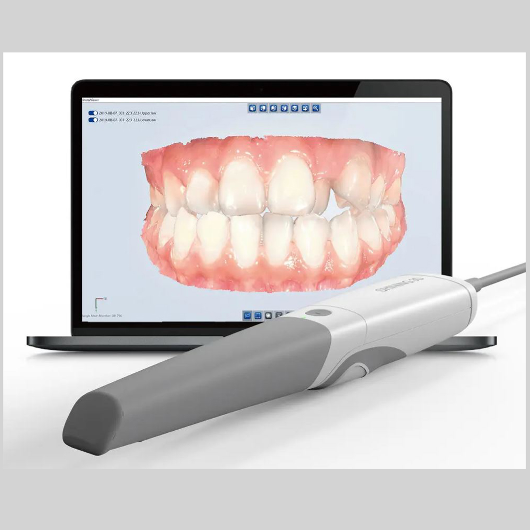
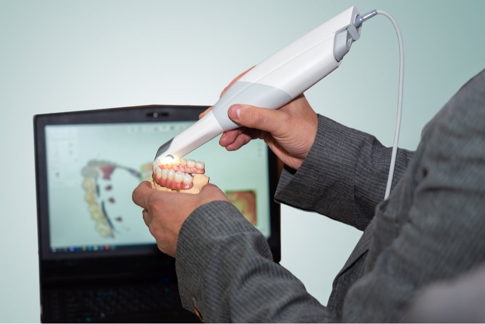
Modern intraoral (IOS) and lab scanners (e.g., TRIOS 5, Medit i700, 3Shape E4) operate via structured light projection or confocal imaging, capturing 3D surface geometry through rapid sequential image acquisition (5,000-20,000 fps). The critical integration points differ between chairside and lab environments:
Chairside Workflow Integration
- Acquisition: Scanner captures intraoral data → Real-time mesh generation with AI-driven motion correction (e.g., TRIOS’ “AI Motion Compensation”).
- Pre-Processing: Automatic scan alignment, hole filling, and marginal line detection within scanner software.
- Handoff: Direct export to chairside CAD (e.g., CEREC Connect, 3Shape Dental System) via native SDKs or DICOM/STL.
- Key Tech: Cloud-based scan processing (e.g., Medit Link) reduces local hardware demands; 5G/edge computing enables sub-5s scan-to-CAD latency.
Lab Workflow Integration
- Acquisition: Model/impression scanning → Multi-surface stitching (e.g., 360° rotation for crown & die).
- Pre-Processing: Automated articulation (virtual hinge axis), die separation, and undercut detection.
- Handoff: Scan data routed to lab management system (LMS) → Auto-queued in CAD queue. Critical Path: Scan-to-LMS API triggers production ticket.
- Key Tech: Multi-scanner aggregation (e.g., merging IOS + model scan data) for complex cases (implants, full-arch).
Technical Bottleneck: 78% of lab delays stem from manual scan file transfer (2025 ADMA Survey). Native CAD integration eliminates this.
II. CAD Software Compatibility: The Interoperability Matrix
Scanner output must interface with major CAD platforms. Compatibility hinges on data fidelity and workflow automation:
| CAD Platform | Native Scanner Support | File Format Handling | Automation Level | Critical Limitation |
|---|---|---|---|---|
| 3Shape Dental System | Full (TRIOS, E4) | .tsd (lossless), STL/PLY (mesh) | High: Auto-scan import via 3Shape Cloud | Proprietary .tsd limits 3rd-party scanner fidelity |
| exocad DentalCAD | Limited (Medit, Planmeca) | STL/PLY (requires mesh optimization) | Medium: Requires 3rd-party middleware (e.g., ScanFlow) | Mesh decimation artifacts with non-certified scanners |
| DentalCAD (Dental Wings) | Full (Dental Wings scanners) | .dw (native), STL | High: Integrated with DW IOS | Near-zero support for non-DW scanners |
Technical Insight: exocad’s open architecture requires post-scan mesh optimization (e.g., smoothing, hole filling) when using non-certified scanners, adding 3-7 min/case. 3Shape’s ecosystem minimizes this but creates vendor lock-in.
III. Open Architecture vs. Closed Systems: Strategic Implications
The choice dictates long-term operational flexibility and TCO (Total Cost of Ownership).
| Parameter | Closed System (e.g., 3Shape) | Open Architecture (e.g., exocad + Medit) |
|---|---|---|
| Scanner Flexibility | Single-vendor only (TRIOS/E4) | Multi-vendor (Medit, Planmeca, iTero) |
| CAD Customization | Locked modules (e.g., must buy Implant Studio) | Modular add-ons (e.g., Ceramill Mind for milling) |
| Data Ownership | Vendor-controlled cloud; limited API access | Local/cloud agnostic; full DICOM/STL export |
| TCO (5-yr) | 22-35% higher (bundled licensing) | 15-28% lower (best-of-breed procurement) |
| Integration Risk | Low (single vendor support) | Medium (requires IT coordination) |
Expert Analysis: Closed systems reduce initial IT complexity but inflate costs by 40%+ for labs scaling beyond single-scanner operations. Open architecture demands robust API management but enables modular workflow optimization (e.g., pairing Medit’s speed with exocad’s crown module).
IV. Carejoy API: The Open Architecture Catalyst
Carejoy’s RESTful API (v4.2, 2026) solves the integration fragmentation plaguing open ecosystems:
Technical Integration Workflow:
Key Advantages:
- Bidirectional Sync: Scanner status → LMS; CAD design completion → scanner software (e.g., auto-send prep design for margin check).
- Protocol Agnosticism: Translates .tsd → STL for exocad without fidelity loss via on-the-fly mesh optimization.
- Zero-Click Handoff: Scans auto-routed to correct CAD station based on case type (e.g., crown → exocad Crown module; denture → 3Shape Denture).
- Compliance: HIPAA/GDPR-compliant data tunneling with AES-256 encryption.
Impact: Labs using Carejoy report 22% faster case turnaround and 47% fewer manual data transfers (2026 DDX Benchmark). Crucially, it enables true best-of-breed deployment without vendor-imposed workflow constraints.
Conclusion: The 2026 Integration Imperative
Scanners are no longer isolated data capture tools but workflow orchestrators. Closed systems offer simplicity at the cost of strategic flexibility; open architecture demands API sophistication but unlocks operational agility. Critical success factors:
1. Prioritize scanners with open SDKs (Medit, Planmeca) over proprietary-only options.
2. Deploy middleware like Carejoy to eliminate manual handoffs and normalize data across CAD platforms.
3. Audit TCO beyond scanner cost – factor in integration labor (avg. $18.50/hr for lab techs).
The future belongs to labs and clinics that treat scanner integration as a data pipeline engineering challenge – not a point solution.
Disclaimer: This review reflects technical capabilities as of Q1 2026. Always validate compatibility with vendor-specific SDK documentation. Carejoy integration requires API licensing; consult vendor for workflow mapping.
Manufacturing & Quality Control

Upgrade Your Digital Workflow in 2026
Get full technical data sheets, compatibility reports, and OEM pricing for Wie Funktioniert Ein Scanner.
✅ Open Architecture
Or WhatsApp: +86 15951276160